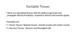
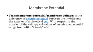
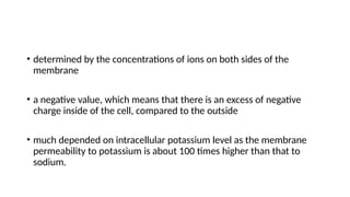
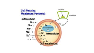
PHS 213- Human Physiology I
L.O. Adegbite
MEMBRANE POTENTIALS AND EXCITABLE TISSUES
Excitable Tissues
• These are specialized tissues with the ability to generate and
propagate electrical impulses, respond to stimuli and transmit signals.
• Examples are:
• 1. Musce Tissues: Skeletal muscle, smooth muscle and cardiac muscle
• 2. Nervous Tissues: Neurons and Neuroglial cells
29-4
Classification of neurons
• The functional classification of neurons is based on the direction in
which they conduct impulses
• Afferent or sensory neurons
• Sensory information (impulse) from environment or inside body
to CNS for interpretation
• Efferent or motor neurons
• Impulses from CNS to PNS to allow for movement or action
• Interneruons
• Interpretive neurons between afferent and efferent nerves in
the CNS
PNS = afferent neurons (their activity
“affects” what will happen
next) into the CNS
+
efferent neurons (“effecting” change:
movement, secretion, etc.)
projecting out of the CNS.
CNS PNS
CNS = brain
+
spinal cord;
all parts of
interneurons
are in the CNS.
Supporting Cells
• Aid the functions of neurons
• About five times more abundant than neurons
• In the CNS, supporting cells are collectively called neuroglia, or simply
glial cells
• There are two types of supporting cells in the PNS:
1. Schwann cells which form myelin sheaths around peripheral axons
2. Satellite cells which support neuron cell bodies within the ganglia of the PNS.
• There are four types of supporting cells in the CNS:
1. Oligodendrocytes which form myelin sheaths around axons of the CNS
2. Microglia which migrate through the CNS and phagocytose foreign and degenerated material
3. Astrocytes which help to regulate the external environment of neurons in the CNS.
4. Ependymal cells, which line the ventricles (cavities) of the brain and the central canal of the
spinal cord.
Membrane Potential
• Transmembrane potential/membrane voltage) is the
difference in electric potential between the interior and
the exterior of a biological cell. With respect to the
exterior of the cell, typical values of membrane potential
range from –40 mV to –80 mV.
Differences in the concentrations of ions on opposite sides of a cellular
membrane lead to a voltage called the membrane potential.
Resting Membrane potential
• Resting membrane potential is:
• the unequal distribution of ions on the both sides of the cell
membrane
• the voltage difference of resting cells
• the membrane potential that would be maintained if there weren’t
any stimuli or conducting impulses across it
• determined by the concentrations of ions on both sides of the
membrane
• a negative value, which means that there is an excess of negative
charge inside of the cell, compared to the outside
• much depended on intracellular potassium level as the membrane
permeability to potassium is about 100 times higher than that to
sodium.
Resting Membrane potential
• Resting membrane potential varies according to types of cells
For example:
• Skeletal muscle cells: −95 mV
• Smooth muscle cells: −50 mV
• Astrocytes: −80/−90 mV
• Neurons: −70 mV
• Erythrocytes: −12 mV
29-17
• Neuron cell membrane at rest is in a polarized state
• Inside of cell membrane is negative
• Outside of cell membrane is positive due to more Na+
and K+
• As Na+
and K+
move into the cell, the membrane becomes
depolarized
• Inside becomes more positive
• Action potential (nerve impulse) is created
• Repolarization occurs when K+
and later Na+
move to the outside of
the cell membrane
• Return of the cell to polarized (resting) state
ACTION POTENTIAL
• Action potential is a short-lasting event in which the
electrical membrane potential of a cell rapidly rises and
falls, following a consistent trajectory.
• Action potentials occur in several types of animal cells,
called excitable cells, which include neurons and muscle
cells
BIOPHYSICAL BASIS OF ACTION POTENTIAL
Action potentials occur due to the presence special types of
voltage-gated ion channels in a cell membrane
A voltage-gated ion channel is a cluster of proteins embedded
in the membrane that has three key properties:
• It is capable of assuming more than one conformation.
• At least one of the conformations creates a channel through
the membrane that is permeable to specific types of ions.
• The transition between conformations is influenced by the
membrane potential.
A plot of an Action Potential
"All-or-none" principle
• The amplitude of an action potential is independent of the
amount of current that produced it. In other words, larger
currents do not create larger action potentials.
• Action potential obeys all-or-none law, since either they
occur fully or they do not occur at all.
• This is in contrast to receptor potentials whose
amplitudes are dependent on the intensity of a stimulus.
Action Potential explanation
• As the membrane potential is increased,
sodium ion channels open, allowing the entry
of sodium ions into the cell.
• This is followed by the opening of potassium
ion channels that permit the exit of potassium
ions from the cell.
• The inward flow of sodium ions increases the
concentration of positively charged cations in
the cell and causes depolarization, where the
potential of the cell is higher than the cell's
resting potential.
Cont’d
• The sodium channels close at the peak of the action
potential, while potassium continues to leave the cell.
• The efflux of potassium ions decreases the membrane
potential or hyperpolarizes the cell.
• For small voltage increases from rest, the potassium
current exceeds the sodium current and the voltage
returns to its normal resting value, typically −70 mV.
Cont’d
• However, if the voltage increases past a
critical threshold, typically 15 mV higher than
the resting value, the sodium current
dominates.
• This results in a runaway condition whereby
the positive feedback from the sodium current
activates even more sodium channels. Thus,
the cell fires, producing an action potential.
• The frequency at which cellular action
potentials are produced is known as its firing
rate.
An action potential
is an “all-or-none”
sequence of changes
in membrane potential.
Action potentials result
from an all-or-none
sequence of changes
in ion permeability
due to the operation
of voltage-gated
Na+
and K +
channels.
The rapid opening of
voltage-gated Na+
channels
allows rapid entry of Na+
,
moving membrane potential
closer to the sodium
equilibrium potential (+60 mv)
The slower opening of
voltage-gated K+
channels
allows K+
exit,
moving membrane potential
closer to the potassium
equilibrium potential (-90 mv)
The rapid opening of voltage-gated Na+
channels
explains the rapid-depolarization phase at the
beginning of the action potential.
The slower opening of voltage-gated K+
channels
explains the repolarization and after hyperpolarization
phases that complete the action potential.

Membrane Potentials and Excitable Tissues.pptx

  • 1.
    PHS 213- HumanPhysiology I L.O. Adegbite
  • 2.
    MEMBRANE POTENTIALS ANDEXCITABLE TISSUES
  • 3.
    Excitable Tissues • Theseare specialized tissues with the ability to generate and propagate electrical impulses, respond to stimuli and transmit signals. • Examples are: • 1. Musce Tissues: Skeletal muscle, smooth muscle and cardiac muscle • 2. Nervous Tissues: Neurons and Neuroglial cells
  • 4.
    29-4 Classification of neurons •The functional classification of neurons is based on the direction in which they conduct impulses • Afferent or sensory neurons • Sensory information (impulse) from environment or inside body to CNS for interpretation • Efferent or motor neurons • Impulses from CNS to PNS to allow for movement or action • Interneruons • Interpretive neurons between afferent and efferent nerves in the CNS
  • 5.
    PNS = afferentneurons (their activity “affects” what will happen next) into the CNS + efferent neurons (“effecting” change: movement, secretion, etc.) projecting out of the CNS. CNS PNS CNS = brain + spinal cord; all parts of interneurons are in the CNS.
  • 7.
    Supporting Cells • Aidthe functions of neurons • About five times more abundant than neurons • In the CNS, supporting cells are collectively called neuroglia, or simply glial cells
  • 8.
    • There aretwo types of supporting cells in the PNS: 1. Schwann cells which form myelin sheaths around peripheral axons 2. Satellite cells which support neuron cell bodies within the ganglia of the PNS. • There are four types of supporting cells in the CNS: 1. Oligodendrocytes which form myelin sheaths around axons of the CNS 2. Microglia which migrate through the CNS and phagocytose foreign and degenerated material 3. Astrocytes which help to regulate the external environment of neurons in the CNS. 4. Ependymal cells, which line the ventricles (cavities) of the brain and the central canal of the spinal cord.
  • 10.
    Membrane Potential • Transmembranepotential/membrane voltage) is the difference in electric potential between the interior and the exterior of a biological cell. With respect to the exterior of the cell, typical values of membrane potential range from –40 mV to –80 mV.
  • 11.
    Differences in theconcentrations of ions on opposite sides of a cellular membrane lead to a voltage called the membrane potential.
  • 12.
    Resting Membrane potential •Resting membrane potential is: • the unequal distribution of ions on the both sides of the cell membrane • the voltage difference of resting cells • the membrane potential that would be maintained if there weren’t any stimuli or conducting impulses across it
  • 13.
    • determined bythe concentrations of ions on both sides of the membrane • a negative value, which means that there is an excess of negative charge inside of the cell, compared to the outside • much depended on intracellular potassium level as the membrane permeability to potassium is about 100 times higher than that to sodium.
  • 16.
    Resting Membrane potential •Resting membrane potential varies according to types of cells For example: • Skeletal muscle cells: −95 mV • Smooth muscle cells: −50 mV • Astrocytes: −80/−90 mV • Neurons: −70 mV • Erythrocytes: −12 mV
  • 17.
    29-17 • Neuron cellmembrane at rest is in a polarized state • Inside of cell membrane is negative • Outside of cell membrane is positive due to more Na+ and K+ • As Na+ and K+ move into the cell, the membrane becomes depolarized • Inside becomes more positive • Action potential (nerve impulse) is created • Repolarization occurs when K+ and later Na+ move to the outside of the cell membrane • Return of the cell to polarized (resting) state
  • 18.
    ACTION POTENTIAL • Actionpotential is a short-lasting event in which the electrical membrane potential of a cell rapidly rises and falls, following a consistent trajectory. • Action potentials occur in several types of animal cells, called excitable cells, which include neurons and muscle cells
  • 19.
    BIOPHYSICAL BASIS OFACTION POTENTIAL Action potentials occur due to the presence special types of voltage-gated ion channels in a cell membrane A voltage-gated ion channel is a cluster of proteins embedded in the membrane that has three key properties: • It is capable of assuming more than one conformation. • At least one of the conformations creates a channel through the membrane that is permeable to specific types of ions. • The transition between conformations is influenced by the membrane potential.
  • 20.
    A plot ofan Action Potential
  • 21.
    "All-or-none" principle • Theamplitude of an action potential is independent of the amount of current that produced it. In other words, larger currents do not create larger action potentials. • Action potential obeys all-or-none law, since either they occur fully or they do not occur at all. • This is in contrast to receptor potentials whose amplitudes are dependent on the intensity of a stimulus.
  • 22.
    Action Potential explanation •As the membrane potential is increased, sodium ion channels open, allowing the entry of sodium ions into the cell. • This is followed by the opening of potassium ion channels that permit the exit of potassium ions from the cell. • The inward flow of sodium ions increases the concentration of positively charged cations in the cell and causes depolarization, where the potential of the cell is higher than the cell's resting potential.
  • 23.
    Cont’d • The sodiumchannels close at the peak of the action potential, while potassium continues to leave the cell. • The efflux of potassium ions decreases the membrane potential or hyperpolarizes the cell. • For small voltage increases from rest, the potassium current exceeds the sodium current and the voltage returns to its normal resting value, typically −70 mV.
  • 24.
    Cont’d • However, ifthe voltage increases past a critical threshold, typically 15 mV higher than the resting value, the sodium current dominates. • This results in a runaway condition whereby the positive feedback from the sodium current activates even more sodium channels. Thus, the cell fires, producing an action potential. • The frequency at which cellular action potentials are produced is known as its firing rate.
  • 25.
    An action potential isan “all-or-none” sequence of changes in membrane potential. Action potentials result from an all-or-none sequence of changes in ion permeability due to the operation of voltage-gated Na+ and K + channels. The rapid opening of voltage-gated Na+ channels allows rapid entry of Na+ , moving membrane potential closer to the sodium equilibrium potential (+60 mv) The slower opening of voltage-gated K+ channels allows K+ exit, moving membrane potential closer to the potassium equilibrium potential (-90 mv)
  • 26.
    The rapid openingof voltage-gated Na+ channels explains the rapid-depolarization phase at the beginning of the action potential. The slower opening of voltage-gated K+ channels explains the repolarization and after hyperpolarization phases that complete the action potential.