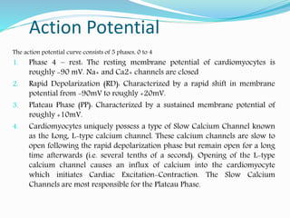
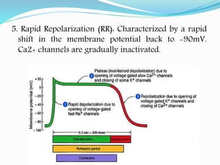
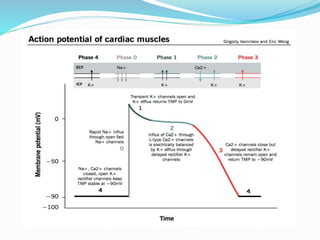
Dr. Haji Bahadar
Assistant professor IPMS-KMU
BASIC TERMS
Electrophysiology: Electrophysiology is the science and branch of
physiology that pertains to the flow of ions (ion current) in
biological tissues.
Cardiac cells types
1. Electrical cells
a) Make up the conduction system of the heart
b) Are distributed in an orderly fashion through the heart
Specific properties
(1) automaticity – the ability to spontaneously generate and
discharge an electrical impulse
(2) excitability – the ability of the cell to respond to an electrical
impulse
(3) conductivity – the ability to transmit an electrical impulse from
one cell to the next
2. Myocardial cells
a) Make up the muscular walls of the atrium and
ventricles of the heart
b) Possess specific properties
(1) contractility – the ability of the cell to shorten and
lengthen its fibers
(2) extensibility – the ability of the cell to stretch
 Cardiac cells at rest are considered polarized, meaning
no electrical activity takes place.
 Depolarization: Once an electrical cell generates an
electrical impulse, this electrical impulse causes the
ions to cross the cell membrane and causes the action
potential, also called depolarization.
Fast, Voltage-gated Sodium Channel: Opening of fast
Sodium Channels is responsible for the initial, rapid
depolarization of the cardiomyocyte. These sodium
channels allow for a rapid influx of positive, Na+ ions
into the cells which depolarize the membrane potential
with incredibly quick kinetics. However, these channels
also quickly close and thus eliminate the Na+ influx
soon after maximum depolarization of +20mV is
achieved.
Repolarization is the return of the ions to their previous
resting state, which corresponds with relaxation of the
myocardial muscle.
Action Potential
The action potential curve consists of 5 phases, 0 to 4
1. Phase 4 – rest: The resting membrane potential of cardiomyocytes is
roughly -90 mV. Na+ and Ca2+ channels are closed
2. Rapid Depolarization (RD): Characterized by a rapid shift in membrane
potential from -90mV to roughly +20mV.
3. Plateau Phase (PP): Characterized by a sustained membrane potential of
roughly +10mV.
4. Cardiomyocytes uniquely possess a type of Slow Calcium Channel known
as the Long, L-type calcium channel. These calcium channels are slow to
open following the rapid depolarization phase but remain open for a long
time afterwards (i.e. several tenths of a second). Opening of the L-type
calcium channel causes an influx of calcium into the cardiomyocyte
which initiates Cardiac Excitation-Contraction. The Slow Calcium
Channels are most responsible for the Plateau Phase.
5. Rapid Repolarization (RR): Characterized by a rapid
shift in the membrane potential back to -90mV.
Ca2+ channels are gradually inactivated.
Action potential in cardiac cell

Action potential in cardiac cell

  • 1.
    Dr. Haji Bahadar Assistantprofessor IPMS-KMU
  • 2.
    BASIC TERMS Electrophysiology: Electrophysiologyis the science and branch of physiology that pertains to the flow of ions (ion current) in biological tissues. Cardiac cells types 1. Electrical cells a) Make up the conduction system of the heart b) Are distributed in an orderly fashion through the heart Specific properties (1) automaticity – the ability to spontaneously generate and discharge an electrical impulse (2) excitability – the ability of the cell to respond to an electrical impulse (3) conductivity – the ability to transmit an electrical impulse from one cell to the next
  • 3.
    2. Myocardial cells a)Make up the muscular walls of the atrium and ventricles of the heart b) Possess specific properties (1) contractility – the ability of the cell to shorten and lengthen its fibers (2) extensibility – the ability of the cell to stretch
  • 4.
     Cardiac cellsat rest are considered polarized, meaning no electrical activity takes place.  Depolarization: Once an electrical cell generates an electrical impulse, this electrical impulse causes the ions to cross the cell membrane and causes the action potential, also called depolarization. Fast, Voltage-gated Sodium Channel: Opening of fast Sodium Channels is responsible for the initial, rapid depolarization of the cardiomyocyte. These sodium channels allow for a rapid influx of positive, Na+ ions into the cells which depolarize the membrane potential with incredibly quick kinetics. However, these channels also quickly close and thus eliminate the Na+ influx soon after maximum depolarization of +20mV is achieved.
  • 5.
    Repolarization is thereturn of the ions to their previous resting state, which corresponds with relaxation of the myocardial muscle.
  • 6.
    Action Potential The actionpotential curve consists of 5 phases, 0 to 4 1. Phase 4 – rest: The resting membrane potential of cardiomyocytes is roughly -90 mV. Na+ and Ca2+ channels are closed 2. Rapid Depolarization (RD): Characterized by a rapid shift in membrane potential from -90mV to roughly +20mV. 3. Plateau Phase (PP): Characterized by a sustained membrane potential of roughly +10mV. 4. Cardiomyocytes uniquely possess a type of Slow Calcium Channel known as the Long, L-type calcium channel. These calcium channels are slow to open following the rapid depolarization phase but remain open for a long time afterwards (i.e. several tenths of a second). Opening of the L-type calcium channel causes an influx of calcium into the cardiomyocyte which initiates Cardiac Excitation-Contraction. The Slow Calcium Channels are most responsible for the Plateau Phase.
  • 7.
    5. Rapid Repolarization(RR): Characterized by a rapid shift in the membrane potential back to -90mV. Ca2+ channels are gradually inactivated.