
Recommended
More Related Content
What's hot
What's hot (20)
Similar to Different types of animal Tissues DMLT .pptx
Similar to Different types of animal Tissues DMLT .pptx (20)
More from PunamSahoo3
More from PunamSahoo3 (6)
Recently uploaded
Recently uploaded (20)
Different types of animal Tissues DMLT .pptx
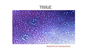
- 1. TISSUE PRESENTED BY PUNAM SAHOO
- 2. What is tissue ?? • It is the group of cells that forms a structure and performs particular functions. • It can be classified according to its shape, size and structure of its cells. There are four types of the tissues. • They are epithelial tissue, connective tissue, muscular tissue and nervous tissue. • They form a cellular organizational level, intermediate between the cells and organ system. Organs are then created by combining the functional groups of tissues. • The study of tissue is known as histology and study of disease-related to tissue is known as histopathology.
- 3. TYPES OF TISSUE Animal tissues are grouped into four types: • Connective Tissue • Muscle Tissue • Nervous Tissue • Epithelial Tissue
- 4. Epithelial Tissue • These are the tissues which are seen in the lining of cavities, hollow organs, and tubular structures and in glands. • The function of epithelial tissue is protection, absorption and secretion. • The types of epithelial tissue include simple epithelium and stratified epithelium. Simple epithelium • It consists of single layer of identical cells. It is found on absorptive and secretory surfaces. • It consists of squamous epithelium, cuboidal epithelium, columnar epithelium and ciliated columnar epithelium.
- 5. Continue… Squamous epithelium: It consists of a layer of flattened cells. Its cells are arranged on the basement membrane. It is found in the inner lining of heart, blood vessels, lymph vessels and lung alveoli. Cuboidal epithelium: It consists of a layer of cube like cells. The cells are arranged on the basement membrane. It forms glands and kidney tubules.
- 6. Continue…. Columnar epithelium: It consists of a layer of column like cells. The cells are arranged on basement membrane. They are seen in the alimentary tract. The specialized columnar cells are goblet cells that secrete mucus. Ciliated columnar epithelium: It consists of microscopic hair like structures called cilia on the free border of columnar cells. It consists of single layer of columnar cells. They are arranged on basement membrane. They are seen in the uterine tubes and airways.
- 7. Stratified Epithelium • The stratified epithelium is formed of different types of cells. They are arranged in many layers. It protects the underlying structure from mechanical wear and tear. • The cells of superficial layer are flat. Superficial layer is formed from the layer below. It has no basement membrane. The deeper layer is columnar. • Superficial layer is continuously been shed off. • Stratified epithelium is of two types. They are stratified squamous epithelium and transitional epithelium.
- 8. Stratified Squamous Epithelium They are of two different types like keratinized epithelium and non- keratinized It epithelium. Keratinized epithelium: They are seen on dry surfaces, e.g., skin, nails, hair. Its superficial layer consists of dead cells and these dead cells contain a protein called keratin. It makes it a hard water proof outer layer. It protects the underlying structures from drying. Non-keratinized epithelium: These are found in wet surfaces, e.g., conjunctiva of eyes, mouth, pharynx, esophagus, vagina.
- 9. Transitional Epithelium Transitional Epithelium It consists of many layers of cells that are pear shaped. They are found in the wall of urinary bladder. It can stretch when the bladder is filled with urine.
- 10. Connective Tissue It is the most widely distributed tissue in our body. It has many functions such as it supports and strengthens the other tissues; acts as a transport medium; covers the internal organ and protects them; functions as energy reserves and it also helps in providing immunity. Various types of connective tissues include loose connective tissue, dense connective tissue, adipose tissue, fibrous tissue, elastic tissue, lymphoid tissue, blood, cartilage and bone.
- 11. Elements of Connective Tissue It has two main elements. They are connective tissue cells and extra cellular matrix. Connective tissue cells Connective tissue cells are embedded in extra cellular matrix. They include fibroblasts, macrophages, plasma cells, mast cells, adipocytes and WBC. Fibroblasts: It is the large, flat structure with branching processes. They secrete fibers of and ground substance for the extra cellular matrix. o A fibroblast is a type of cell that contributes to the formation of connective tissue, a fibrous cellular material that supports and connects other tissues or organs in the body.
- 12. Continue… Macrophages: These are small irregular shaped structure with many small branching. It destroys bacteria and cell debris by the process of phagocytosis. Plasma Cells: They are developed from B-lymphocytes. They produce antibodies against antigens. They help in providing immunity. Adipocytes: They are fat cells or adipose cell that store fats. They are seen covering the internal organs. White Blood Cells: They are present in small numbers in all healthy connective tissues. During infection or injury WBC migrate to the site of infection or injury. Mast Cells: They produce histamine, Histamine is released in response to allergic reaction
- 14. Continue.. Extra Cellular Matrix -It is a part of connective tissue in which connective tissue cells are embedded. Extra cellular matrix consists of fibers and ground substance. Fibers: They are present in the extra cellular matrix between the connective tissue cells. They are secreted by fibroblasts. It support and strengthen the connective tissue. The fibers include collagen fibers, elastic fibers and reticular fibers Ground substance: It is present in the extra cellular matrix between connective tissue cells and fibers. It is made of water, polysaccharide, fat and protein. It may be liquid, semisolid or calcified. They support the connective tissue and bind the cells together. It strengthens the connective tissue. It is a medium for exchange of substances. It is a medium for multiplication, growth and migration of connective tissue cells.
- 15. Muscular Tissue It has the ability to contract and relax and it help in the movement within the body and of the body itself. It has blood supply to it and the blood provide oxygen, calcium and nutrients to the muscle tissue. There are three types of specialized muscle cells. They are: skeletal muscle, smooth muscle and cardiac muscle. Skeletal Muscle-These are attached to bones. They help in the movement of our body. They are voluntary and their activity can be controlled by us. They are cylindrical and have multiple nuclei. It has many striations and is striated muscles. They are stimulated by motor nerves impulses
- 17. Continue.. Smooth muscle -These muscles are seen mainly on the walls of the internal organ. They are involuntary. They have no striations and they are non- striated muscle. Each cell of the smooth muscle fibre contains a nucleus. They are stimulated by autonomic nervous system. Cardiac Muscle -These are muscle fibers that are only seen in heart. It has many branching fibers and the fibers are interconnected. Each cell has a nucleus. The junctions between the cells are called intercalated discs. They are involuntary muscle fibers. It has an intrinsic system that controls the contraction and relaxation of heart.
- 18. Nervous Tissue These are the tissues that makes up the nervous system. It is includes excitable cells and non- excitable cells. Excitable cells: It includes neurons. They initiate, transmit, conduct and receive nerve impulses. Non-excitable cells: It includes glial cells. They support the cells of the nervous system.