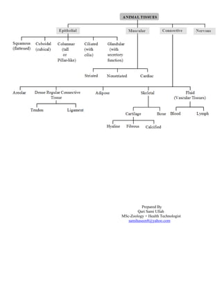
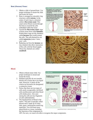
This document provides an introduction to the four basic types of tissues in the human body: epithelium, connective, muscular, and nervous tissues. It then focuses on epithelium tissues, describing their main characteristics and classifications. The classifications are based on cell shape and arrangement, with examples including simple squamous, simple cuboidal, simple columnar, pseudostratified columnar, stratified squamous, and transitional epithelium. Specific epithelium tissues are then described, along with instructions to observe slides of lung, kidney, intestine, trachea, skin, and bladder tissue under a microscope.
NCERT Solutions | Class IX | Science (Biology) | Chapter 6 | TissuesBiswarup Majumder
NCERT Solutions for Class 9 Biology is available in PDF format which you can download easily. Here is the most accurate and detailed Biology NCERT solutions for Class 9th CBSE textbook for free of cost.
I hope this document is helpful to you. Please share the document with your friends if you think this will benefit them. Get ready for the next solution. Thanks.
A tissue may be defined as an aggregate or collection of same type of cells performing the same general functions of the body. For example: Blood, bone, muscle etc.
this ppt is all about tissues it contains full information and pictures about plant tissues and animal tissues
hope the people who download it will not get any time wasted cause it is fully containing info n pics but u guys have to do the animataions......
:):):):):):):):):):):):):):):):):):):):):):):):):):):):):):):):):):):):):):):):):):):):):):):):):):):):):):):):):):):):):):):):):):)):):):):):):):):)::):):):):):):)):):):)
What structural characteristics did you observe for each type of tis.pdfnaveenkumar29100
What is the probability of having a boy, then a girl, then a boy, and then two girls in that birth
order?
Solution
Probability of boy P(b) = 1/2
and probability of girl is also P(g) = 1/2
as they are the only 2 outcome possible and are equally likely
So probability of having a boy, then a girl, then a boy, and then two girls =
P(b)*P(g)*P(b)*P(g)*P(g)
= 1/2 * 1/2 * 1/2 * 1/2 *1/2 = 1/32 =0.03125.
Tissues definition and classification, function & location of of epithelium tissues, connective tissues, muscular tissues, nervous tissue etc.. it is usefull for the diploma in pharmacy students , bachelor of pharmacy students and doctor of pharmacy students
of layers- The or an epithelial tissue includes two descriptive terms-.pdfacrylicBangles
of layers. The or an epithelial tissue includes two descriptive terms: the epithelial tissue mossible
shapes are squamos, cuboidal, and columt means that cell lay be simple or stratified. Simple
means that there is meaning that cell layers are placed on top of each other. Some epithelial t or
hairlike that they only appear to be layered. Epithelium may also have joins an epithelium to
underlying connective tissue. Observation: Simple and Stratified Squamous Epithelium Simple
Squamous Epithellum Simple squamous epithelium is a single layer of thin, flat, many-sided
cells, each with a central nucleus. It lines internal cavities, the heart, and all the blood vessels. It
also lines parts of the urinary, respiratory, and male reproductive tracts. 1. Study a model or
diagram of simple squamous epithelium. What does squamous mean? 2. Examine a prepared
slide of squamous epithelium. Under low power, note the close packing of the flat cells. What
shapes are the cells? 3. Under high power, examine an individual cell, and identify the plasma
membrane, cytoplasm, and nucleus. 4. Add a sketch of this tissue to Table 6.1. Stratiffed
Squamous Epithelium As would be expected from its name, stratified squamous epithelium
consists of many layers of cells. The innermost. layer produces cells that are first cuboidal or
columnar in shape, but as the cells push toward the surface, they become flattened. The outer
region of the skin, called the epidermis, is stratified squamous epithelium. As the cells move
toward the surface, th protein called keratin, and eventually die. Keratin makes the outer layet
able to repel water. 1. Either now or when you are studying skin in section 6.2, examine the slide
that is stratified squamous epithelium. 2. Approximately how many layers of cells make up this
portion of st 3. Which layers of cells best represent squamous epithelium? 4. Add a sketch of this
tissue to Table 6.1. The linings of the mouth, throat, anal canal, and vagina are stratifi cells
surrounding the cavity is simple squamous epithelium. In these org moist, and alive. 58
Laboratory 6 Body Tissues Observation: Simple Cuboidol Epithellum Simple cuboidal
epithelium is a single layer of cube-shaped cells. each with a central nucleus. It is found in
tubules of the kidney and in the ducts of many glands, where it has a protective function. It also
occurs in the secretory portions of some glands - that is, where the tissue produces and releases
secretions. 1. Study a model or diagram of simple cuboidal epithelium. 2. Examine a prepared
slide of simple cuboidal epithelium. Move the slide until you locate cube-shaped cells that line a
lumen (cavity). Are these cells ciliated? 3. Add a sketch of this tissue to Table 6.1. Observation:
Simple Columnar Epithellum mple columnar epithelium is a single layer of tall, cylindrical lls,
each with a nucleus near the base. This tissue, which lines digestive tract from the stomach to the
anus, protects, cretes, and allows absorption of .
A tissue is a group of connected cells that have a similar function within an organism. There are four basic types of tissue in the body of all animals, including the human body. These make up all the organs, structures and other contents of the body.
Beautiful Slide Presentation on Different orders of Entomology with the help of Pictures. Download it and see the magic, it is highly dynamic presentation. you will must like it.
Introduction to AI for Nonprofits with Tapp NetworkTechSoup
Dive into the world of AI! Experts Jon Hill and Tareq Monaur will guide you through AI's role in enhancing nonprofit websites and basic marketing strategies, making it easy to understand and apply.
How to Make a Field invisible in Odoo 17Celine George
It is possible to hide or invisible some fields in odoo. Commonly using “invisible” attribute in the field definition to invisible the fields. This slide will show how to make a field invisible in odoo 17.
Operation “Blue Star” is the only event in the history of Independent India where the state went into war with its own people. Even after about 40 years it is not clear if it was culmination of states anger over people of the region, a political game of power or start of dictatorial chapter in the democratic setup.
The people of Punjab felt alienated from main stream due to denial of their just demands during a long democratic struggle since independence. As it happen all over the word, it led to militant struggle with great loss of lives of military, police and civilian personnel. Killing of Indira Gandhi and massacre of innocent Sikhs in Delhi and other India cities was also associated with this movement.
2024.06.01 Introducing a competency framework for languag learning materials ...Sandy Millin
http://sandymillin.wordpress.com/iateflwebinar2024
Published classroom materials form the basis of syllabuses, drive teacher professional development, and have a potentially huge influence on learners, teachers and education systems. All teachers also create their own materials, whether a few sentences on a blackboard, a highly-structured fully-realised online course, or anything in between. Despite this, the knowledge and skills needed to create effective language learning materials are rarely part of teacher training, and are mostly learnt by trial and error.
Knowledge and skills frameworks, generally called competency frameworks, for ELT teachers, trainers and managers have existed for a few years now. However, until I created one for my MA dissertation, there wasn’t one drawing together what we need to know and do to be able to effectively produce language learning materials.
This webinar will introduce you to my framework, highlighting the key competencies I identified from my research. It will also show how anybody involved in language teaching (any language, not just English!), teacher training, managing schools or developing language learning materials can benefit from using the framework.
A Strategic Approach: GenAI in EducationPeter Windle
Artificial Intelligence (AI) technologies such as Generative AI, Image Generators and Large Language Models have had a dramatic impact on teaching, learning and assessment over the past 18 months. The most immediate threat AI posed was to Academic Integrity with Higher Education Institutes (HEIs) focusing their efforts on combating the use of GenAI in assessment. Guidelines were developed for staff and students, policies put in place too. Innovative educators have forged paths in the use of Generative AI for teaching, learning and assessments leading to pockets of transformation springing up across HEIs, often with little or no top-down guidance, support or direction.
This Gasta posits a strategic approach to integrating AI into HEIs to prepare staff, students and the curriculum for an evolving world and workplace. We will highlight the advantages of working with these technologies beyond the realm of teaching, learning and assessment by considering prompt engineering skills, industry impact, curriculum changes, and the need for staff upskilling. In contrast, not engaging strategically with Generative AI poses risks, including falling behind peers, missed opportunities and failing to ensure our graduates remain employable. The rapid evolution of AI technologies necessitates a proactive and strategic approach if we are to remain relevant.
The Roman Empire A Historical Colossus.pdfkaushalkr1407
The Roman Empire, a vast and enduring power, stands as one of history's most remarkable civilizations, leaving an indelible imprint on the world. It emerged from the Roman Republic, transitioning into an imperial powerhouse under the leadership of Augustus Caesar in 27 BCE. This transformation marked the beginning of an era defined by unprecedented territorial expansion, architectural marvels, and profound cultural influence.
The empire's roots lie in the city of Rome, founded, according to legend, by Romulus in 753 BCE. Over centuries, Rome evolved from a small settlement to a formidable republic, characterized by a complex political system with elected officials and checks on power. However, internal strife, class conflicts, and military ambitions paved the way for the end of the Republic. Julius Caesar’s dictatorship and subsequent assassination in 44 BCE created a power vacuum, leading to a civil war. Octavian, later Augustus, emerged victorious, heralding the Roman Empire’s birth.
Under Augustus, the empire experienced the Pax Romana, a 200-year period of relative peace and stability. Augustus reformed the military, established efficient administrative systems, and initiated grand construction projects. The empire's borders expanded, encompassing territories from Britain to Egypt and from Spain to the Euphrates. Roman legions, renowned for their discipline and engineering prowess, secured and maintained these vast territories, building roads, fortifications, and cities that facilitated control and integration.
The Roman Empire’s society was hierarchical, with a rigid class system. At the top were the patricians, wealthy elites who held significant political power. Below them were the plebeians, free citizens with limited political influence, and the vast numbers of slaves who formed the backbone of the economy. The family unit was central, governed by the paterfamilias, the male head who held absolute authority.
Culturally, the Romans were eclectic, absorbing and adapting elements from the civilizations they encountered, particularly the Greeks. Roman art, literature, and philosophy reflected this synthesis, creating a rich cultural tapestry. Latin, the Roman language, became the lingua franca of the Western world, influencing numerous modern languages.
Roman architecture and engineering achievements were monumental. They perfected the arch, vault, and dome, constructing enduring structures like the Colosseum, Pantheon, and aqueducts. These engineering marvels not only showcased Roman ingenuity but also served practical purposes, from public entertainment to water supply.
1. Introduction:
Tissues
As mentioned earlier, cells are the smallest units of life. In complex organisms, cells group together with one another
based on similar structure and function to form tissues. Tissues provide the numerous functions of organs necessary to
maintain biological life. This lab exercise seeks to introduce the various tissues found in the human body and to
familiarize you with their composition and function. The study of tissues is called histology or, in connection with
disease, histopathology, and is important to the understanding of how the human body is able to function as a unit.
Classifications of Tissues:
The human body is composed of four basic types of tissues; epithelium, connective, muscular, and nervous tissues.
These tissues vary in their composition and their function. A basic understanding of the role of each tissue makes
understanding the specific functions easier.
1. Epithelium- lines and covers surfaces
2. Connective tissue- protect, support, and bind together
3. Muscular tissue- produces movement
4. Nervous tissue- receive stimuli and conduct impulses
Now that we have an understanding of the basic roles of the tissues, we turn our focus to a more thorough investigation of
the form and function of the different types of tissues in the human body.
Epithelium:
Epithelium forms the coverings of surfaces of the body. As such, it serves many purposes, including protection,
adsorption, excretion, secretion, filtration, and sensory reception. When considering the characteristics that make a tissue
epithelium, it is important to think about the following:
Polarity- Epithelium is arranged so there is one free surface (apical surface) and one attached surface (basal
surface)
Cellular nature- Cells in epithelium fit closely together side by side and sometimes atop each other to formsheets
of cells. These sheets are held together by specialized junctions.
Supported by connective tissue- Attachment to a layer of connective tissue at the basal surface forms a layer
called the basement membrane, an adhesive layer formed by secretions from the epithelial cells and the
connective tissue cells.
Avascular- Epithelium typically lacks its own blood supply.
Regeneration- Epithelium cells can regenerate if proper nourished.
Classification of epithelium is based on the shape of the cells and the arrangement of the cells within the tissue. Typically,
the arrangement of the cells is stated first, then the shape, and is followed by “epithelium” to complete the naming (Ex.
Simple Squamous Epithelium).
2. Arrangements:
Simple- Cells are found in a single layer
attached to the basement membrane
Stratified- Cells are found in 2 or more
layers stacked atop each other
Pseudo stratified- a single layer of cells
that appears to be multiple layers due to
variance in height and location of the nuclei
in the cells.
Transitional- cells are rounded and can
slide across one another to allow stretching
Shapes:
Squamous- (Latin, squama- scale)- flat,
thin, scale-like cells
Cuboidal- cells that have a basic cube
shape. Typically the cell's height and width
are about equal.
Columnar- tall, rectangular or column-
shaped cells. Typically taller than they are
wide.
Special Features of Epithelium:
Cilia- (singular= cilium, Latin= eyelash)- hair-like appendages attached to the apical surface of cells that act as
sensory structures or to produce movement.
Goblet cells- specialized cells that produce mucus to lubricate and protect the surface of an organ
Villi- (singular= villus, Latin= shaggy hair)- finger-like projections that arise from the epithelial layer in some
organs. They help to increase surface area allowing for faster and more efficient adsorption.
Microvilli- smaller projections that arise from the cell's surface that also increase surface area. Due to the bushy
appearance that they sometimes produce, they are sometimes referred to as the brush border of an organ.
Observing Epithelial Tissue:
Simple Squamous Epithelium:
1. Obtain a slide of the lung.
2. Using proper technique, mount the slide on
the stage and locate the tissue.
3. The lungs contain small air sacs called
alveoli and these structures are composed of
a single layer of flattened cells called simple
squamous epithelium.
4. Notice that one surface is facing an open
space to the inside of the air sac. This open
space is referred to as the lumen (used to
refer to any space or opening inside an organ
or structure) and the cell surface facing it is
the apical surface.
3. Simple Cuboidal Epithelium:
1. Obtain a slide of the kidney.
2. Using proper technique, mount the
slide on the stage and locate the tissue.
3. The kidney tubules are composed of
simple columnar epithelium and will
appear as thick-walled circular
structures scattered throughout the
slide.
4. Again, notice the lumen in the center
of each tubule and take note of the
single layer of square cells
surrounding it.
Simple Columnar Epithelium:
1. Obtain a slide of the intestine.
2. Using proper technique, mount the
slide on the stage and locate the tissue.
3. Notice that the lining of the intestine is
marked with finger-like projections
hanging into the lumen. These
structures are called villi and they act
to increase the surface area of the
intestine to encourage absorption.
4. The tissue lining the inside of the
organ is simple columnar epithelium.
Notice the large rectangular cells. You
might also notice clear looking cells
which are goblet cells. These cells
produce mucus to lubricate and protect
the lining of the intestine.
Pseudo stratified Columnar Epithelium:
1. Obtain a slide of the trachea.
2. Using proper technique, mount the slide
on the stage and locate the tissue.
3. The tissue lining the trachea is marked by
the presence of goblet cells and is covered
with tiny hair-like structures called cilia.
The cilia act to trap debris and to distribute
the mucus secreted by the goblet cells
across the surface of the tissue.
4. Notice that the cells are irregularly shaped
and that the nuclei of the cells are found in
differing spots within the cells. This is
what gives the tissue its false layered
appearance. If you examine the tissue
closely, you will see that a single cell
stretches from the apical surface to the
basement membrane.
4. Stratified Squamous Epithelium:
1. Obtain a slide of the skin.
2. Using proper technique, mount the
slide on the stage and locate the
tissue.
3. The outer layer of the skin, the
epidermis, is composed of multiple
layers of flattened cells. As the cells
are pushed higher, they flatten more
and eventually die, composing the
outer dead layers of the epidermis.
4. The cells die due to the loss of
nutrients, whose source is located in
the connective tissue underneath.
Transitional Epithelium:
1. Obtain a slide of the bladder.
2. Using proper technique, mount the
slide on the stage and locate the
tissue.
3. The inner lining of the bladder is
composed of rounded cells that
resemble a mix of cuboidal and
columnar shaped cells. This is
transitional epithelium. This tissue
can stretch and distend when
necessary without causing gaps or
perforations in the lining.
Connective Tissue:
Connective tissue is the most abundant and widely distributed tissue type found in the human body. The role of
connective tissue is to protect, support, and bind together parts of the body. While other functions are attributed to
connective tissues, these are the main ones and the ones we should focus on in our study. When considering the
characteristics that make a tissue a connective tissue, we should consider the following:
Connective tissues tend to be very vascular (have a rich blood supply). Some exceptions, such as tendons,
ligaments, and cartilages, are less vascularized, but overall, connective tissues possess a great blood supply than
the epithelial tissue previously discussed.
Connective tissues are made up of many types of specialized cells.
Connective tissues contain a large amount of non-living material referred to as the matrix (composed of ground
substance and fibers). Typically, this material is manufactured and secreted by the cells of the specific
connective tissues.
5. There are four types of connective tissues found in the human body:
1. Connective tissue proper
a. Loose Connective Tissue
i. Areolar
ii. Adipose
iii. Reticular
b. Dense Connective Tissue
i. Dense regular
ii. Dense irregular
iii. Elastic
2. Cartilage
a. Hyaline
b. Elastic
c. Fibrocartilage
3. Bone (osseous tissue)
4. Blood
Observing Connective Tissues:
Areolar Tissue:
1. Obtain a slide of areolar tissue and
proper mount it on the slide.
2. Notice as you examine the tissue that
it is composed of a jumble of fibers
and cells. Areolar tissue is a loose
connective tissue type and has a very
diffuse arrangement.
3. The cells (dark spots within the
tissue) are called fibroblasts (fibro=
fiber, blast= to make or create). These
are the cells responsible for secreting
the fibers present. The larger fibers
(typically pink) are called collagen
fibers. The small black fibers are
elastic fibers. Collectively, the fibers
and the rest of the substance
surrounding the cells would be
referred to as the matrix.
Adipose Tissue:
1. Obtain a slide of adipose tissue (or
use a kidney or trachea slide).
2. Use proper technique to mount the
slide and locate the tissue.
3. Adipose tissue appears as large,
irregular, bubble-like cells lumped
together.
4. The cells that you are observing are
suspended in a matrix similar to the
previous slide. The cells themselves
are filled with fat, which is used as
stored energy, insulation, and protection.
6. Reticular Tissue:
1. Obtain a slide of reticular tissue.
Use proper technique to place the
slide on the stage and locate the
tissue.
2. Notice the evident cells (reticular
cells) and the large, dark fibers
(reticular fibers).
3. These fibers form a soft internal
skeleton that support the cells of
certain organs, such as the spleen,
lymph nodes, and bone marrow.
Dense Regular Connective Tissue:
1. Obtain a slide of a tendon. Use
proper technique to mount the
slide and to locate the tissue.
2. Notice the orderly fibers running
throughout the tissue. These
fibers are collagen fibers.
3. Embedded within the fibers are
the fiber-producing cells called
fibroblasts.
4. This tissue forms tendons and
ligaments needed to connect
muscles to bones and bones to
other bones.
Dense Irregular Connective Tissue:
1. Obtain a slide of skin. Use proper
technique to mount the slide and
to locate the tissue.
2. The middle layer of the skin is
called the dermis, and it is
composed of dense irregular
connective tissue. The
composition of the tissue is the
same as that of dense regular
tissue, but the components are not
arranged as orderly as in that
tissue.
7. Hyaline Cartilage:
1. Obtain a trachea slide or a slide
labeled hyaline cartilage. Use proper
technique to mount the slide and
locate the tissue.
2. Earlier, we observed the lining of the
trachea. Below that layer of tissue,
we will find rings of hyaline
cartilage embedded within the organ
to provide structure and support.
3. The hyaline cartilage will have a
smooth, amorphous (no form)
matrix with cells suspended within
it. The matrix is compact fibers of
collagen and the cells producing the
matrix are called chondrocytes.
They are contained within tiny
chambers called lacunae (“little
lakes”).
Elastic Cartilage:
1. Obtain a slide of elastic cartilage.
Use proper technique to mount the
slide and locate the tissue.
2. Notice the large chondrocytes
located in the lacunae.
3. Also notice the presence of elastic
fibers within the matrix of the tissue.
While the tissue is similar in
composition to hyaline cartilage, the
addition of more elastic fibers allows
it to stretch and maintain its shape.
This type of tissue is found forming
most of the structure of the external
ear.
Fibrocartilage:
1. Obtain a slide of fibrocartilage. Use
proper technique to mount the slide
and locate the tissue
2. Fibrocartilage is found forming the
intervertebral discs in the spinal
column, the pubic symphysis, and in
the discs of the knee. While its
composition is similar to that of
hyaline cartilage, it is not as firm and
possesses more noticeable collagen
fibers.
3. The cells present are still referred to
as chondrocytes and are found in
lacunae in this tissue as well.
8. Bone (Osseous) Tissue:
1. Obtain a slide of ground bone. Use
proper technique to mount the slide
and locate the tissue.
2. Bone is arranged in concentric ring
structures called osteons. In the
center of each ring is a structure
called a Haversian canal. This
canal carries blood vessels through
the bone to nourish the cells
embedded within the tissue.
3. Around the Haversian canal, rings
of bone tissue from called lamellae.
Within these rings are the chambers
called lacunae that would contain
the cells. The cells themselves are
called osteocytes (osteo= bone,
cyte= cell).
4. Radiating out from the lacunae are
tiny channels that allow interaction
of the cells with the blood supply.
These tiny channels are called
canaliculi.
Blood:
1. Obtain a blood smear slide. Use
proper technique to mount and
locate the tissue.
2. Students typically do not consider
blood to be a tissue due to its liquid
state. However, it meets all the
criteria set forward to be designated
a connective tissue.
3. Notice that there are two types of
cells easily recognizable; red blood
cells (erythrocytes) and white blood
cells (leukocytes). Also seen
among the cells are tiny fragments
of cells called platelets.
4. The liquid portion of blood is the
matrix. This part is typically called
plasma and is made up of water,
salts, gases, and proteins (nonliving
materials). You will learn more
about blood as a tissue, including
the functions of the components in
AP2. For now, we want to be able to recognize the major components.
9. The Integument:
The body is protected externally by one of its largest organs, the skin or integument. While protection is the main function
of the skin, it performs main other functions, such as providing insulation, helping with temperature regulation, and
provides tactility (sense of touch). It evens functions in the production of vitamin D needed for proper body function.
Skin Composition:
The skin is formed by three distinctive layers; the epidermis (outer layer), the dermis (middle layer), and hypodermis
{Subcutaneous layer} (Innermost layer).
[Bracketed numbers next to structures correspond to the numbers on the following image]
Epidermis- composed of keratinized stratified squamous epithelium
o Arranged into five layers called strata (singular = stratum)
Stratum corneum- outermost layer of flattened, dead cells [1]
Stratum lucidum- thin, translucent layer found only in thick areas of the skin. [2]
Stratum granulosum- names for the abundance of granules present. Upper boundary of this layer
is where cells begin to die [3]
Stratum spinosum- layer where cells divide rapidly. Usually one of the thicker layers ofthe
epidermis. [4]
Stratum basale- the lowest layer of the skin. Attached to the dermis where it forms a basement
membrane. Cells are constantly dividing to produce new cells. [5]
Dermis- composed of dense irregular connective tissue [6]
o Dermal papillae- projections or ridges that arise from the dermis that serve as attachment points for the
epidermis
Hypodermis- composed of adipose tissue [7]
Skin also has unique structures that perform various functions:
Gland Structures
o Eccrine gland (sudoriferous gland)- produces sweat (mixture of water, salts, and urea) that acts to
cool the body. [22]
o Sebaceous gland- produces sebum (oil) to help keep the skin soft and pliable. [10]
Nervous Structures
o Free nerve endings- associated with pain sensation; located in near dermal papillae [29]
o Meissner’s corpuscle- touch receptors- associated with tactility; located in near dermal papillae [28]
o Pacinian corpuscle- pressure receptors; located deep within dermis at the boundary of the dermisand
hypodermis.[31, 32, 33]
Muscle Structures
o Arrector pili- muscle that pulls up hair follicle leading to goose flesh or “goose bumps” [20]
Appendages
o Hair shaft [8]
o Hair follicle [12]