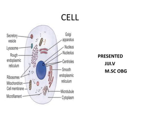
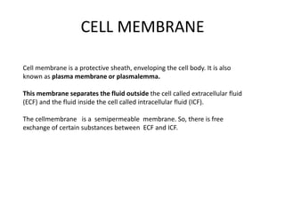
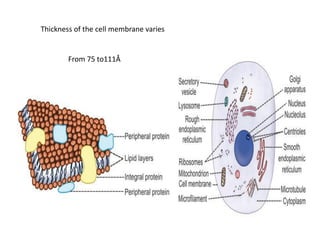
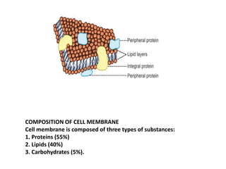
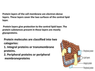
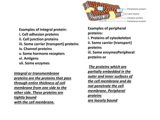
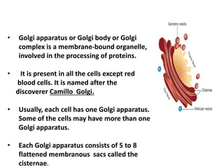
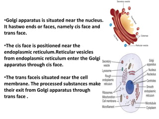
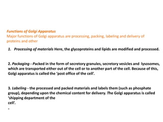
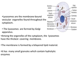
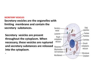
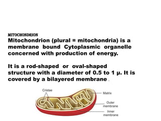
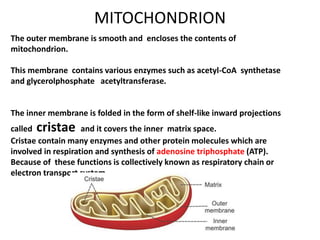
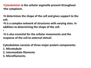
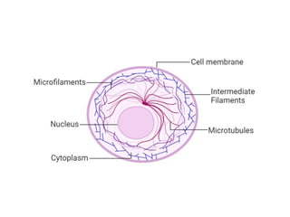
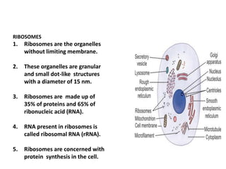
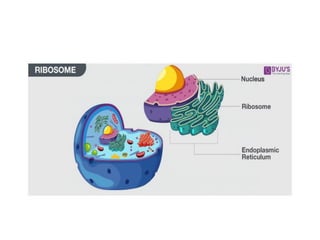
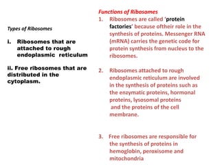
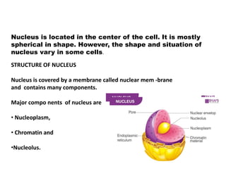
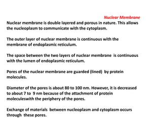
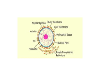
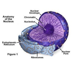
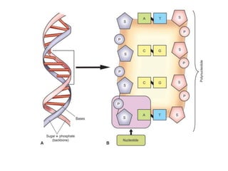
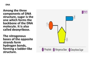
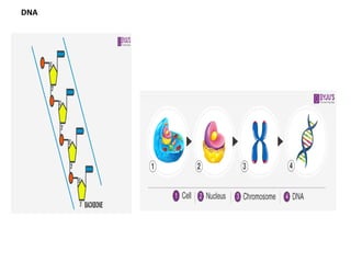
SlideShare a Scribd company logo
1 of 74
Download to read offline
CELL
PRESENTED
JIJI.V
M.SC OBG
CELL
INTRODUCTION
„
•All the living things are composed of cells.
• A single cell is the smallest unit that has
all the characteristics of life.
•Cell is defined as the structural and
functional unit of the
living body.
General Characteristics of Cell
• Each cell in the body:
• 1. Needs nutrition and oxygen
• 2. Produces its own energy necessary for its growth,
repair and other activities
• 3. Eliminates carbon dioxide and other metabolic
wastes,
• 4. Maintains the medium, i.e. the environment for its
survival
• 5. Shows immediate response to the entry of
invaders like bacteria or toxic substances
into the body
• 6. Reproduces by division. There are some
exceptions like neuron, which do not
reproduce
STRUCTURE OF THE CELL
• Each cell is formed by a cell body and a
membrane covering the cell body called the cell
membrane.
Cellbody has two parts, namely nucleus and
cytoplasm surrounding the nucleus .
• Thus, the structureof the cell is studied under
three headings:
• 1. Cell membrane
• 2. Cytoplasm
• 3. Nucleus.
Cell membrane is a protective sheath, enveloping the cell body. It is also
known as plasma membrane or plasmalemma.
This membrane separates the fluid outside the cell called extracellular fluid
(ECF) and the fluid inside the cell called intracellular fluid (ICF).
The cellmembrane is a semipermeable membrane. So, there is free
exchange of certain substances between ECF and ICF.
CELL MEMBRANE
Thickness of the cell membrane varies
From 75 to111Å
COMPOSITION OF CELL MEMBRANE
Cell membrane is composed of three types of substances:
1. Proteins (55%)
2. Lipids (40%)
3. Carbohydrates (5%).
1.The materials dissolved in lipid layer also move to all
areas of the cell membrane.
Major lipids are:
1. Phospholipids
2. Cholesterol. 2.Lipid layer of the cell membrane is a
semipermeablemembrane and allows only the fat-
soluble substances to pass through it.
Thus, the fat-soluble substances like oxygen, carbon
dioxide and alcohol can pass through this lipid layer.
3.The water-soluble substances such as
glucose, urea and electrolytes cannot pass
through this.
Protein layers of the cell membrane are electron-dense
layers. These layers cover the two surfaces of the central lipid
layer.
Protein layers give protection to the central lipid layer. The
protein substances present in these layers are mostly
glycoproteins.
Protein molecules are classified into two
categories:
1. Integral proteins or transmembrane
proteins.
2. Peripheral proteins or peripheral
membraneproteins
Examples of integral protein:
i. Cell adhesion proteins
ii. Cell junction proteins
iii. Some carrier (transport) proteins
iv. Channel proteins
v. Some hormone receptors
vi. Antigens
vii. Some enzymes
Examples of peripheral
proteins:
i. Proteins of cytoskeleton
ii. Some carrier (transport)
proteins
iii. Some enzymesPeripheral
proteins or
The proteins which are
partially embedded in the
outer and inner surfaces of
the cell membrane and do
not penetrate the cell
membrane. Peripheral
proteins
are loosely bound
Integral or transmembrane
proteins are the proteins that pass
through entire thickness of cell
membrane from one side to the
other side. These proteins are
tightly bound
with the cell membrane.
Some of the carbohydrate molecules present in
cell membrane are attached to proteins and form
glycoproteins (proteoglycans).
Some carbohydrate molecules are attached to lipids and form
glycolipids.
Carbohydrate molecules form a thin and loose covering over the
entire surface of the cell membrane called glycocalyx.
1. Carbohydrate molecules are negatively charged and
do not permit the negatively charged substances to
move in and out of the cell
2. Glycocalyx from the neighboring cells helps in the
tight fixation of cells with one another
FUNCTIONS OF CELL MEMBRANE
1. Protective function: Cell membrane protects the
cytoplasm and the organelles present in the
cytoplasm
2. Selective permeability: Cell membrane acts as a
semipermeable membrane, which allows only some
substances to pass through it and acts as a barrier
for other substances
3. Absorptive function:
Nutrients are absorbed into the
cell through the cell membrane
4. Excretory function: Metabolites and
other waste products from the cell are
excreted out through thecell membrane
5. Exchange of gases: Oxygen enters the
cell from theblood and carbon dioxide
leaves the cell and enters the blood
through the cell membrane
6. Maintenance of shape and size of the
cell: Cell membraneis responsible for
the maintenance of shapeand size of
the cell
Cytoplasm of the cell is the jelly like material formed
by80% of water. It contains a clear liquid portion
called cytosol and various particles of different shape
and size.
These particles are proteins, carbohydrates, lipidsor
electrolytes in nature.
Cytoplasm also contains manyorganelles with distinct
structure and function.
Cytoplasm is made up of two zones:
1. Ectoplasm: Peripheral part of cytoplasm,
situatedjust beneath the cell membrane
2. Endoplasm: Inner part of cytoplasm, interposed
between the ectoplasm and the nucleus.
Organelles with limiting
membrane
1. Endoplasmic reticulum
2. Golgi apparatus
3. Lysosome
4. Peroxisome
5. Centrosome and centrioles
6. Secretory vesicles
7. Mitochondria
8. Nucleus
Cytoplasmic organelles are the cellular structures
embedded in the cytoplasm.
Organelles are consideredas small organs of the cell.
Some organelles are bound
1, limiting membrane and 2.not have limitingmembrane
Organelles without limiting membrane
1. Ribosomes
2. Cytoskeleton
„a.ENDOPLASMIC RETICULUM
•Endoplasmic reticulum is a network of tubular and
microsomal vesicular structures which are interconnected
with one another.
•It is covered by a limiting membrane which is formed by
proteins and bilayered lipids.
•The lumenof endoplasmic reticulum contains a fluid medium
called endoplasmic matrix.
•The endoplasmic reticulum forms the link between nucleus
and cell membrane by connecting the cell membrane with
the nuclear membrane.
Types of Endoplasmic Reticulum
Endoplasmic reticulum is of two types:
1.Rough endoplasmic reticulum
2.Smooth endoplasmic reticulum.
Both the types are interconnected and continuous
with one another.
1.Rough Endoplasmic Reticulum
It is the endoplasmic reticulum with rough, bumpy or
bead-like appearance. Rough appearance is due to the
attachment of granular ribosomes to its outer surface.
Hence, it is also called the granular endoplasmic
reticulum.
Functions of Rough Endoplasmic
Reticulum
• Synthesis of proteins
•Degradation of worn-out organelles
It is the endoplasmic reticulum with smooth appearance. It is also called a
granular reticulum.
It is formed by many interconnected tubules. So, it is also called tubular
endoplasmic reticulum.
Functions of Smooth Endoplasmic Reticulum
1. Synthesis of non-protein proteins such as cholesterol
and steroid.
2.Storage and metabolism of calcium
3.Catabolism and detoxification
• Golgi apparatus or Golgi body or Golgi
complex is a membrane-bound organelle,
involved in the processing of proteins.
• It is present in all the cells except red
blood cells. It is named after the
discoverer Camillo Golgi.
• Usually, each cell has one Golgi apparatus.
Some of the cells may have more than one
Golgi apparatus.
• Each Golgi apparatus consists of 5 to 8
flattened membranous sacs called the
cisternae.
•Golgi apparatus is situated near the nucleus.
It hastwo ends or faces, namely cis face and
trans face.
•The cis face is positioned near the
endoplasmic reticulum.Reticular vesicles
from endoplasmic reticulum enter the Golgi
apparatus through cis face.
•The trans faceis situated near the cell
membrane. The processed substances make
their exit from Golgi apparatus through
trans face .
Functions of Golgi Apparatus
Major functions of Golgi apparatus are processing, packing, labeling and delivery of
proteins and other
1. Processing of materials Here, the glycoproteins and lipids are modified and processed.
2. Packaging - Packed in the form of secretory granules, secretory vesicles and lysosomes,
which are transported either out of the cell or to another part of the cell. Because of this,
Golgi apparatus is called the ‘post office of the cell’.
3. Labeling - the processed and packed materials and labels them (such as phosphate
group), depending upon the chemical content for delivery. The Golgi apparatus is called
‘shipping department of the
cell’.
„
•Lysosomes are the membrane-bound
vesicular organelles found throughout the
cytoplasm.
• The lysosomes are formed by Golgi
apparatus.
•Among the organelles of the cytoplasm, the lysosomes
have the thickest covering membrane.
•The membrane is formed by a bilayered lipid material.
•It has many small granules which contain hydrolytic
enzymes
Lysosomes are of two types:
1. Primary lysosome, which is pinched off from Golgi
apparatus. It is inactive in spite of having hydrolytic
Enzymes
2. Secondary lysosome, which is the active lyso some.
It is formed by the fusion of a primary lysosome
PEROXISOMES
Peroxisomes or microbodies are the membrane limited
vesicles like the lysosomes
Peroxisomes contain some oxidative enzymes such as catalase, urate oxidase
and Damino acid oxidase.
Functions of Peroxisomes
•Breakdown the fatty acids by means of a process called betaoxidation:
•Large number of peroxisomes are present in the cells of liver, which is the
major organ for detoxification
•Form the major site of oxygen utilization in the cells
• Accelerate gluconeogenesis from fats
• Degrade purine to uric acid
• Participate in the formation of myelin
• Play a role in the formation of bile acids.
„
Centrosome is the membrane-bound
cellular organelle situated almost in the
center of cell, close to nucleus.
It consists of two cylindrical structures
called centrioles
which are made up of proteins.
Centrioles are responsiblefor the
movement of chromosomes during cell
division
SECRETORY VESICLES
Secretory vesicles are the organelles with
limiting membrane and contain the
secretory substances.
Secretory vesicles are present
throughout the cytoplasm. When
necessary, these vesicles are ruptured
and secretory substances are released
into the cytoplasm.
„
MITOCHONDRION
Mitochondrion (plural = mitochondria) is a
membrane bound Cytoplasmic organelle
concerned with production of energy.
It is a rod-shaped or oval-shaped
structure with a diameter of 0.5 to 1 μ. It is
covered by a bilayered membrane .
The outer membrane is smooth and encloses the contents of
mitochondrion.
This membrane contains various enzymes such as acetyl-CoA synthetase
and glycerolphosphate acetyltransferase.
The inner membrane is folded in the form of shelf-like inward projections
called cristae and it covers the inner matrix space.
Cristae contain many enzymes and other protein molecules which are
involved in respiration and synthesis of adenosine triphosphate (ATP).
Because of these functions is collectively known as respiratory chain or
electron transport system.
MITOCHONDRION
Mitochondrion moves freely
in the cytoplasm of the cell. It
is capable of reproducing
itself.
Mitochondrion contains its
own deoxyribonucleic
acid (DNA), which is
responsible for many
enzymatic actions.
Functions of Mitochondrion
1. Production of energy
Mitochondrion is called the ‘power
house’ or ‘power plant’ of the cell
because it produces the energy
required for cellular functions.
2. Synthesis of ATP
The components of respiratory chain in
mitochondrion are responsible for the
synthesis of ATP by utilizing the
energy by oxidative phosphorylation
3.Other functions
Other functions of mitochondria include
storage of
calcium and detoxification of ammonia in liver
•Cytoskeleton is the cellular organelle present throughout
•the cytoplasm.
•It determines the shape of the cell and gives support to the
cell.
•It is a complex network of structures with varying sizes. In
addition to determining the shape of the cell.
•
•it is also essential for the cellular movements and the
response of the cell to external stimuli.
Cytoskeleton consists of three major protein components:
1. Microtubule
2. Intermediate filaments
3. Microfilaments.
RIBOSOMES
1. Ribosomes are the organelles
without limiting membrane.
2. These organelles are granular
and small dot-like structures
with a diameter of 15 nm.
3. Ribosomes are made up of
35% of proteins and 65% of
ribonucleic acid (RNA).
4. RNA present in ribosomes is
called ribosomal RNA (rRNA).
5. Ribosomes are concerned with
protein synthesis in the cell.
Types of Ribosomes
i. Ribosomes that are
attached to rough
endoplasmic reticulum
ii. Free ribosomes that are
distributed in the
cytoplasm.
Functions of Ribosomes
1. Ribosomes are called ‘protein
factories’ because oftheir role in the
synthesis of proteins. Messenger RNA
(mRNA) carries the genetic code for
protein synthesis from nucleus to the
ribosomes.
2. Ribosomes attached to rough
endoplasmic reticulum are involved
in the synthesis of proteins such as
the enzymatic proteins, hormonal
proteins, lysosomal proteins
and the proteins of the cell
membrane.
3. Free ribosomes are responsible for
the synthesis of proteins in
hemoglobin, peroxisome and
mitochondria
Nucleus is the most prominent and the largest cellular organelle. It
has a diameter of 10 μ to 22 μ and occupies about 10% of total
volume of the cell
•Nucleus is present in all the cells in the body except the red blood
cells.
•The cells with nucleus are called eukaryotes and those without
nucleus are known as prokaryotes.
•Presence of nucleus is necessary for cell division.
•Most of the cells have only one nucleus (uninucleated
•cells).
•Few types of cells like skeletal muscle cells have
•many nuclei (multinucleated cells). „
Nucleus is located in the center of the cell. It is mostly
spherical in shape. However, the shape and situation of
nucleus vary in some cells.
STRUCTURE OF NUCLEUS
Nucleus is covered by a membrane called nuclear mem -brane
and contains many components.
Major compo nents of nucleus are
• Nucleoplasm,
• Chromatin and
•Nucleolus.
Nuclear Membrane
Nuclear membrane is double layered and porous in nature. This allows
the nucleoplasm to communicate with the cytoplasm.
The outer layer of nuclear membrane is continuous with the
membrane of endoplasmic reticulum.
The space between the two layers of nuclear membrane is continuous
with the lumen of endoplasmic reticulum.
Pores of the nuclear membrane are guarded (lined) by protein
molecules.
Diameter of the pores is about 80 to 100 nm. However, it is decreased
to about 7 to 9 nm because of the attachment of protein
moleculeswith the periphery of the pores.
Exchange of materials between nucleoplasm and cytoplasm occurs
through these pores.
1. Nucleoplasm is a highly viscous fluid that forms the
ground substance of the nucleus. It is similar to
cytoplasm present outside the nucleus.
2. Nucleoplasm surrounds chromatin and nucleolus.
3. It contains dense fibrillar network of proteins called
the nuclear matrix and many substances such as
nucleotides and enzymes.
4. The nuclear matrix forms the structural framework
for organizing chromatin.
5. The soluble liquid part of nucleoplasm is known as
nuclear hyaloplasm
.DNA is a double helix which wraps around central core of eight histone
molecules to form the fundamental packing unit of chromatin called
nucleosome.
Nucleosomes are packed together tightly with the help of a histone molecule to
form a chromatin fiber.
.Just before cell division, the
chromatin condenses to form
chromosome.
Chromatin is a thread-like material made up of
large molecules of DNA.
Chromosomes
Chromosome is the rod-shaped nuclear structure that carries a complete
blueprint of all the hereditary characteristics of that species
.
A chromosome is formed from a single DNA molecule coiled around histone
molecules. Each DNA contains many genes.
The chromosomes are not visible in the
nucleus under microscope. Only during cell division,
the chromosomes are visible under microscope. This is
because DNA becomes more tightly packed just before
cell division.
All the dividing cells of the body except
reproductive cells contain 23 pairs of
chromosomes
The cells with 23 pairs of chromosomes are called
diploid cells.
The reproductive cells called gametes or
sex cells contain only 23 single chromosomes. These
cells are called haploid cells.
CHROMOSOME
•Nucleolus is a small, round granular structure of the nucleus.
•Each nucleus contains one or more nucleoli.
•The nucleolus contains RNA and some proteins, which are similar
to those found in ribosomes.
•The RNA is synthesized by five different pairs of chromosomes and
stored in the nucleolus. Later, it is condensed to form the subunits
of ribosomes.
•All the subunits formed in the nucleolus are transported to
cytoplasm through the pores of nuclear membrane.
•In the cytoplasm, these subunits fuse to form ribosomes, which
play an essential role in the formation of proteins.
1. Control of all the cell activities that include
metabolism, protein synthesis, growth and
reproduction (cell division)
2. Synthesis of RNA
3. Formation of subunits of ribosomes.
4. Sending genetic instruction to the cytoplasm for
protein synthesis through messenger RNA (mRNA)
5. Control of the cell division through genes
6. Storage of hereditary information (in genes)
and transformation of this information from one
generation of the species to the next.
DNA is present in the nucleus
(chromosome)
and mitochondria of the cell.
The DNA present in the nucleus
is responsible for the formation
of RNA.
RNA regulates the synthesis of
proteins by ribosomes. DNA in
mitochondria is called non-
chromosomal DNA
DNA
Among the three
components of DNA
structure, sugar is the
one which forms the
backbone of the DNA
molecule. It is also
called deoxyribose.
The nitrogenous
bases of the opposite
strands form
hydrogen bonds,
forming a ladder-like
structure.
DNA
Each chain of DNA molecule consists of many
nucleotides. Each nucleotide is formed by:
1. Deoxyribose – sugar
2. Phosphate
3. One of the following organic (nitrogenous) bases:
Purines – Adenine (A) – Guanine (G)
Pyrimidines – Thymine (T) – Cytosine (C)
•The adenine of one strand binds specifically
with thymine of opposite strand.
•Similarly, the cytosine of one strand binds
with guanine of the other strand.
•DNA forms the component of chromosomes,
which carries the hereditary information. The
hereditary information that is encoded in DNA
is called genome.
•Each DNA molecule is divided into discrete
units called genes.
DNA
• The two strands of DNA run in opposite directions.
These strands are held together by the hydrogen
bond that is present between the two
complementary bases.
• The strands are helically twisted, where each strand
forms a right-handed coil and ten nucleotides make
up a single turn.
• The pitch of each helix is 3.4 nm. Hence, the distance
between two consecutive base pairs (i.e., hydrogen-
bonded bases of the opposite strands) is 0.34 nm.
STRUCTURE OF DNA
DNA is a doublestranded complex nucleic acid. It
is formed by deoxyribose, phosphoric acid and four
types of bases.
Each DNA molecule consists of two polynucleotide chains,
which are twisted around one another in the form of a
double helix.
The two chains are formed by the sugar deoxyribose and
phosphate.
These two substances form the backbone of DNA molecule.
Both chains of DNA are connected with each other by
some organic bases
Each chain of DNA molecule consists of many
nucleotides. Each nucleotide is formed by:
1. Deoxyribose – sugar
2. Phosphate
3. One of the following organic (nitrogenous) bases:
Purines – Adenine (A) – Guanine (G)
Pyrimidines – Thymine (T) – Cytosine (C)
•The adenine of one strand binds specifically
with thymine of opposite strand.
•Similarly, the cytosine of one strand binds
with guanine of the other strand.
•DNA forms the component of chromosomes,
which carries the hereditary information. The
hereditary information that is encoded in DNA
is called genome.
•Each DNA molecule is divided into discrete
units called genes.
Gene is a portion of DNA molecule that contains the message or code for
the synthesis of a specific proteinfrom amino acids.
It is like a book that contains the information necessary for protein
synthesis.
Gene is considered as the basic hereditary unit of the cell.
In the nucleotide of DNA, three of the successive base pairs are together
called a triplet or a codon.
„
30000 genes
DNA
DNA
Gene is a portion of DNA molecule that contains the message or code for
the synthesis of a specific proteinfrom amino acids.
It is like a book that contains the information necessary for protein
synthesis.
Gene is considered as the basic hereditary unit of the cell.
In the nucleotide of DNA, three of the successive base pairs are together
called a triplet or a codon.
„
30000 genes
Each codon codes or forms code word (information) for one
amino acid.
There are 20 amino acids and there is
separate code for each amino acid. For example, the
triplet CCA is the code for glycine and GGC is the code
for proline.
Thus, each gene forms the code word for a particular
protein to be synthesized in ribosome (outside the
nucleus) from amino acids.
GENETIC DISORDERS
Causes of Gene Disorders
Genetic disorders occur due to
two causes:
1. Genetic variation: Presence of a
different form of gene
2. Genetic mutation: Generally,
mutation means an alteration
or a change in nature, form, or
quality.
Genetic mutation refers to change
of the DNA sequence within a
gene or chromosome of an
organism, which results in the
creation of a new character.
„
1. A genetic disorder
is a disorder that
occurs because of
the abnormalities
in an individual’s
genetic material
(genome).
2. Genetic disorders
are either
hereditary
disorders or due to
defect in genes.
RIBONUCLEIC ACID
1. Ribonucleic acid (RNA) is a nucleic acid that
contains a long chain of nucleotide units. It
is similar to DNA but contains ribose
instead of deoxyribose.
2. Various functions coded in the genes are
carried out in the cytoplasm of the cell by
RNA.
RNA is formed from DNA.
RNA
STRUCTURE OF RNA
Each RNA molecule consists of a single strand
of polynucleotide unlike the double
stranded DNA.
Eachnucleotide in RNA is formed by:
1. Ribose – sugar.
2. Phosphate.
3. One of the following organic bases:
Purines – Adenine (A) – Guanine (G)
Pyrimidines – Uracil (U)– Cytosine (C).
Uracil replaces the thymine of DNA and it has
similar structure of thymine.
„
rna
RNA
1. Messenger RNA (mRNA)
Messenger RNA carries the genetic
code of the aminoacid sequence for
synthesis of protein from the DNA to
the cytoplasm.
2. Transfer RNA (tRNA)
Transfer RNA is responsible for
decoding the genetic message present
in mRNA.
3. Ribosomal RNA (rRNA)
Ribosomal RNA is present within the
ribosome and forms a part of the
structure of ribosome. It is responsible
for the assembly of protein from amino
acids in the ribosome
TYPES OF RNA
•RNA is of three
types.
• Each type of RNA
plays a specific
role in protein
synthesis
Functions of RNA
• Facilitate the translation of
DNA into proteins
• Functions as an adapter
molecule in protein synthesis
• Serves as a messenger
between the DNA and the
ribosomes.
• They are the carrier of genetic
information in all living cells
• Promotes the ribosomes to
choose the right amino acid
which is required in building
up of new proteins in the
body.

More Related Content

What's hot

What's hot (20)

Prokaryotes and Eukaryotes
Prokaryotes and EukaryotesProkaryotes and Eukaryotes
Prokaryotes and Eukaryotes
 
Endoplasmic reticulum
Endoplasmic reticulumEndoplasmic reticulum
Endoplasmic reticulum
 
7-2: Eukaryotic Cell Structure
7-2: Eukaryotic Cell Structure7-2: Eukaryotic Cell Structure
7-2: Eukaryotic Cell Structure
 
Cilia and flagella ppt by ramana babu
Cilia and flagella ppt by ramana babuCilia and flagella ppt by ramana babu
Cilia and flagella ppt by ramana babu
 
Cytoskeleton
CytoskeletonCytoskeleton
Cytoskeleton
 
Cell structure & function
Cell structure & functionCell structure & function
Cell structure & function
 
Centrioles[1]
Centrioles[1]Centrioles[1]
Centrioles[1]
 
Cell structure and function
Cell structure and functionCell structure and function
Cell structure and function
 
Cell
CellCell
Cell
 
Cell, the unit of life
Cell, the unit of lifeCell, the unit of life
Cell, the unit of life
 
Cell motility cilia and flagella
Cell motility  cilia and flagellaCell motility  cilia and flagella
Cell motility cilia and flagella
 
Endomembrane system
Endomembrane systemEndomembrane system
Endomembrane system
 
Plant & animal cells
Plant & animal cellsPlant & animal cells
Plant & animal cells
 
Structure and composition of Biomembranes
Structure and composition of BiomembranesStructure and composition of Biomembranes
Structure and composition of Biomembranes
 
Cell organelle presentation
Cell organelle presentationCell organelle presentation
Cell organelle presentation
 
Cell & Its Orgenells
Cell & Its OrgenellsCell & Its Orgenells
Cell & Its Orgenells
 
Cell: it’s structure and functions
Cell: it’s structure and functionsCell: it’s structure and functions
Cell: it’s structure and functions
 
Cell : Structure and Function Part 01
Cell : Structure and Function Part 01Cell : Structure and Function Part 01
Cell : Structure and Function Part 01
 
Cell structure and its function
Cell structure and its functionCell structure and its function
Cell structure and its function
 
Cytoplasm & cell organelles By Manoj Dhital (M.Sc Medical Microbiology))
Cytoplasm & cell organelles By Manoj Dhital (M.Sc Medical Microbiology))Cytoplasm & cell organelles By Manoj Dhital (M.Sc Medical Microbiology))
Cytoplasm & cell organelles By Manoj Dhital (M.Sc Medical Microbiology))
 

Similar to human cell and function

human cell anatomy and function
human cell anatomy and functionhuman cell anatomy and function
human cell anatomy and functionjiji jiya
 
The cell its organells and their functions
The cell its organells and their functionsThe cell its organells and their functions
The cell its organells and their functionsSumama Shakir
 
Functions of cell organells nursery
Functions of cell organells nurseryFunctions of cell organells nursery
Functions of cell organells nurserydina merzeban
 
About the Cell & Physiology of the cell.
About the Cell & Physiology of the cell.About the Cell & Physiology of the cell.
About the Cell & Physiology of the cell.MedicTalks
 
CELL STRUCTURE AND ITS FUNCTION pptx
CELL STRUCTURE  AND ITS FUNCTION pptxCELL STRUCTURE  AND ITS FUNCTION pptx
CELL STRUCTURE AND ITS FUNCTION pptxMagarani Balachandar
 
cell structure.pptx
cell structure.pptxcell structure.pptx
cell structure.pptxKowsikaS8
 
Chapter 4th cell and tissues
Chapter 4th cell and tissues Chapter 4th cell and tissues
Chapter 4th cell and tissues TehminaNaz8
 
Cell and its Physiology.pdf
Cell and its Physiology.pdfCell and its Physiology.pdf
Cell and its Physiology.pdfRAMDAS BHAT
 
Lecture 2 (cell and it's function)
Lecture 2 (cell and it's function)Lecture 2 (cell and it's function)
Lecture 2 (cell and it's function)Ayub Abdi
 
the cell and its functions
the cell and its functionsthe cell and its functions
the cell and its functionsFatimaSundus1
 
introduction to Biochemistry.pptx
introduction to Biochemistry.pptxintroduction to Biochemistry.pptx
introduction to Biochemistry.pptxKomalNanaware6
 
Cellular level of organization
Cellular level of organizationCellular level of organization
Cellular level of organizationSheetal Patil
 
CELL Biochemistry.ppt
CELL Biochemistry.pptCELL Biochemistry.ppt
CELL Biochemistry.pptAnnaKhurshid
 
CELL Biochemistry - new.ppt
CELL Biochemistry - new.pptCELL Biochemistry - new.ppt
CELL Biochemistry - new.pptAnnaKhurshid
 

Similar to human cell and function (20)

human cell anatomy and function
human cell anatomy and functionhuman cell anatomy and function
human cell anatomy and function
 
The cell its organells and their functions
The cell its organells and their functionsThe cell its organells and their functions
The cell its organells and their functions
 
Functions of cell organells nursery
Functions of cell organells nurseryFunctions of cell organells nursery
Functions of cell organells nursery
 
About the Cell & Physiology of the cell.
About the Cell & Physiology of the cell.About the Cell & Physiology of the cell.
About the Cell & Physiology of the cell.
 
CELL STRUCTURE AND ITS FUNCTION pptx
CELL STRUCTURE  AND ITS FUNCTION pptxCELL STRUCTURE  AND ITS FUNCTION pptx
CELL STRUCTURE AND ITS FUNCTION pptx
 
cell structure.pptx
cell structure.pptxcell structure.pptx
cell structure.pptx
 
cell physiology 1.pptx
cell physiology 1.pptxcell physiology 1.pptx
cell physiology 1.pptx
 
Chapter 4th cell and tissues
Chapter 4th cell and tissues Chapter 4th cell and tissues
Chapter 4th cell and tissues
 
Cell and its Physiology.pdf
Cell and its Physiology.pdfCell and its Physiology.pdf
Cell and its Physiology.pdf
 
Seminar on cell
Seminar on cellSeminar on cell
Seminar on cell
 
Lecture 2 (cell and it's function)
Lecture 2 (cell and it's function)Lecture 2 (cell and it's function)
Lecture 2 (cell and it's function)
 
CELL PHYSIOLOGY
CELL PHYSIOLOGYCELL PHYSIOLOGY
CELL PHYSIOLOGY
 
Unit II
Unit IIUnit II
Unit II
 
the cell and its functions
the cell and its functionsthe cell and its functions
the cell and its functions
 
introduction to Biochemistry.pptx
introduction to Biochemistry.pptxintroduction to Biochemistry.pptx
introduction to Biochemistry.pptx
 
Cellular level of organization
Cellular level of organizationCellular level of organization
Cellular level of organization
 
CELL Biochemistry.ppt
CELL Biochemistry.pptCELL Biochemistry.ppt
CELL Biochemistry.ppt
 
Cells
CellsCells
Cells
 
CELL Biochemistry - new.ppt
CELL Biochemistry - new.pptCELL Biochemistry - new.ppt
CELL Biochemistry - new.ppt
 
Class 1 Human cell physiology
Class 1 Human cell physiology Class 1 Human cell physiology
Class 1 Human cell physiology
 

Recently uploaded

Russian Escort Service in Delhi 11k Hotel Foreigner Russian Call Girls in Delhi
Russian Escort Service in Delhi 11k Hotel Foreigner Russian Call Girls in DelhiRussian Escort Service in Delhi 11k Hotel Foreigner Russian Call Girls in Delhi
Russian Escort Service in Delhi 11k Hotel Foreigner Russian Call Girls in Delhikauryashika82
 
The basics of sentences session 2pptx copy.pptx
The basics of sentences session 2pptx copy.pptxThe basics of sentences session 2pptx copy.pptx
The basics of sentences session 2pptx copy.pptxheathfieldcps1
 
Call Girls in Dwarka Mor Delhi Contact Us 9654467111
Call Girls in Dwarka Mor Delhi Contact Us 9654467111Call Girls in Dwarka Mor Delhi Contact Us 9654467111
Call Girls in Dwarka Mor Delhi Contact Us 9654467111Sapana Sha
 
Grant Readiness 101 TechSoup and Remy Consulting
Grant Readiness 101 TechSoup and Remy ConsultingGrant Readiness 101 TechSoup and Remy Consulting
Grant Readiness 101 TechSoup and Remy ConsultingTechSoup
 
social pharmacy d-pharm 1st year by Pragati K. Mahajan
social pharmacy d-pharm 1st year by Pragati K. Mahajansocial pharmacy d-pharm 1st year by Pragati K. Mahajan
social pharmacy d-pharm 1st year by Pragati K. Mahajanpragatimahajan3
 
Web & Social Media Analytics Previous Year Question Paper.pdf
Web & Social Media Analytics Previous Year Question Paper.pdfWeb & Social Media Analytics Previous Year Question Paper.pdf
Web & Social Media Analytics Previous Year Question Paper.pdfJayanti Pande
 
Arihant handbook biology for class 11 .pdf
Arihant handbook biology for class 11 .pdfArihant handbook biology for class 11 .pdf
Arihant handbook biology for class 11 .pdfchloefrazer622
 
Measures of Central Tendency: Mean, Median and Mode
Measures of Central Tendency: Mean, Median and ModeMeasures of Central Tendency: Mean, Median and Mode
Measures of Central Tendency: Mean, Median and ModeThiyagu K
 
Unit-IV- Pharma. Marketing Channels.pptx
Unit-IV- Pharma. Marketing Channels.pptxUnit-IV- Pharma. Marketing Channels.pptx
Unit-IV- Pharma. Marketing Channels.pptxVishalSingh1417
 
Holdier Curriculum Vitae (April 2024).pdf
Holdier Curriculum Vitae (April 2024).pdfHoldier Curriculum Vitae (April 2024).pdf
Holdier Curriculum Vitae (April 2024).pdfagholdier
 
Software Engineering Methodologies (overview)
Software Engineering Methodologies (overview)Software Engineering Methodologies (overview)
Software Engineering Methodologies (overview)eniolaolutunde
 
1029 - Danh muc Sach Giao Khoa 10 . pdf
1029 -  Danh muc Sach Giao Khoa 10 . pdf1029 -  Danh muc Sach Giao Khoa 10 . pdf
1029 - Danh muc Sach Giao Khoa 10 . pdfQucHHunhnh
 
BAG TECHNIQUE Bag technique-a tool making use of public health bag through wh...
BAG TECHNIQUE Bag technique-a tool making use of public health bag through wh...BAG TECHNIQUE Bag technique-a tool making use of public health bag through wh...
BAG TECHNIQUE Bag technique-a tool making use of public health bag through wh...Sapna Thakur
 
Q4-W6-Restating Informational Text Grade 3
Q4-W6-Restating Informational Text Grade 3Q4-W6-Restating Informational Text Grade 3
Q4-W6-Restating Informational Text Grade 3JemimahLaneBuaron
 
SOCIAL AND HISTORICAL CONTEXT - LFTVD.pptx
SOCIAL AND HISTORICAL CONTEXT - LFTVD.pptxSOCIAL AND HISTORICAL CONTEXT - LFTVD.pptx
SOCIAL AND HISTORICAL CONTEXT - LFTVD.pptxiammrhaywood
 
BASLIQ CURRENT LOOKBOOK LOOKBOOK(1) (1).pdf
BASLIQ CURRENT LOOKBOOK  LOOKBOOK(1) (1).pdfBASLIQ CURRENT LOOKBOOK  LOOKBOOK(1) (1).pdf
BASLIQ CURRENT LOOKBOOK LOOKBOOK(1) (1).pdfSoniaTolstoy
 
Key note speaker Neum_Admir Softic_ENG.pdf
Key note speaker Neum_Admir Softic_ENG.pdfKey note speaker Neum_Admir Softic_ENG.pdf
Key note speaker Neum_Admir Softic_ENG.pdfAdmir Softic
 

Recently uploaded (20)

Russian Escort Service in Delhi 11k Hotel Foreigner Russian Call Girls in Delhi
Russian Escort Service in Delhi 11k Hotel Foreigner Russian Call Girls in DelhiRussian Escort Service in Delhi 11k Hotel Foreigner Russian Call Girls in Delhi
Russian Escort Service in Delhi 11k Hotel Foreigner Russian Call Girls in Delhi
 
The basics of sentences session 2pptx copy.pptx
The basics of sentences session 2pptx copy.pptxThe basics of sentences session 2pptx copy.pptx
The basics of sentences session 2pptx copy.pptx
 
Call Girls in Dwarka Mor Delhi Contact Us 9654467111
Call Girls in Dwarka Mor Delhi Contact Us 9654467111Call Girls in Dwarka Mor Delhi Contact Us 9654467111
Call Girls in Dwarka Mor Delhi Contact Us 9654467111
 
Grant Readiness 101 TechSoup and Remy Consulting
Grant Readiness 101 TechSoup and Remy ConsultingGrant Readiness 101 TechSoup and Remy Consulting
Grant Readiness 101 TechSoup and Remy Consulting
 
social pharmacy d-pharm 1st year by Pragati K. Mahajan
social pharmacy d-pharm 1st year by Pragati K. Mahajansocial pharmacy d-pharm 1st year by Pragati K. Mahajan
social pharmacy d-pharm 1st year by Pragati K. Mahajan
 
Web & Social Media Analytics Previous Year Question Paper.pdf
Web & Social Media Analytics Previous Year Question Paper.pdfWeb & Social Media Analytics Previous Year Question Paper.pdf
Web & Social Media Analytics Previous Year Question Paper.pdf
 
Arihant handbook biology for class 11 .pdf
Arihant handbook biology for class 11 .pdfArihant handbook biology for class 11 .pdf
Arihant handbook biology for class 11 .pdf
 
Measures of Central Tendency: Mean, Median and Mode
Measures of Central Tendency: Mean, Median and ModeMeasures of Central Tendency: Mean, Median and Mode
Measures of Central Tendency: Mean, Median and Mode
 
Unit-IV- Pharma. Marketing Channels.pptx
Unit-IV- Pharma. Marketing Channels.pptxUnit-IV- Pharma. Marketing Channels.pptx
Unit-IV- Pharma. Marketing Channels.pptx
 
Holdier Curriculum Vitae (April 2024).pdf
Holdier Curriculum Vitae (April 2024).pdfHoldier Curriculum Vitae (April 2024).pdf
Holdier Curriculum Vitae (April 2024).pdf
 
Código Creativo y Arte de Software | Unidad 1
Código Creativo y Arte de Software | Unidad 1Código Creativo y Arte de Software | Unidad 1
Código Creativo y Arte de Software | Unidad 1
 
Software Engineering Methodologies (overview)
Software Engineering Methodologies (overview)Software Engineering Methodologies (overview)
Software Engineering Methodologies (overview)
 
1029 - Danh muc Sach Giao Khoa 10 . pdf
1029 -  Danh muc Sach Giao Khoa 10 . pdf1029 -  Danh muc Sach Giao Khoa 10 . pdf
1029 - Danh muc Sach Giao Khoa 10 . pdf
 
BAG TECHNIQUE Bag technique-a tool making use of public health bag through wh...
BAG TECHNIQUE Bag technique-a tool making use of public health bag through wh...BAG TECHNIQUE Bag technique-a tool making use of public health bag through wh...
BAG TECHNIQUE Bag technique-a tool making use of public health bag through wh...
 
Q4-W6-Restating Informational Text Grade 3
Q4-W6-Restating Informational Text Grade 3Q4-W6-Restating Informational Text Grade 3
Q4-W6-Restating Informational Text Grade 3
 
SOCIAL AND HISTORICAL CONTEXT - LFTVD.pptx
SOCIAL AND HISTORICAL CONTEXT - LFTVD.pptxSOCIAL AND HISTORICAL CONTEXT - LFTVD.pptx
SOCIAL AND HISTORICAL CONTEXT - LFTVD.pptx
 
BASLIQ CURRENT LOOKBOOK LOOKBOOK(1) (1).pdf
BASLIQ CURRENT LOOKBOOK  LOOKBOOK(1) (1).pdfBASLIQ CURRENT LOOKBOOK  LOOKBOOK(1) (1).pdf
BASLIQ CURRENT LOOKBOOK LOOKBOOK(1) (1).pdf
 
Advance Mobile Application Development class 07
Advance Mobile Application Development class 07Advance Mobile Application Development class 07
Advance Mobile Application Development class 07
 
Key note speaker Neum_Admir Softic_ENG.pdf
Key note speaker Neum_Admir Softic_ENG.pdfKey note speaker Neum_Admir Softic_ENG.pdf
Key note speaker Neum_Admir Softic_ENG.pdf
 
Mattingly "AI & Prompt Design: Structured Data, Assistants, & RAG"
Mattingly "AI & Prompt Design: Structured Data, Assistants, & RAG"Mattingly "AI & Prompt Design: Structured Data, Assistants, & RAG"
Mattingly "AI & Prompt Design: Structured Data, Assistants, & RAG"
 

human cell and function

  • 2. CELL INTRODUCTION „ •All the living things are composed of cells. • A single cell is the smallest unit that has all the characteristics of life. •Cell is defined as the structural and functional unit of the living body.
  • 3. General Characteristics of Cell • Each cell in the body: • 1. Needs nutrition and oxygen • 2. Produces its own energy necessary for its growth, repair and other activities • 3. Eliminates carbon dioxide and other metabolic wastes, • 4. Maintains the medium, i.e. the environment for its survival
  • 4. • 5. Shows immediate response to the entry of invaders like bacteria or toxic substances into the body • 6. Reproduces by division. There are some exceptions like neuron, which do not reproduce
  • 5. STRUCTURE OF THE CELL • Each cell is formed by a cell body and a membrane covering the cell body called the cell membrane. Cellbody has two parts, namely nucleus and cytoplasm surrounding the nucleus . • Thus, the structureof the cell is studied under three headings: • 1. Cell membrane • 2. Cytoplasm • 3. Nucleus.
  • 6. Cell membrane is a protective sheath, enveloping the cell body. It is also known as plasma membrane or plasmalemma. This membrane separates the fluid outside the cell called extracellular fluid (ECF) and the fluid inside the cell called intracellular fluid (ICF). The cellmembrane is a semipermeable membrane. So, there is free exchange of certain substances between ECF and ICF. CELL MEMBRANE
  • 7. Thickness of the cell membrane varies From 75 to111Å
  • 8. COMPOSITION OF CELL MEMBRANE Cell membrane is composed of three types of substances: 1. Proteins (55%) 2. Lipids (40%) 3. Carbohydrates (5%).
  • 9. 1.The materials dissolved in lipid layer also move to all areas of the cell membrane. Major lipids are: 1. Phospholipids 2. Cholesterol. 2.Lipid layer of the cell membrane is a semipermeablemembrane and allows only the fat- soluble substances to pass through it. Thus, the fat-soluble substances like oxygen, carbon dioxide and alcohol can pass through this lipid layer. 3.The water-soluble substances such as glucose, urea and electrolytes cannot pass through this.
  • 10. Protein layers of the cell membrane are electron-dense layers. These layers cover the two surfaces of the central lipid layer. Protein layers give protection to the central lipid layer. The protein substances present in these layers are mostly glycoproteins. Protein molecules are classified into two categories: 1. Integral proteins or transmembrane proteins. 2. Peripheral proteins or peripheral membraneproteins
  • 11. Examples of integral protein: i. Cell adhesion proteins ii. Cell junction proteins iii. Some carrier (transport) proteins iv. Channel proteins v. Some hormone receptors vi. Antigens vii. Some enzymes Examples of peripheral proteins: i. Proteins of cytoskeleton ii. Some carrier (transport) proteins iii. Some enzymesPeripheral proteins or The proteins which are partially embedded in the outer and inner surfaces of the cell membrane and do not penetrate the cell membrane. Peripheral proteins are loosely bound Integral or transmembrane proteins are the proteins that pass through entire thickness of cell membrane from one side to the other side. These proteins are tightly bound with the cell membrane.
  • 12. Some of the carbohydrate molecules present in cell membrane are attached to proteins and form glycoproteins (proteoglycans). Some carbohydrate molecules are attached to lipids and form glycolipids. Carbohydrate molecules form a thin and loose covering over the entire surface of the cell membrane called glycocalyx.
  • 13. 1. Carbohydrate molecules are negatively charged and do not permit the negatively charged substances to move in and out of the cell 2. Glycocalyx from the neighboring cells helps in the tight fixation of cells with one another
  • 14. FUNCTIONS OF CELL MEMBRANE 1. Protective function: Cell membrane protects the cytoplasm and the organelles present in the cytoplasm 2. Selective permeability: Cell membrane acts as a semipermeable membrane, which allows only some substances to pass through it and acts as a barrier for other substances 3. Absorptive function: Nutrients are absorbed into the cell through the cell membrane
  • 15. 4. Excretory function: Metabolites and other waste products from the cell are excreted out through thecell membrane 5. Exchange of gases: Oxygen enters the cell from theblood and carbon dioxide leaves the cell and enters the blood through the cell membrane 6. Maintenance of shape and size of the cell: Cell membraneis responsible for the maintenance of shapeand size of the cell
  • 16. Cytoplasm of the cell is the jelly like material formed by80% of water. It contains a clear liquid portion called cytosol and various particles of different shape and size. These particles are proteins, carbohydrates, lipidsor electrolytes in nature. Cytoplasm also contains manyorganelles with distinct structure and function. Cytoplasm is made up of two zones: 1. Ectoplasm: Peripheral part of cytoplasm, situatedjust beneath the cell membrane 2. Endoplasm: Inner part of cytoplasm, interposed between the ectoplasm and the nucleus.
  • 17. Organelles with limiting membrane 1. Endoplasmic reticulum 2. Golgi apparatus 3. Lysosome 4. Peroxisome 5. Centrosome and centrioles 6. Secretory vesicles 7. Mitochondria 8. Nucleus Cytoplasmic organelles are the cellular structures embedded in the cytoplasm. Organelles are consideredas small organs of the cell. Some organelles are bound 1, limiting membrane and 2.not have limitingmembrane Organelles without limiting membrane 1. Ribosomes 2. Cytoskeleton
  • 18. „a.ENDOPLASMIC RETICULUM •Endoplasmic reticulum is a network of tubular and microsomal vesicular structures which are interconnected with one another. •It is covered by a limiting membrane which is formed by proteins and bilayered lipids. •The lumenof endoplasmic reticulum contains a fluid medium called endoplasmic matrix. •The endoplasmic reticulum forms the link between nucleus and cell membrane by connecting the cell membrane with the nuclear membrane.
  • 19.
  • 20. Types of Endoplasmic Reticulum Endoplasmic reticulum is of two types: 1.Rough endoplasmic reticulum 2.Smooth endoplasmic reticulum. Both the types are interconnected and continuous with one another. 1.Rough Endoplasmic Reticulum It is the endoplasmic reticulum with rough, bumpy or bead-like appearance. Rough appearance is due to the attachment of granular ribosomes to its outer surface. Hence, it is also called the granular endoplasmic reticulum.
  • 21. Functions of Rough Endoplasmic Reticulum • Synthesis of proteins •Degradation of worn-out organelles
  • 22. It is the endoplasmic reticulum with smooth appearance. It is also called a granular reticulum. It is formed by many interconnected tubules. So, it is also called tubular endoplasmic reticulum. Functions of Smooth Endoplasmic Reticulum 1. Synthesis of non-protein proteins such as cholesterol and steroid. 2.Storage and metabolism of calcium 3.Catabolism and detoxification
  • 23. • Golgi apparatus or Golgi body or Golgi complex is a membrane-bound organelle, involved in the processing of proteins. • It is present in all the cells except red blood cells. It is named after the discoverer Camillo Golgi. • Usually, each cell has one Golgi apparatus. Some of the cells may have more than one Golgi apparatus. • Each Golgi apparatus consists of 5 to 8 flattened membranous sacs called the cisternae.
  • 24. •Golgi apparatus is situated near the nucleus. It hastwo ends or faces, namely cis face and trans face. •The cis face is positioned near the endoplasmic reticulum.Reticular vesicles from endoplasmic reticulum enter the Golgi apparatus through cis face. •The trans faceis situated near the cell membrane. The processed substances make their exit from Golgi apparatus through trans face .
  • 25. Functions of Golgi Apparatus Major functions of Golgi apparatus are processing, packing, labeling and delivery of proteins and other 1. Processing of materials Here, the glycoproteins and lipids are modified and processed. 2. Packaging - Packed in the form of secretory granules, secretory vesicles and lysosomes, which are transported either out of the cell or to another part of the cell. Because of this, Golgi apparatus is called the ‘post office of the cell’. 3. Labeling - the processed and packed materials and labels them (such as phosphate group), depending upon the chemical content for delivery. The Golgi apparatus is called ‘shipping department of the cell’. „
  • 26. •Lysosomes are the membrane-bound vesicular organelles found throughout the cytoplasm. • The lysosomes are formed by Golgi apparatus. •Among the organelles of the cytoplasm, the lysosomes have the thickest covering membrane. •The membrane is formed by a bilayered lipid material. •It has many small granules which contain hydrolytic enzymes
  • 27. Lysosomes are of two types: 1. Primary lysosome, which is pinched off from Golgi apparatus. It is inactive in spite of having hydrolytic Enzymes 2. Secondary lysosome, which is the active lyso some. It is formed by the fusion of a primary lysosome
  • 28. PEROXISOMES Peroxisomes or microbodies are the membrane limited vesicles like the lysosomes Peroxisomes contain some oxidative enzymes such as catalase, urate oxidase and Damino acid oxidase. Functions of Peroxisomes •Breakdown the fatty acids by means of a process called betaoxidation: •Large number of peroxisomes are present in the cells of liver, which is the major organ for detoxification •Form the major site of oxygen utilization in the cells • Accelerate gluconeogenesis from fats • Degrade purine to uric acid • Participate in the formation of myelin • Play a role in the formation of bile acids. „
  • 29.
  • 30. Centrosome is the membrane-bound cellular organelle situated almost in the center of cell, close to nucleus. It consists of two cylindrical structures called centrioles which are made up of proteins. Centrioles are responsiblefor the movement of chromosomes during cell division
  • 31. SECRETORY VESICLES Secretory vesicles are the organelles with limiting membrane and contain the secretory substances. Secretory vesicles are present throughout the cytoplasm. When necessary, these vesicles are ruptured and secretory substances are released into the cytoplasm. „
  • 32. MITOCHONDRION Mitochondrion (plural = mitochondria) is a membrane bound Cytoplasmic organelle concerned with production of energy. It is a rod-shaped or oval-shaped structure with a diameter of 0.5 to 1 μ. It is covered by a bilayered membrane .
  • 33. The outer membrane is smooth and encloses the contents of mitochondrion. This membrane contains various enzymes such as acetyl-CoA synthetase and glycerolphosphate acetyltransferase. The inner membrane is folded in the form of shelf-like inward projections called cristae and it covers the inner matrix space. Cristae contain many enzymes and other protein molecules which are involved in respiration and synthesis of adenosine triphosphate (ATP). Because of these functions is collectively known as respiratory chain or electron transport system. MITOCHONDRION
  • 34. Mitochondrion moves freely in the cytoplasm of the cell. It is capable of reproducing itself. Mitochondrion contains its own deoxyribonucleic acid (DNA), which is responsible for many enzymatic actions. Functions of Mitochondrion 1. Production of energy Mitochondrion is called the ‘power house’ or ‘power plant’ of the cell because it produces the energy required for cellular functions. 2. Synthesis of ATP The components of respiratory chain in mitochondrion are responsible for the synthesis of ATP by utilizing the energy by oxidative phosphorylation 3.Other functions Other functions of mitochondria include storage of calcium and detoxification of ammonia in liver
  • 35. Mitochondrion moves freely in the cytoplasm of the cell. It is capable of reproducing itself. Mitochondrion contains its own deoxyribonucleic acid (DNA), which is responsible for many enzymatic actions. Functions of Mitochondrion 1. Production of energy Mitochondrion is called the ‘power house’ or ‘power plant’ of the cell because it produces the energy required for cellular functions. 2. Synthesis of ATP The components of respiratory chain in mitochondrion are responsible for the synthesis of ATP by utilizing the energy by oxidative phosphorylation 3.Other functions Other functions of mitochondria include storage of calcium and detoxification of ammonia in liver
  • 36. •Cytoskeleton is the cellular organelle present throughout •the cytoplasm. •It determines the shape of the cell and gives support to the cell. •It is a complex network of structures with varying sizes. In addition to determining the shape of the cell. • •it is also essential for the cellular movements and the response of the cell to external stimuli. Cytoskeleton consists of three major protein components: 1. Microtubule 2. Intermediate filaments 3. Microfilaments.
  • 37.
  • 38. RIBOSOMES 1. Ribosomes are the organelles without limiting membrane. 2. These organelles are granular and small dot-like structures with a diameter of 15 nm. 3. Ribosomes are made up of 35% of proteins and 65% of ribonucleic acid (RNA). 4. RNA present in ribosomes is called ribosomal RNA (rRNA). 5. Ribosomes are concerned with protein synthesis in the cell.
  • 39.
  • 40. Types of Ribosomes i. Ribosomes that are attached to rough endoplasmic reticulum ii. Free ribosomes that are distributed in the cytoplasm. Functions of Ribosomes 1. Ribosomes are called ‘protein factories’ because oftheir role in the synthesis of proteins. Messenger RNA (mRNA) carries the genetic code for protein synthesis from nucleus to the ribosomes. 2. Ribosomes attached to rough endoplasmic reticulum are involved in the synthesis of proteins such as the enzymatic proteins, hormonal proteins, lysosomal proteins and the proteins of the cell membrane. 3. Free ribosomes are responsible for the synthesis of proteins in hemoglobin, peroxisome and mitochondria
  • 41. Nucleus is the most prominent and the largest cellular organelle. It has a diameter of 10 μ to 22 μ and occupies about 10% of total volume of the cell •Nucleus is present in all the cells in the body except the red blood cells. •The cells with nucleus are called eukaryotes and those without nucleus are known as prokaryotes. •Presence of nucleus is necessary for cell division. •Most of the cells have only one nucleus (uninucleated •cells). •Few types of cells like skeletal muscle cells have •many nuclei (multinucleated cells). „
  • 42. Nucleus is located in the center of the cell. It is mostly spherical in shape. However, the shape and situation of nucleus vary in some cells. STRUCTURE OF NUCLEUS Nucleus is covered by a membrane called nuclear mem -brane and contains many components. Major compo nents of nucleus are • Nucleoplasm, • Chromatin and •Nucleolus.
  • 43. Nuclear Membrane Nuclear membrane is double layered and porous in nature. This allows the nucleoplasm to communicate with the cytoplasm. The outer layer of nuclear membrane is continuous with the membrane of endoplasmic reticulum. The space between the two layers of nuclear membrane is continuous with the lumen of endoplasmic reticulum. Pores of the nuclear membrane are guarded (lined) by protein molecules. Diameter of the pores is about 80 to 100 nm. However, it is decreased to about 7 to 9 nm because of the attachment of protein moleculeswith the periphery of the pores. Exchange of materials between nucleoplasm and cytoplasm occurs through these pores.
  • 44.
  • 45. 1. Nucleoplasm is a highly viscous fluid that forms the ground substance of the nucleus. It is similar to cytoplasm present outside the nucleus. 2. Nucleoplasm surrounds chromatin and nucleolus. 3. It contains dense fibrillar network of proteins called the nuclear matrix and many substances such as nucleotides and enzymes. 4. The nuclear matrix forms the structural framework for organizing chromatin. 5. The soluble liquid part of nucleoplasm is known as nuclear hyaloplasm
  • 46.
  • 47.
  • 48. .DNA is a double helix which wraps around central core of eight histone molecules to form the fundamental packing unit of chromatin called nucleosome. Nucleosomes are packed together tightly with the help of a histone molecule to form a chromatin fiber. .Just before cell division, the chromatin condenses to form chromosome. Chromatin is a thread-like material made up of large molecules of DNA.
  • 49. Chromosomes Chromosome is the rod-shaped nuclear structure that carries a complete blueprint of all the hereditary characteristics of that species . A chromosome is formed from a single DNA molecule coiled around histone molecules. Each DNA contains many genes. The chromosomes are not visible in the nucleus under microscope. Only during cell division, the chromosomes are visible under microscope. This is because DNA becomes more tightly packed just before cell division. All the dividing cells of the body except reproductive cells contain 23 pairs of chromosomes
  • 50. The cells with 23 pairs of chromosomes are called diploid cells. The reproductive cells called gametes or sex cells contain only 23 single chromosomes. These cells are called haploid cells.
  • 52. •Nucleolus is a small, round granular structure of the nucleus. •Each nucleus contains one or more nucleoli. •The nucleolus contains RNA and some proteins, which are similar to those found in ribosomes. •The RNA is synthesized by five different pairs of chromosomes and stored in the nucleolus. Later, it is condensed to form the subunits of ribosomes. •All the subunits formed in the nucleolus are transported to cytoplasm through the pores of nuclear membrane. •In the cytoplasm, these subunits fuse to form ribosomes, which play an essential role in the formation of proteins.
  • 53. •Nucleolus is a small, round granular structure of the nucleus. •Each nucleus contains one or more nucleoli. •The nucleolus contains RNA and some proteins, which are similar to those found in ribosomes. •The RNA is synthesized by five different pairs of chromosomes and stored in the nucleolus. Later, it is condensed to form the subunits of ribosomes. •All the subunits formed in the nucleolus are transported to cytoplasm through the pores of nuclear membrane. •In the cytoplasm, these subunits fuse to form ribosomes, which play an essential role in the formation of proteins.
  • 54. 1. Control of all the cell activities that include metabolism, protein synthesis, growth and reproduction (cell division) 2. Synthesis of RNA 3. Formation of subunits of ribosomes. 4. Sending genetic instruction to the cytoplasm for protein synthesis through messenger RNA (mRNA)
  • 55. 5. Control of the cell division through genes 6. Storage of hereditary information (in genes) and transformation of this information from one generation of the species to the next.
  • 56. DNA is present in the nucleus (chromosome) and mitochondria of the cell. The DNA present in the nucleus is responsible for the formation of RNA. RNA regulates the synthesis of proteins by ribosomes. DNA in mitochondria is called non- chromosomal DNA
  • 57.
  • 58. DNA Among the three components of DNA structure, sugar is the one which forms the backbone of the DNA molecule. It is also called deoxyribose. The nitrogenous bases of the opposite strands form hydrogen bonds, forming a ladder-like structure.
  • 59. DNA
  • 60. Each chain of DNA molecule consists of many nucleotides. Each nucleotide is formed by: 1. Deoxyribose – sugar 2. Phosphate 3. One of the following organic (nitrogenous) bases: Purines – Adenine (A) – Guanine (G) Pyrimidines – Thymine (T) – Cytosine (C) •The adenine of one strand binds specifically with thymine of opposite strand. •Similarly, the cytosine of one strand binds with guanine of the other strand. •DNA forms the component of chromosomes, which carries the hereditary information. The hereditary information that is encoded in DNA is called genome. •Each DNA molecule is divided into discrete units called genes.
  • 61. DNA • The two strands of DNA run in opposite directions. These strands are held together by the hydrogen bond that is present between the two complementary bases. • The strands are helically twisted, where each strand forms a right-handed coil and ten nucleotides make up a single turn. • The pitch of each helix is 3.4 nm. Hence, the distance between two consecutive base pairs (i.e., hydrogen- bonded bases of the opposite strands) is 0.34 nm.
  • 62. STRUCTURE OF DNA DNA is a doublestranded complex nucleic acid. It is formed by deoxyribose, phosphoric acid and four types of bases. Each DNA molecule consists of two polynucleotide chains, which are twisted around one another in the form of a double helix. The two chains are formed by the sugar deoxyribose and phosphate. These two substances form the backbone of DNA molecule. Both chains of DNA are connected with each other by some organic bases
  • 63. Each chain of DNA molecule consists of many nucleotides. Each nucleotide is formed by: 1. Deoxyribose – sugar 2. Phosphate 3. One of the following organic (nitrogenous) bases: Purines – Adenine (A) – Guanine (G) Pyrimidines – Thymine (T) – Cytosine (C) •The adenine of one strand binds specifically with thymine of opposite strand. •Similarly, the cytosine of one strand binds with guanine of the other strand. •DNA forms the component of chromosomes, which carries the hereditary information. The hereditary information that is encoded in DNA is called genome. •Each DNA molecule is divided into discrete units called genes.
  • 64. Gene is a portion of DNA molecule that contains the message or code for the synthesis of a specific proteinfrom amino acids. It is like a book that contains the information necessary for protein synthesis. Gene is considered as the basic hereditary unit of the cell. In the nucleotide of DNA, three of the successive base pairs are together called a triplet or a codon. „ 30000 genes
  • 66. Gene is a portion of DNA molecule that contains the message or code for the synthesis of a specific proteinfrom amino acids. It is like a book that contains the information necessary for protein synthesis. Gene is considered as the basic hereditary unit of the cell. In the nucleotide of DNA, three of the successive base pairs are together called a triplet or a codon. „ 30000 genes
  • 67.
  • 68. Each codon codes or forms code word (information) for one amino acid. There are 20 amino acids and there is separate code for each amino acid. For example, the triplet CCA is the code for glycine and GGC is the code for proline. Thus, each gene forms the code word for a particular protein to be synthesized in ribosome (outside the nucleus) from amino acids.
  • 69. GENETIC DISORDERS Causes of Gene Disorders Genetic disorders occur due to two causes: 1. Genetic variation: Presence of a different form of gene 2. Genetic mutation: Generally, mutation means an alteration or a change in nature, form, or quality. Genetic mutation refers to change of the DNA sequence within a gene or chromosome of an organism, which results in the creation of a new character. „ 1. A genetic disorder is a disorder that occurs because of the abnormalities in an individual’s genetic material (genome). 2. Genetic disorders are either hereditary disorders or due to defect in genes.
  • 70. RIBONUCLEIC ACID 1. Ribonucleic acid (RNA) is a nucleic acid that contains a long chain of nucleotide units. It is similar to DNA but contains ribose instead of deoxyribose. 2. Various functions coded in the genes are carried out in the cytoplasm of the cell by RNA. RNA is formed from DNA.
  • 71. RNA STRUCTURE OF RNA Each RNA molecule consists of a single strand of polynucleotide unlike the double stranded DNA. Eachnucleotide in RNA is formed by: 1. Ribose – sugar. 2. Phosphate. 3. One of the following organic bases: Purines – Adenine (A) – Guanine (G) Pyrimidines – Uracil (U)– Cytosine (C). Uracil replaces the thymine of DNA and it has similar structure of thymine. „ rna
  • 72.
  • 73. RNA 1. Messenger RNA (mRNA) Messenger RNA carries the genetic code of the aminoacid sequence for synthesis of protein from the DNA to the cytoplasm. 2. Transfer RNA (tRNA) Transfer RNA is responsible for decoding the genetic message present in mRNA. 3. Ribosomal RNA (rRNA) Ribosomal RNA is present within the ribosome and forms a part of the structure of ribosome. It is responsible for the assembly of protein from amino acids in the ribosome TYPES OF RNA •RNA is of three types. • Each type of RNA plays a specific role in protein synthesis
  • 74. Functions of RNA • Facilitate the translation of DNA into proteins • Functions as an adapter molecule in protein synthesis • Serves as a messenger between the DNA and the ribosomes. • They are the carrier of genetic information in all living cells • Promotes the ribosomes to choose the right amino acid which is required in building up of new proteins in the body.