Download as PDF, PPTX
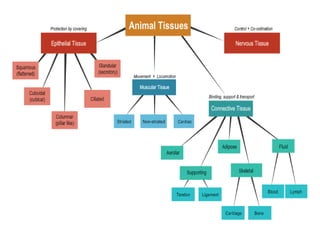


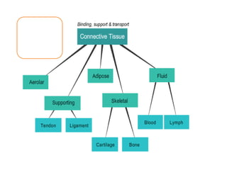
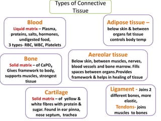
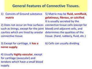
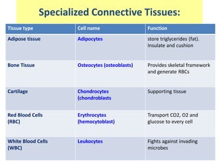
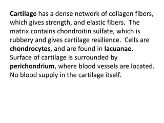
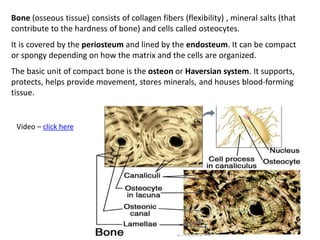
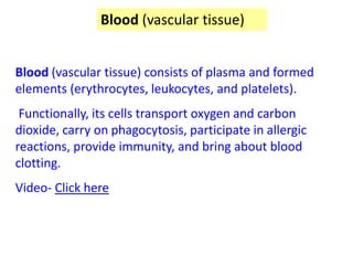
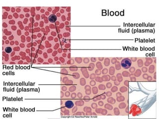
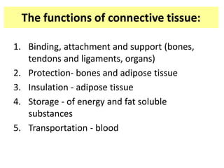


Connective tissues connect and support other tissues in the body. The main types are blood, bone, cartilage, dense connective tissue, adipose tissue, and blood. Blood transports nutrients and oxygen, removes waste, and has red blood cells, white blood cells and platelets. Bone provides structure, support and protection and contains collagen fibers and mineral salts. Cartilage contains collagen fibers and glycosaminoglycans to connect tissues and provide flexibility. Connective tissues come together to form the framework that holds the entire body together.
![29 [chapter 29 development and inheritance]](https://cdn.slidesharecdn.com/ss_thumbnails/29chapter29developmentandinheritance-170828044352-thumbnail.jpg?width=640&height=640&fit=bounds)


![11 [chapter 11 the muscular system]](https://cdn.slidesharecdn.com/ss_thumbnails/11chapter11themuscularsystem-170828041038-thumbnail.jpg?width=640&height=640&fit=bounds)


