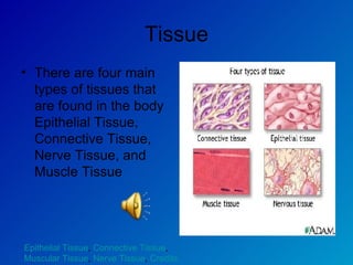
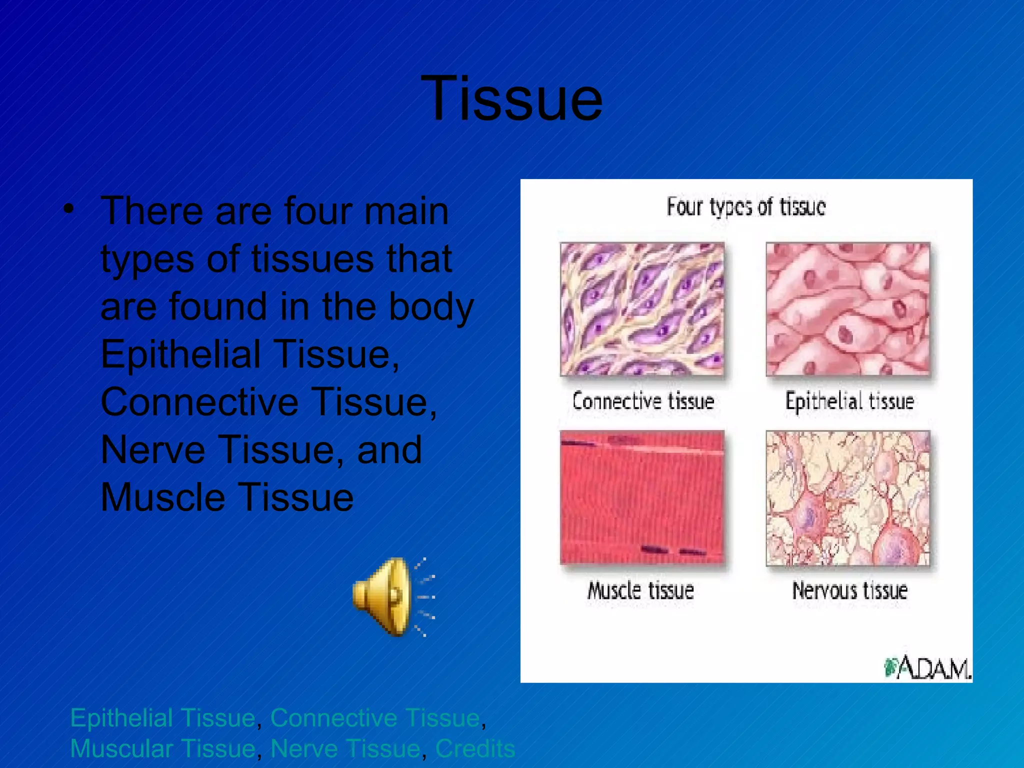
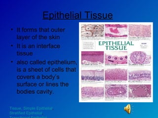
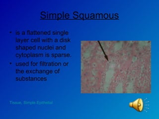
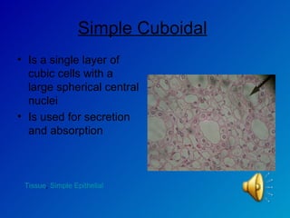
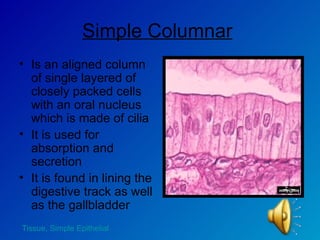
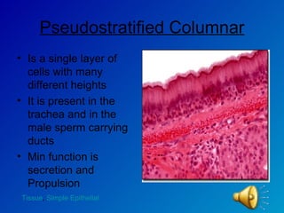
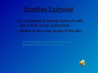
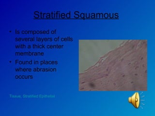
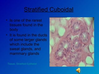
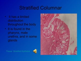
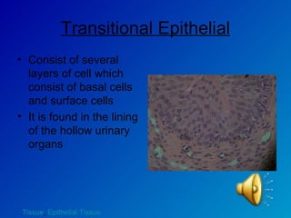
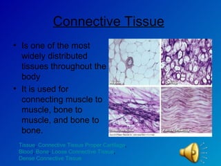
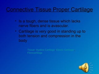
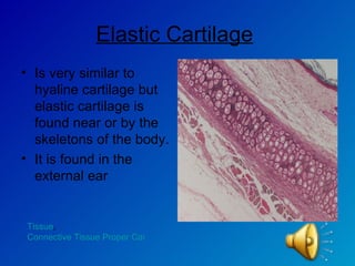
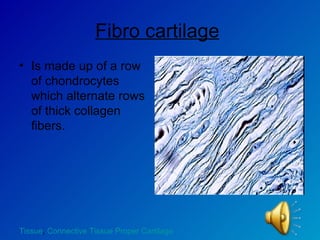
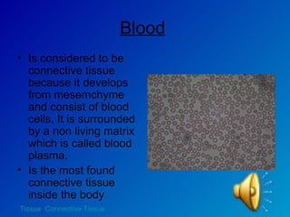
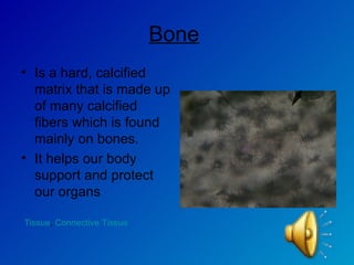
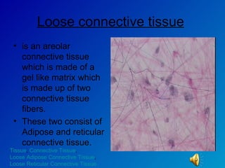
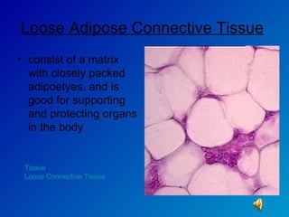
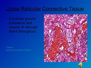
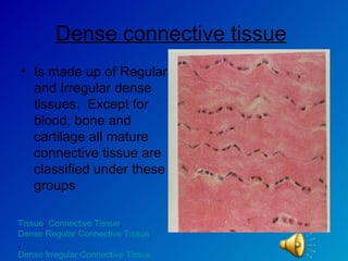
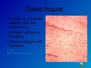
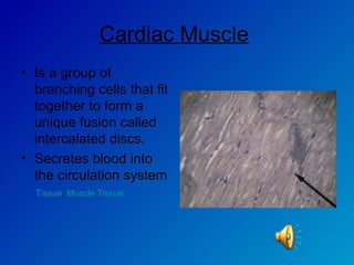
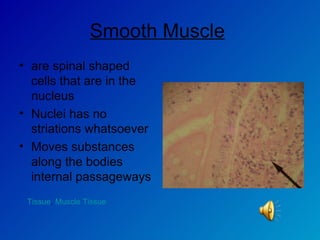
Tissue There are four main types of tissues that are found in the body Epithelial Tissue, Connective Tissue, Nerve Tissue, and Muscle Tissue Epithelial Tissue ,  Connective Tissue ,  Muscular Tissue ,  Nerve Tissue ,  Credits
Epithelial Tissue   It forms that outer layer of the skin  It is an interface tissue  also called epithelium, is a sheet of cells that covers a body’s surface or lines the bodies cavity. Tissue , Simple Epithelial ,  Stratified Epithelial ,  Transitional Epithelial
Simple Epithelial Is composed of single cell layer They are found where thin barriers are needed in the body as well as where absorption and filtration occur at  Tissue ,  Epithelial Tissue ,  Simple Squamous ,  Simple Cuboidal ,  Simple Columnar ,  Pseudostratified Columnar
Simple Squamous is a flattened single layer cell with a disk shaped nuclei and cytoplasm is sparse. used for filtration or the exchange of substances  Tissue,  Simple Epithelial
Simple Cuboidal   Is a single layer of cubic cells with a large spherical central nuclei  Is used for secretion and absorption  Tissue ,  Simple Epithelial
Simple Columnar   Is an aligned column of single layered of closely packed cells with an oral nucleus which is made of cilia It is used for absorption and secretion  It is found in lining the digestive track as well as the gallbladder  Tissue,  Simple Epithelial
Pseudostratified Columnar Is a single layer of cells with many different heights  It is present in the trachea and in the male sperm carrying ducts  Min function is secretion and Propulsion Tissue ,  Simple Epithelial
Stratified Epithelial   Is composed of several layers of cells with a thick center membrane  Makes up the outer layers of the skin Tissue ,  Epithelial Tissue ,  Stratified Squamous ,  Stratified Cuboidal ,  Stratified Columnar
Stratified Squamous   Is composed of several layers of cells with a thick center membrane  Found in places where abrasion occurs Tissue,  Stratified Epithelial
Stratified Cuboidal   Is one of the rarest tissues found in the body  It is found in the ducts of some larger glands which include the sweat glands, and mammary glands  Tissue,  Stratified Epithelial
Stratified Columnar It has a limited distribution throughout the body It is found in the pharynx, male urethra, and in some glands  Tissue ,  Stratified Epithelial
Transitional Epithelial Consist of several layers of cell which consist of basal cells and surface cells It is found in the lining of the hollow urinary organs  Tissue ,  Epithelial Tissue
Connective Tissue   Is one of the most widely distributed tissues throughout the body It is used for connecting muscle to muscle, bone to muscle, and bone to bone. Tissue ,  Connective Tissue Proper Cartilage ,  Blood ,  Bone ,  Loose Connective Tissue ,  Dense Connective Tissue
Connective Tissue Proper Cartilage   Is a tough, dense tissue which lacks nerve fibers and is avascular. Cartilage is very good in standing up to both tension and compression in the body  Tissue ,  Hyaline Cartilage ,  Elastic Cartilage ,  Fibrocartilage
Hyaline Cartilage   Is the most abundant cartilage found in the body  It mainly covers long bones and is mostly found in young children Tissue ,  Connective Tissue Proper Cartilage
Elastic Cartilage   Is very similar to hyaline cartilage but elastic cartilage is found near or by the skeletons of the body. It is found in the external ear  Tissue ,  Connective Tissue Proper Cartilage
Fibro cartilage   Is made up of a row of chondrocytes which alternate rows of thick collagen fibers. Tissue ,  Connective Tissue Proper Cartilage
Blood   Is considered to be connective tissue because it develops from mesemchyme and consist of blood cells. It is surrounded by a non living matrix which is called blood plasma.  Is the most found connective tissue inside the body Tissue ,  Connective Tissue
Bone   Is a hard, calcified matrix that is made up of many calcified fibers which is found mainly on bones. It helps our body support and protect our organs Tissue ,  Connective Tissue
Loose connective tissue   is an areolar connective tissue which is made of a gel like matrix which is made up of two connective tissue fibers.  These two consist of Adipose and reticular connective tissue.  Tissue ,  Connective Tissue ,  Loose Adipose Connective Tissue ,  Loose Reticular Connective Tissue
Loose Adipose Connective Tissue   consist of a matrix with closely packed adipoetyes, and is good for supporting and protecting organs in the body Tissue ,  Loose Connective Tissue
Loose Reticular Connective Tissue   is a loose ground substance and consist of reticular fibers throughout.  Tissue ,  loose Connective Tissue
Dense connective tissue   Is made up of Regular and Irregular dense tissues.  Except for blood, bone and cartilage all mature connective tissue are classified under these groups  Tissue ,  Connective Tissue ,  Dense Regular Connective Tissue ,  Dense Irregular Connective Tissue
Dense Regular   is made up of parallel collagen with few elastic fibers.  Its major cell type is fibroblast. Parallel collagen with few fibers Tissue ,  Dense Connective Tissue
Dense Irregular   Gets its name form the irregularly arranged collagen fibers mixed with some elastic fibers that are distributed throughout he body.  Tissue ,  Dense Connective Tissue
Nervous Tissue   Is the tissue in our body that allows us to be able to feel and sense things all around us. Nervous tissue also allows our body to respond to the simplest and smallest feel or presser.  Tissue
Muscle Tissue Are highly cellular tissues that are responsible for most of our bodies movement. Muscle cells produce myofilaments that enable contractions and movement in all cell types throughout the body.  Tissue ,  Skeletal Muscle ,  Cardiac Muscle ,  Smooth Muscle
Skeletal Muscle   Is a tightly compact connection of tissue sheets into organs that attack the bones of the skeleton together.  Tissue ,  Muscle Tissue
Cardiac Muscle   Is a group of branching cells that fit together to form a unique fusion called intercalated discs. Secretes blood into the circulation system Tissue ,  Muscle Tissue
Smooth Muscle   are spinal shaped cells that are in the nucleus  Nuclei has no striations whatsoever Moves substances along the bodies internal passageways Tissue ,  Muscle Tissue
Credits All Slides  Information by Justin Owen Voice Voice of Justin Owen
Credits Pictures Slide 1. By 2007 University of Maryland Medical Center (UMMC). 2007 University of Maryland Medical Center (UMMC). http://images.search.yahoo.com/search/images/view?back=http%3A%2F%2Fimages.search.yahoo.com%2Fsearch%2Fimages%3Fp%3Dtissue%26ei%3DUTF-8%26fr%3Dyfp-t-501%26x%3Dwrt%26js%3D1%26ni%3D21&w=400&h=320&imgurl=www.umm.edu%2Fgraphics%2Fimages%2Fen%2F8682.jpg&rurl=http%3A%2F%2Fwww.umm.edu%2Fimagepages%2F8682.htm&size=21.9kB&name=8682.jpg&p=tissue&type=jpeg&no=1&tt=507,329&oid=7ffec963eee56adc&ei=UTF-8 Slide 2. By author unknown. http://images.search.yahoo.com/search/images/view?back=http%3A%2F%2Fimages.search.yahoo.com%2Fsearch%2Fimages%3Fp%3Depithelial%2Btissue%26ei%3DUTF-8%26fr%3Dyfp-t-501%26x%3Dwrt%26js%3D1%26ni%3D21&w=449&h=600&imgurl=www.biocam.com%2Fimages%2Fwcepith2.jpg&rurl=http%3A%2F%2Fwww.biocam.com%2Fwcepith.htm&size=88.2kB&name=wcepith2.jpg&p=epithelial+tissue&type=jpeg&no=1&tt=774&oid=7b44db72e18c0560&ei=UTF-8
Credits Slide 3 By Stasinos Stavrianeas, Ph.D.  http://images.search.yahoo.com/search/images/view?back=http%3A%2F%2Fimages.search.yahoo.com%2Fsearch%2Fimages%3Fp%3Dsimple%2Bepithelial%26ei%3DUTF-8%26fr%3Dyfp-t-501%26x%3Dwrt%26js%3D1%26ni%3D21&w=270&h=504&imgurl=www.willamette.edu%2F%7Estas%2Fphysiology%2Flabs%2Flab1%2Fepithelial1a.jpg&rurl=http%3A%2F%2Fwww.willamette.edu%2F%7Estas%2Fphysiology%2Flabs%2Flab1%2Flab1.htm&size=40.4kB&name=epithelial1a.jpg&p=simple+epithelial&type=jpeg&no=13&tt=202 &oid=f3bde798eb8b7cd2&ei=UTF-8 Slide 4. By Calida  Justin and Amanda Slide 5.By  Calida Justin and Amanda
Credits Slide 6.  By Author Unknown http://images.search.yahoo.com/search/images/view?back=http%3A%2F%2Fimages.search.yahoo.com%2Fsearch%2Fimages%3Fp%3DSimple%2BColumnar%26ei%3DUTF-8%26fr%3Dyfp-t-501%26x%3Dwrt%26js%3D1%26ni%3D21&w=800&h=600&imgurl=www.technion.ac.il%2F%7Emdcourse%2F274203%2Fslides%2FEpithelium%2F4-Simple%2520Columnar%2520Epithelium.jpg&rurl=http%3A%2F%2Fwww.technion.ac.il%2F%7Emdcourse%2F274203%2Flect2.html&size=79.4kB&name=4-Simple+Columnar+Epithelium.jpg&p=Simple+Columnar&type= jpeg&no=1&tt=553&oid=c024af6db1879cb0&ei=UTF-8 Slide 7  By 2000-2007 Olympus, America Inc http://images.search.yahoo.com/search/images/view?back=http%3A%2F%2Fimages.search.yahoo.com%2Fsearch%2Fimages%3Fp%3DPseudostratified%2BEpithelial%26ei%3DUTF-8%26fr%3Dyfp-t-501%26x%3Dwrt%26js%3D1%26ni%3D21&w=508&h=368&imgurl=www.olympusmicro.com%2Fprimer%2Fanatomy%2Fbrightfieldgallery%2Fimages%2Fpseudostratifiedcilialarge.jpg&rurl=http%3A%2F%2Fwww.olympusmicro.com%2Fprimer%2Fanatomy%2Fbrightfieldgallery%2Fpseudostratifiedcilialarge.html&size=57.5kB&name=pseudostratifiedcilialarge.jpg&p=Pseudostratified+Epithelial&type=jpeg&no=5&tt=44&oid=01030af7f8b8a13c&ei=UTF-8
Credits Slide 9 By Calida, Justin and Amanda Slide 10 By  BIO-LCV (Physiology) - Photos of slides http://images.search.yahoo.com/search/images/view?back=http%3A%2F%2Fimages.search.yahoo.com%2Fsearch%2Fimages%3Fp%3DStratified%2BCuboidal%26ei%3DUTF-8%26fr%3Dyfp-t-501%26x%3Dwrt%26js%3D1%26ni%3D21&w=640&h=480&imgurl=www2.marianopolis.edu%2Fbio-lcv%2Fepithelials%2Fstratifiedcuboidalepithelium_sweatglandducts400x.jpg&rurl=http%3A%2F%2Fwww2.marianopolis.edu%2Fbio-lcv&size=69.7kB&name=stratifiedcuboidalepithelium_sweatglandducts400x.jpg&p=Stratified+Cuboidal&type=jpeg&no=10&tt=104&oid=df72b0e8341d4b3a&ei=UTF-8
Credits Slide 11  By  BY30 – Histology http://images.search.yahoo.com/search/images/view?back=http%3A%2F%2Fimages.search.yahoo.com%2Fsearch%2Fimages%3Fp%3DStratified%2BColumnar%26ei%3DUTF-8%26fr%3Dyfp-t-501%26x%3Dwrt%26js%3D1%26ni%3D21&w=1280&h=960&imgurl=www2.sunysuffolk.edu%2Fpickenc%2FStratified%2520Columnar%2520Epithelium%2520400X.JPG&rurl=http%3A%2F%2Fwww2.sunysuffolk.edu%2Fpickenc%2FBY30%2520Histology.htm&size=428.9kB&name=Stratified+Columnar+Epithelium+400X.JPG&p=Stratified+Columnar&type=jpeg&no=1&tt=150&oid=17e586175e4dc 0a6&ei=UTF-8 Slide 12  By Calida, Justin and Amanda
Credits Slide 13 By Author Unknown http://images.search.yahoo.com/search/images/view?back=http%3A%2F%2Fimages.search.yahoo.com%2Fsearch%2Fimages%3Fp%3DThe%2BConnective%2BTissues%26ei%3DUTF-8%26fr%3Dyfp-t-501%26x%3Dwrt%26js%3D1%26ni%3D21&w=609&h=425&imgurl=fig.cox.miami.edu%2F%7Ecmallery%2F150%2Fphysiol%2Fc40x2connective-pics.jpg&rurl=http%3A%2F%2Ffig.cox.miami.edu%2F%7Ecmallery%2F150%2Fphysiol%2Fphysiology.htm&size=84.8kB&name=c40x2connective-pics.jpg&p=The+Connective+Tissues&type =jpeg&no=2&tt=875&oid=db4db85f3479d828&ei=UTF-8 Slide 15  By Author Unknown  http://washington.uwc.edu/about/faculty/schaefer_w/TissuesPage.htm
Credits Slide 16 By  BY30 – Histology http://images.search.yahoo.com/search/images/view?back=http%3A%2F%2Fimages.search.yahoo.com%2Fsearch%2Fimages%3Fp%3DElastic%2BCartilage%26ei%3DUTF-8%26fr%3Dyfp-t-501%26x%3Dwrt%26js%3D1%26ni%3D21&w=1280&h=960&imgurl=www2.sunysuffolk.edu%2Fpickenc%2FElastic%2520Cartilage%2520100X.JPG&rurl=http%3A%2F%2Fwww2.sunysuffolk.edu%2Fpickenc%2FBY30%2520Histology.htm&size=453.7kB&name=Elastic+Cartilage+100X.JPG&p=Elastic+Cartilage&type=jpeg&no=12&tt=290&oid=32434dface27f1ba&ei=UTF- 8 Slide 17 By  K. Wynne  http://images.search.yahoo.com/search/images/view?back=http%3A%2F%2Fimages.search.yahoo.com%2Fsearch%2Fimages%3Fp%3DFibroCartilage%26sp%3D1%26fr2%3Dsp-top%26ei%3DUTF-8%26fr%3Dyfp-t-501%26x%3Dwrt%26js%3D1%26ni%3D21%26ei%3DUTF-8%26SpellState%3Dn-3517502926_q-jwcFP6Dwg9dFBn2N0pWbfwAAAA%40%40&w=800&h=600&imgurl=science.tjc.edu%2FCourse%2FBIOLOGY%2F1409%2Ffibrocartilage1.6-11.jpg&rurl=http%3A%2F%2Fscience.tjc.edu%2FCourse%2FBIOLOGY%2F1409%2F1409connective.htm&size=214.2kB&name=fibrocartilage1.6-11.jpg&p=FibroCartilage&type=jpeg&no=6&tt=236&oid=edcb0609530f8048&ei=UTF-8
Credits Slide 18  By  Calida, Justin and Amanda Slide 19  By  Calida, Justin and Amanda Slide 20 By  acpcommunity.acp.edu http://images.search.yahoo.com/search/images/view?back=http%3A%2F%2Fimages.search.yahoo.com%2Fsearch%2Fimages%3Fp%3DLoose%2BConnective%2BTissue%26ei%3DUTF-8%26fr%3Dyfp-t-501%26x%3Dwrt%26js%3D1%26ni%3D21&w=640&h=480&imgurl=acpcommunity.acp.edu%2FFacultystaff%2Fmoon%2FBiology2%2FImages%2FConnective%2FLoose%2520%28areolar%29%2520connective%2520tissue%2C%2520200x.JPG&rurl=http%3A%2F%2Facpcommunity.acp.edu%2FFacultystaff%2Fmoon%2FBiology2%2FImages%2FConnective&size=42.3kB&name=Loose+%28areolar%29+connective+tissue%2C+200x.JPG&p=Loose+Connective+Tissue&type=jpeg&no=5&tt=281&oid=1124d5ec7569f638&ei=UTF-8
Credits Slide 21  By Author Unknown http://images.search.yahoo.com/search/images/view?back=http%3A%2F%2Fimages.search.yahoo.com%2Fsearch%2Fimages%3Fp%3DLoose%2BAdipose%2BConnective%2BTissue%26ei%3DUTF-8%26fr%3Dyfp-t-501%26x%3Dwrt%26js%3D1%26ni%3D21&w=200&h=188&imgurl=pharyngula.org%2F%7Epzmyers%2FMyersLab%2Fteaching%2FBi104%2Fl04%2Fimg%2Fx-ct-loose-adipose.jpg&rurl=http%3A%2F%2Fpharyngula.org%2F%7Epzmyers%2FMyersLab%2Fteaching%2FBi104%2Fl04%2Floose.html&size=4.6kB&name=x-ct-loose-adipose .jpg&p=Loose+Adipose+Connective+Tissue&type=jpeg&no=7&tt=34&oid=1a62fd0292fb831a&ei=UTF-8
Credits Slide 22  By University of Texas Medical Branch   Cell Biology Graduate Program http://images.search.yahoo.com/search/images/view?back=http%3A%2F%2Fimages.search.yahoo.com%2Fsearch%2Fimages%3Fp%3DLoose%2BReticular%2BConnective%2BTissue%26ei%3DUTF-8%26fr%3Dyfp-t-501%26x%3Dwrt%26js%3D1%26ni%3D21&w=640&h=480&imgurl=cellbio.utmb.edu%2Fmicroanatomy%2Fconnective_tissue%2F00004518.jpg&rurl=http%3A%2F%2Fcellbio.utmb.edu%2Fmicroanatomy%2Fconnective_tissue%2Floose_connective_tissue.htm&size=208.3kB&name=00004518.jpg&p=Loose+Reticular+Connective+Tissue&type=jpeg&no=4&tt=12&oid=c858c 65cb0d517e4&ei=UTF-8 Slide 23 By Author Unknown http://images.search.yahoo.com/search/images/view?back=http%3A%2F%2Fimages.search.yahoo.com%2Fsearch%2Fimages%3Fp%3DDense%2BConnective%2BTissue%26ei%3DUTF-8%26fr%3Dyfp-t-501%26x%3Dwrt%26js%3D1%26ni%3D21&w=900&h=600&imgurl=www.public.coe.edu%2F%7Ekredborg%2FHumanslides%2Fdense.jpg&rurl=http%3A%2F%2Fwww.public.coe.edu%2F%7Ekredborg%2Fphys%2FExercise1.htm&size=290.8kB&name=dense.jpg&p=Dense+Connective+Tissue&type=jpeg&no=3&tt=367&oid=bfbff8a5add1475c&ei=UTF-8
Credits Slide 24  By Carols Classroom http://images.search.yahoo.com/search/images/view?back=http%3A%2F%2Fimages.search.yahoo.com%2Fsearch%2Fimages%3Fp%3Ddense%2Bregular%2Bconnective%2Btissue%26rs%3D0%26ei%3DUTF-8%26js%3D1%26ni%3D21%26fr%3Dyfp-t-501%26vf%3D&w=360&h=241&imgurl=www.carolguze.com%2Fimages%2Ftissues%2Fdense_regular_connective_tissue.jpg&rurl=http%3A%2F%2Fwww.carolguze.com%2Ftext%2F102-19-tissuesorgansystems.shtml&size=24.2kB&name=dense_regular_connective_tissue.jpg&p=dense+regular+connective+tissue&type=jpeg&no =9&tt=110&oid=fab5758e0b16f488&ei=UTF-8 Slide 25 By Author Unknownhttp://images.search.yahoo.com/search/images/view?back=http%3A%2F%2Fimages.search.yahoo.com%2Fsearch%2Fimages%3Fp%3Ddense%2Birregular%2Bconnective%2Btissue%26ei%3DUTF-8%26js%3D1%26ni%3D20%26fr%3Dyfp-t-501%26b%3D1&w=478&h=325&imgurl=www.cytochemistry.net%2Fcell-biology%2Fmedical%2Fpracti11.jpg&rurl=http%3A%2F%2Fwww.cytochemistry.net%2Fcell-biology%2Fmedical%2Fpractice_exam_connective_tissue.htm&size=31.6kB&name=practi11.jpg&p=dense+irregular+connective+tissue&type=jpeg&no=15&tt=102&oid=ae44334c54793152&ei=UTF-8
Credits Slide 26  By Calida, Justin and Amanda Slide 27  By Index of /~abs/Biowulf http://images.search.yahoo.com/search/images/view?back=http%3A%2F%2Fimages.search.yahoo.com%2Fsearch%2Fimages%3Fp%3DNeurons%26ei%3DUTF-8%26fr%3Dyfp-t-501%26x%3Dwrt%26js%3D1%26ni%3D20&w=640&h=480&imgurl=www.well.com%2F%7Eabs%2FBiowulf%2Fneurons.jpg&rurl=http%3A%2F%2Fwww.well.com%2F%7Eabs%2FBiowulf&size=75.3kB&name=neurons.jpg&p=Neurons&type=jpeg&no=14&tt=39,597&oid=8449e2d95563be3a&ei=UTF-8 Slide 29  By Author Unknown http://images.search.yahoo.com/search/images/view?back=http%3A%2F%2Fimages.search.yahoo.com%2Fsearch%2Fimages%3Fp%3Dskeletal%2Bmuscle%2Btissue%26rs%3D0%26ei%3DUTF-8%26js%3D1%26ni%3D20%26fr%3Dyfp-t-501%26vf%3D&w=825&h=508&imgurl=iupucbio2.iupui.edu%2Fanatomy%2Fimages%2FSlide1_16_0400a%2520Skeletal%2520Muscle%2520LS.jpg&rurl=http%3A%2F%2Fiupucbio2.iupui.edu%2Fanatomy%2Flab_chapt_10_skeletal_muscle_tissue.htm&size=140.4kB&name=Slide1_16_0400a+Skeletal+Muscle+LS.jpg&p=skeletal+muscle+tissue&type=jpeg&no=5&tt=396&oid=f66d45a7eec57d16&ei=UTF-8
Credits Slide 30  By Calida, Justin and Amanda Slide 31  By Calida, Justin and Amanda

Tissue Slide Show

  • 1.
    Tissue There arefour main types of tissues that are found in the body Epithelial Tissue, Connective Tissue, Nerve Tissue, and Muscle Tissue Epithelial Tissue , Connective Tissue , Muscular Tissue , Nerve Tissue , Credits
  • 2.
    Epithelial Tissue It forms that outer layer of the skin It is an interface tissue also called epithelium, is a sheet of cells that covers a body’s surface or lines the bodies cavity. Tissue , Simple Epithelial , Stratified Epithelial , Transitional Epithelial
  • 3.
    Simple Epithelial Iscomposed of single cell layer They are found where thin barriers are needed in the body as well as where absorption and filtration occur at Tissue , Epithelial Tissue , Simple Squamous , Simple Cuboidal , Simple Columnar , Pseudostratified Columnar
  • 4.
    Simple Squamous isa flattened single layer cell with a disk shaped nuclei and cytoplasm is sparse. used for filtration or the exchange of substances Tissue, Simple Epithelial
  • 5.
    Simple Cuboidal Is a single layer of cubic cells with a large spherical central nuclei Is used for secretion and absorption Tissue , Simple Epithelial
  • 6.
    Simple Columnar Is an aligned column of single layered of closely packed cells with an oral nucleus which is made of cilia It is used for absorption and secretion It is found in lining the digestive track as well as the gallbladder Tissue, Simple Epithelial
  • 7.
    Pseudostratified Columnar Isa single layer of cells with many different heights It is present in the trachea and in the male sperm carrying ducts Min function is secretion and Propulsion Tissue , Simple Epithelial
  • 8.
    Stratified Epithelial Is composed of several layers of cells with a thick center membrane Makes up the outer layers of the skin Tissue , Epithelial Tissue , Stratified Squamous , Stratified Cuboidal , Stratified Columnar
  • 9.
    Stratified Squamous Is composed of several layers of cells with a thick center membrane Found in places where abrasion occurs Tissue, Stratified Epithelial
  • 10.
    Stratified Cuboidal Is one of the rarest tissues found in the body It is found in the ducts of some larger glands which include the sweat glands, and mammary glands Tissue, Stratified Epithelial
  • 11.
    Stratified Columnar Ithas a limited distribution throughout the body It is found in the pharynx, male urethra, and in some glands Tissue , Stratified Epithelial
  • 12.
    Transitional Epithelial Consistof several layers of cell which consist of basal cells and surface cells It is found in the lining of the hollow urinary organs Tissue , Epithelial Tissue
  • 13.
    Connective Tissue Is one of the most widely distributed tissues throughout the body It is used for connecting muscle to muscle, bone to muscle, and bone to bone. Tissue , Connective Tissue Proper Cartilage , Blood , Bone , Loose Connective Tissue , Dense Connective Tissue
  • 14.
    Connective Tissue ProperCartilage Is a tough, dense tissue which lacks nerve fibers and is avascular. Cartilage is very good in standing up to both tension and compression in the body Tissue , Hyaline Cartilage , Elastic Cartilage , Fibrocartilage
  • 15.
    Hyaline Cartilage Is the most abundant cartilage found in the body It mainly covers long bones and is mostly found in young children Tissue , Connective Tissue Proper Cartilage
  • 16.
    Elastic Cartilage Is very similar to hyaline cartilage but elastic cartilage is found near or by the skeletons of the body. It is found in the external ear Tissue , Connective Tissue Proper Cartilage
  • 17.
    Fibro cartilage Is made up of a row of chondrocytes which alternate rows of thick collagen fibers. Tissue , Connective Tissue Proper Cartilage
  • 18.
    Blood Is considered to be connective tissue because it develops from mesemchyme and consist of blood cells. It is surrounded by a non living matrix which is called blood plasma. Is the most found connective tissue inside the body Tissue , Connective Tissue
  • 19.
    Bone Is a hard, calcified matrix that is made up of many calcified fibers which is found mainly on bones. It helps our body support and protect our organs Tissue , Connective Tissue
  • 20.
    Loose connective tissue is an areolar connective tissue which is made of a gel like matrix which is made up of two connective tissue fibers. These two consist of Adipose and reticular connective tissue. Tissue , Connective Tissue , Loose Adipose Connective Tissue , Loose Reticular Connective Tissue
  • 21.
    Loose Adipose ConnectiveTissue consist of a matrix with closely packed adipoetyes, and is good for supporting and protecting organs in the body Tissue , Loose Connective Tissue
  • 22.
    Loose Reticular ConnectiveTissue is a loose ground substance and consist of reticular fibers throughout. Tissue , loose Connective Tissue
  • 23.
    Dense connective tissue Is made up of Regular and Irregular dense tissues. Except for blood, bone and cartilage all mature connective tissue are classified under these groups Tissue , Connective Tissue , Dense Regular Connective Tissue , Dense Irregular Connective Tissue
  • 24.
    Dense Regular is made up of parallel collagen with few elastic fibers. Its major cell type is fibroblast. Parallel collagen with few fibers Tissue , Dense Connective Tissue
  • 25.
    Dense Irregular Gets its name form the irregularly arranged collagen fibers mixed with some elastic fibers that are distributed throughout he body. Tissue , Dense Connective Tissue
  • 26.
    Nervous Tissue Is the tissue in our body that allows us to be able to feel and sense things all around us. Nervous tissue also allows our body to respond to the simplest and smallest feel or presser. Tissue
  • 27.
    Muscle Tissue Arehighly cellular tissues that are responsible for most of our bodies movement. Muscle cells produce myofilaments that enable contractions and movement in all cell types throughout the body. Tissue , Skeletal Muscle , Cardiac Muscle , Smooth Muscle
  • 28.
    Skeletal Muscle Is a tightly compact connection of tissue sheets into organs that attack the bones of the skeleton together. Tissue , Muscle Tissue
  • 29.
    Cardiac Muscle Is a group of branching cells that fit together to form a unique fusion called intercalated discs. Secretes blood into the circulation system Tissue , Muscle Tissue
  • 30.
    Smooth Muscle are spinal shaped cells that are in the nucleus Nuclei has no striations whatsoever Moves substances along the bodies internal passageways Tissue , Muscle Tissue
  • 31.
    Credits All Slides Information by Justin Owen Voice Voice of Justin Owen
  • 32.
    Credits Pictures Slide1. By 2007 University of Maryland Medical Center (UMMC). 2007 University of Maryland Medical Center (UMMC). http://images.search.yahoo.com/search/images/view?back=http%3A%2F%2Fimages.search.yahoo.com%2Fsearch%2Fimages%3Fp%3Dtissue%26ei%3DUTF-8%26fr%3Dyfp-t-501%26x%3Dwrt%26js%3D1%26ni%3D21&w=400&h=320&imgurl=www.umm.edu%2Fgraphics%2Fimages%2Fen%2F8682.jpg&rurl=http%3A%2F%2Fwww.umm.edu%2Fimagepages%2F8682.htm&size=21.9kB&name=8682.jpg&p=tissue&type=jpeg&no=1&tt=507,329&oid=7ffec963eee56adc&ei=UTF-8 Slide 2. By author unknown. http://images.search.yahoo.com/search/images/view?back=http%3A%2F%2Fimages.search.yahoo.com%2Fsearch%2Fimages%3Fp%3Depithelial%2Btissue%26ei%3DUTF-8%26fr%3Dyfp-t-501%26x%3Dwrt%26js%3D1%26ni%3D21&w=449&h=600&imgurl=www.biocam.com%2Fimages%2Fwcepith2.jpg&rurl=http%3A%2F%2Fwww.biocam.com%2Fwcepith.htm&size=88.2kB&name=wcepith2.jpg&p=epithelial+tissue&type=jpeg&no=1&tt=774&oid=7b44db72e18c0560&ei=UTF-8
  • 33.
    Credits Slide 3By Stasinos Stavrianeas, Ph.D. http://images.search.yahoo.com/search/images/view?back=http%3A%2F%2Fimages.search.yahoo.com%2Fsearch%2Fimages%3Fp%3Dsimple%2Bepithelial%26ei%3DUTF-8%26fr%3Dyfp-t-501%26x%3Dwrt%26js%3D1%26ni%3D21&w=270&h=504&imgurl=www.willamette.edu%2F%7Estas%2Fphysiology%2Flabs%2Flab1%2Fepithelial1a.jpg&rurl=http%3A%2F%2Fwww.willamette.edu%2F%7Estas%2Fphysiology%2Flabs%2Flab1%2Flab1.htm&size=40.4kB&name=epithelial1a.jpg&p=simple+epithelial&type=jpeg&no=13&tt=202 &oid=f3bde798eb8b7cd2&ei=UTF-8 Slide 4. By Calida Justin and Amanda Slide 5.By Calida Justin and Amanda
  • 34.
    Credits Slide 6. By Author Unknown http://images.search.yahoo.com/search/images/view?back=http%3A%2F%2Fimages.search.yahoo.com%2Fsearch%2Fimages%3Fp%3DSimple%2BColumnar%26ei%3DUTF-8%26fr%3Dyfp-t-501%26x%3Dwrt%26js%3D1%26ni%3D21&w=800&h=600&imgurl=www.technion.ac.il%2F%7Emdcourse%2F274203%2Fslides%2FEpithelium%2F4-Simple%2520Columnar%2520Epithelium.jpg&rurl=http%3A%2F%2Fwww.technion.ac.il%2F%7Emdcourse%2F274203%2Flect2.html&size=79.4kB&name=4-Simple+Columnar+Epithelium.jpg&p=Simple+Columnar&type= jpeg&no=1&tt=553&oid=c024af6db1879cb0&ei=UTF-8 Slide 7 By 2000-2007 Olympus, America Inc http://images.search.yahoo.com/search/images/view?back=http%3A%2F%2Fimages.search.yahoo.com%2Fsearch%2Fimages%3Fp%3DPseudostratified%2BEpithelial%26ei%3DUTF-8%26fr%3Dyfp-t-501%26x%3Dwrt%26js%3D1%26ni%3D21&w=508&h=368&imgurl=www.olympusmicro.com%2Fprimer%2Fanatomy%2Fbrightfieldgallery%2Fimages%2Fpseudostratifiedcilialarge.jpg&rurl=http%3A%2F%2Fwww.olympusmicro.com%2Fprimer%2Fanatomy%2Fbrightfieldgallery%2Fpseudostratifiedcilialarge.html&size=57.5kB&name=pseudostratifiedcilialarge.jpg&p=Pseudostratified+Epithelial&type=jpeg&no=5&tt=44&oid=01030af7f8b8a13c&ei=UTF-8
  • 35.
    Credits Slide 9By Calida, Justin and Amanda Slide 10 By BIO-LCV (Physiology) - Photos of slides http://images.search.yahoo.com/search/images/view?back=http%3A%2F%2Fimages.search.yahoo.com%2Fsearch%2Fimages%3Fp%3DStratified%2BCuboidal%26ei%3DUTF-8%26fr%3Dyfp-t-501%26x%3Dwrt%26js%3D1%26ni%3D21&w=640&h=480&imgurl=www2.marianopolis.edu%2Fbio-lcv%2Fepithelials%2Fstratifiedcuboidalepithelium_sweatglandducts400x.jpg&rurl=http%3A%2F%2Fwww2.marianopolis.edu%2Fbio-lcv&size=69.7kB&name=stratifiedcuboidalepithelium_sweatglandducts400x.jpg&p=Stratified+Cuboidal&type=jpeg&no=10&tt=104&oid=df72b0e8341d4b3a&ei=UTF-8
  • 36.
    Credits Slide 11 By BY30 – Histology http://images.search.yahoo.com/search/images/view?back=http%3A%2F%2Fimages.search.yahoo.com%2Fsearch%2Fimages%3Fp%3DStratified%2BColumnar%26ei%3DUTF-8%26fr%3Dyfp-t-501%26x%3Dwrt%26js%3D1%26ni%3D21&w=1280&h=960&imgurl=www2.sunysuffolk.edu%2Fpickenc%2FStratified%2520Columnar%2520Epithelium%2520400X.JPG&rurl=http%3A%2F%2Fwww2.sunysuffolk.edu%2Fpickenc%2FBY30%2520Histology.htm&size=428.9kB&name=Stratified+Columnar+Epithelium+400X.JPG&p=Stratified+Columnar&type=jpeg&no=1&tt=150&oid=17e586175e4dc 0a6&ei=UTF-8 Slide 12 By Calida, Justin and Amanda
  • 37.
    Credits Slide 13By Author Unknown http://images.search.yahoo.com/search/images/view?back=http%3A%2F%2Fimages.search.yahoo.com%2Fsearch%2Fimages%3Fp%3DThe%2BConnective%2BTissues%26ei%3DUTF-8%26fr%3Dyfp-t-501%26x%3Dwrt%26js%3D1%26ni%3D21&w=609&h=425&imgurl=fig.cox.miami.edu%2F%7Ecmallery%2F150%2Fphysiol%2Fc40x2connective-pics.jpg&rurl=http%3A%2F%2Ffig.cox.miami.edu%2F%7Ecmallery%2F150%2Fphysiol%2Fphysiology.htm&size=84.8kB&name=c40x2connective-pics.jpg&p=The+Connective+Tissues&type =jpeg&no=2&tt=875&oid=db4db85f3479d828&ei=UTF-8 Slide 15 By Author Unknown http://washington.uwc.edu/about/faculty/schaefer_w/TissuesPage.htm
  • 38.
    Credits Slide 16By BY30 – Histology http://images.search.yahoo.com/search/images/view?back=http%3A%2F%2Fimages.search.yahoo.com%2Fsearch%2Fimages%3Fp%3DElastic%2BCartilage%26ei%3DUTF-8%26fr%3Dyfp-t-501%26x%3Dwrt%26js%3D1%26ni%3D21&w=1280&h=960&imgurl=www2.sunysuffolk.edu%2Fpickenc%2FElastic%2520Cartilage%2520100X.JPG&rurl=http%3A%2F%2Fwww2.sunysuffolk.edu%2Fpickenc%2FBY30%2520Histology.htm&size=453.7kB&name=Elastic+Cartilage+100X.JPG&p=Elastic+Cartilage&type=jpeg&no=12&tt=290&oid=32434dface27f1ba&ei=UTF- 8 Slide 17 By K. Wynne http://images.search.yahoo.com/search/images/view?back=http%3A%2F%2Fimages.search.yahoo.com%2Fsearch%2Fimages%3Fp%3DFibroCartilage%26sp%3D1%26fr2%3Dsp-top%26ei%3DUTF-8%26fr%3Dyfp-t-501%26x%3Dwrt%26js%3D1%26ni%3D21%26ei%3DUTF-8%26SpellState%3Dn-3517502926_q-jwcFP6Dwg9dFBn2N0pWbfwAAAA%40%40&w=800&h=600&imgurl=science.tjc.edu%2FCourse%2FBIOLOGY%2F1409%2Ffibrocartilage1.6-11.jpg&rurl=http%3A%2F%2Fscience.tjc.edu%2FCourse%2FBIOLOGY%2F1409%2F1409connective.htm&size=214.2kB&name=fibrocartilage1.6-11.jpg&p=FibroCartilage&type=jpeg&no=6&tt=236&oid=edcb0609530f8048&ei=UTF-8
  • 39.
    Credits Slide 18 By Calida, Justin and Amanda Slide 19 By Calida, Justin and Amanda Slide 20 By acpcommunity.acp.edu http://images.search.yahoo.com/search/images/view?back=http%3A%2F%2Fimages.search.yahoo.com%2Fsearch%2Fimages%3Fp%3DLoose%2BConnective%2BTissue%26ei%3DUTF-8%26fr%3Dyfp-t-501%26x%3Dwrt%26js%3D1%26ni%3D21&w=640&h=480&imgurl=acpcommunity.acp.edu%2FFacultystaff%2Fmoon%2FBiology2%2FImages%2FConnective%2FLoose%2520%28areolar%29%2520connective%2520tissue%2C%2520200x.JPG&rurl=http%3A%2F%2Facpcommunity.acp.edu%2FFacultystaff%2Fmoon%2FBiology2%2FImages%2FConnective&size=42.3kB&name=Loose+%28areolar%29+connective+tissue%2C+200x.JPG&p=Loose+Connective+Tissue&type=jpeg&no=5&tt=281&oid=1124d5ec7569f638&ei=UTF-8
  • 40.
    Credits Slide 21 By Author Unknown http://images.search.yahoo.com/search/images/view?back=http%3A%2F%2Fimages.search.yahoo.com%2Fsearch%2Fimages%3Fp%3DLoose%2BAdipose%2BConnective%2BTissue%26ei%3DUTF-8%26fr%3Dyfp-t-501%26x%3Dwrt%26js%3D1%26ni%3D21&w=200&h=188&imgurl=pharyngula.org%2F%7Epzmyers%2FMyersLab%2Fteaching%2FBi104%2Fl04%2Fimg%2Fx-ct-loose-adipose.jpg&rurl=http%3A%2F%2Fpharyngula.org%2F%7Epzmyers%2FMyersLab%2Fteaching%2FBi104%2Fl04%2Floose.html&size=4.6kB&name=x-ct-loose-adipose .jpg&p=Loose+Adipose+Connective+Tissue&type=jpeg&no=7&tt=34&oid=1a62fd0292fb831a&ei=UTF-8
  • 41.
    Credits Slide 22 By University of Texas Medical Branch   Cell Biology Graduate Program http://images.search.yahoo.com/search/images/view?back=http%3A%2F%2Fimages.search.yahoo.com%2Fsearch%2Fimages%3Fp%3DLoose%2BReticular%2BConnective%2BTissue%26ei%3DUTF-8%26fr%3Dyfp-t-501%26x%3Dwrt%26js%3D1%26ni%3D21&w=640&h=480&imgurl=cellbio.utmb.edu%2Fmicroanatomy%2Fconnective_tissue%2F00004518.jpg&rurl=http%3A%2F%2Fcellbio.utmb.edu%2Fmicroanatomy%2Fconnective_tissue%2Floose_connective_tissue.htm&size=208.3kB&name=00004518.jpg&p=Loose+Reticular+Connective+Tissue&type=jpeg&no=4&tt=12&oid=c858c 65cb0d517e4&ei=UTF-8 Slide 23 By Author Unknown http://images.search.yahoo.com/search/images/view?back=http%3A%2F%2Fimages.search.yahoo.com%2Fsearch%2Fimages%3Fp%3DDense%2BConnective%2BTissue%26ei%3DUTF-8%26fr%3Dyfp-t-501%26x%3Dwrt%26js%3D1%26ni%3D21&w=900&h=600&imgurl=www.public.coe.edu%2F%7Ekredborg%2FHumanslides%2Fdense.jpg&rurl=http%3A%2F%2Fwww.public.coe.edu%2F%7Ekredborg%2Fphys%2FExercise1.htm&size=290.8kB&name=dense.jpg&p=Dense+Connective+Tissue&type=jpeg&no=3&tt=367&oid=bfbff8a5add1475c&ei=UTF-8
  • 42.
    Credits Slide 24 By Carols Classroom http://images.search.yahoo.com/search/images/view?back=http%3A%2F%2Fimages.search.yahoo.com%2Fsearch%2Fimages%3Fp%3Ddense%2Bregular%2Bconnective%2Btissue%26rs%3D0%26ei%3DUTF-8%26js%3D1%26ni%3D21%26fr%3Dyfp-t-501%26vf%3D&w=360&h=241&imgurl=www.carolguze.com%2Fimages%2Ftissues%2Fdense_regular_connective_tissue.jpg&rurl=http%3A%2F%2Fwww.carolguze.com%2Ftext%2F102-19-tissuesorgansystems.shtml&size=24.2kB&name=dense_regular_connective_tissue.jpg&p=dense+regular+connective+tissue&type=jpeg&no =9&tt=110&oid=fab5758e0b16f488&ei=UTF-8 Slide 25 By Author Unknownhttp://images.search.yahoo.com/search/images/view?back=http%3A%2F%2Fimages.search.yahoo.com%2Fsearch%2Fimages%3Fp%3Ddense%2Birregular%2Bconnective%2Btissue%26ei%3DUTF-8%26js%3D1%26ni%3D20%26fr%3Dyfp-t-501%26b%3D1&w=478&h=325&imgurl=www.cytochemistry.net%2Fcell-biology%2Fmedical%2Fpracti11.jpg&rurl=http%3A%2F%2Fwww.cytochemistry.net%2Fcell-biology%2Fmedical%2Fpractice_exam_connective_tissue.htm&size=31.6kB&name=practi11.jpg&p=dense+irregular+connective+tissue&type=jpeg&no=15&tt=102&oid=ae44334c54793152&ei=UTF-8
  • 43.
    Credits Slide 26 By Calida, Justin and Amanda Slide 27 By Index of /~abs/Biowulf http://images.search.yahoo.com/search/images/view?back=http%3A%2F%2Fimages.search.yahoo.com%2Fsearch%2Fimages%3Fp%3DNeurons%26ei%3DUTF-8%26fr%3Dyfp-t-501%26x%3Dwrt%26js%3D1%26ni%3D20&w=640&h=480&imgurl=www.well.com%2F%7Eabs%2FBiowulf%2Fneurons.jpg&rurl=http%3A%2F%2Fwww.well.com%2F%7Eabs%2FBiowulf&size=75.3kB&name=neurons.jpg&p=Neurons&type=jpeg&no=14&tt=39,597&oid=8449e2d95563be3a&ei=UTF-8 Slide 29 By Author Unknown http://images.search.yahoo.com/search/images/view?back=http%3A%2F%2Fimages.search.yahoo.com%2Fsearch%2Fimages%3Fp%3Dskeletal%2Bmuscle%2Btissue%26rs%3D0%26ei%3DUTF-8%26js%3D1%26ni%3D20%26fr%3Dyfp-t-501%26vf%3D&w=825&h=508&imgurl=iupucbio2.iupui.edu%2Fanatomy%2Fimages%2FSlide1_16_0400a%2520Skeletal%2520Muscle%2520LS.jpg&rurl=http%3A%2F%2Fiupucbio2.iupui.edu%2Fanatomy%2Flab_chapt_10_skeletal_muscle_tissue.htm&size=140.4kB&name=Slide1_16_0400a+Skeletal+Muscle+LS.jpg&p=skeletal+muscle+tissue&type=jpeg&no=5&tt=396&oid=f66d45a7eec57d16&ei=UTF-8
  • 44.
    Credits Slide 30 By Calida, Justin and Amanda Slide 31 By Calida, Justin and Amanda