Downloaded 176 times


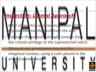
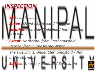


The examination of the thyroid gland involves inspection to observe the size, shape, and movement of the gland, palpation to evaluate the texture, mobility, and presence of nodules, and synthesizing the findings from inspection and palpation to characterize the condition of the gland. Symptoms of thyroid disorders vary depending on whether the gland is underactive, overactive, or cancerous and include changes in heart rate, weight, mood, and appearance of the eyes and skin. The sex, age, occupation, and place of residence of the patient provide clues about their risk for developing different thyroid conditions.


![Goiters 120223070355-phpapp01[1]](https://cdn.slidesharecdn.com/ss_thumbnails/goiters-120223070355-phpapp011-120227054943-phpapp02-thumbnail.jpg?width=640&height=640&fit=bounds)