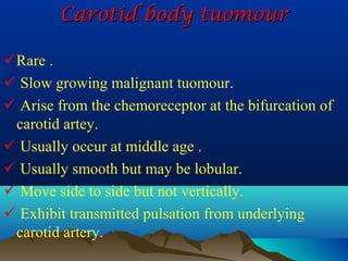
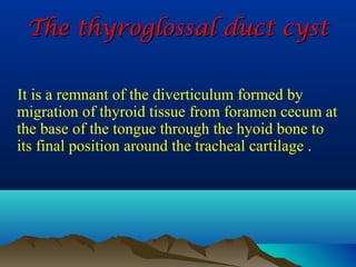
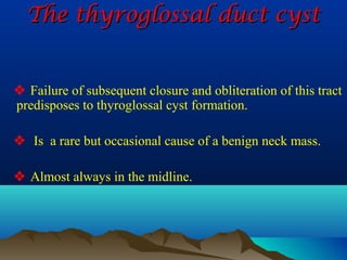
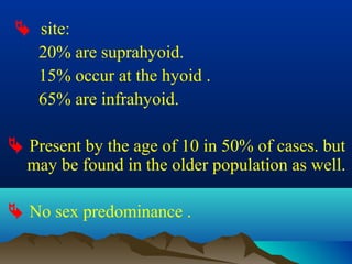
Downloaded 2,421 times


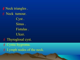

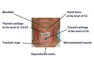
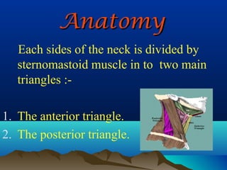
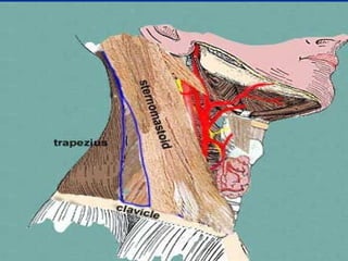


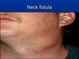
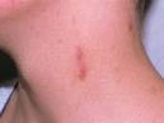
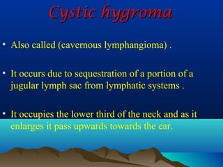
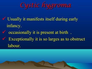


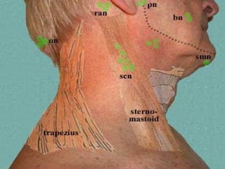


The document discusses the anatomy and triangles of the neck, describing boundaries, contents, and clinical significance. It also covers common neck masses including cysts, sinuses, fistulas, ulcers, tumors, and infections. Lymphatic drainage is described for deep cervical nodes along vertical and circular chains.


![Triangles of the_neck[1]](https://cdn.slidesharecdn.com/ss_thumbnails/trianglesoftheneck1-180125150132-thumbnail.jpg?width=640&height=640&fit=bounds)


