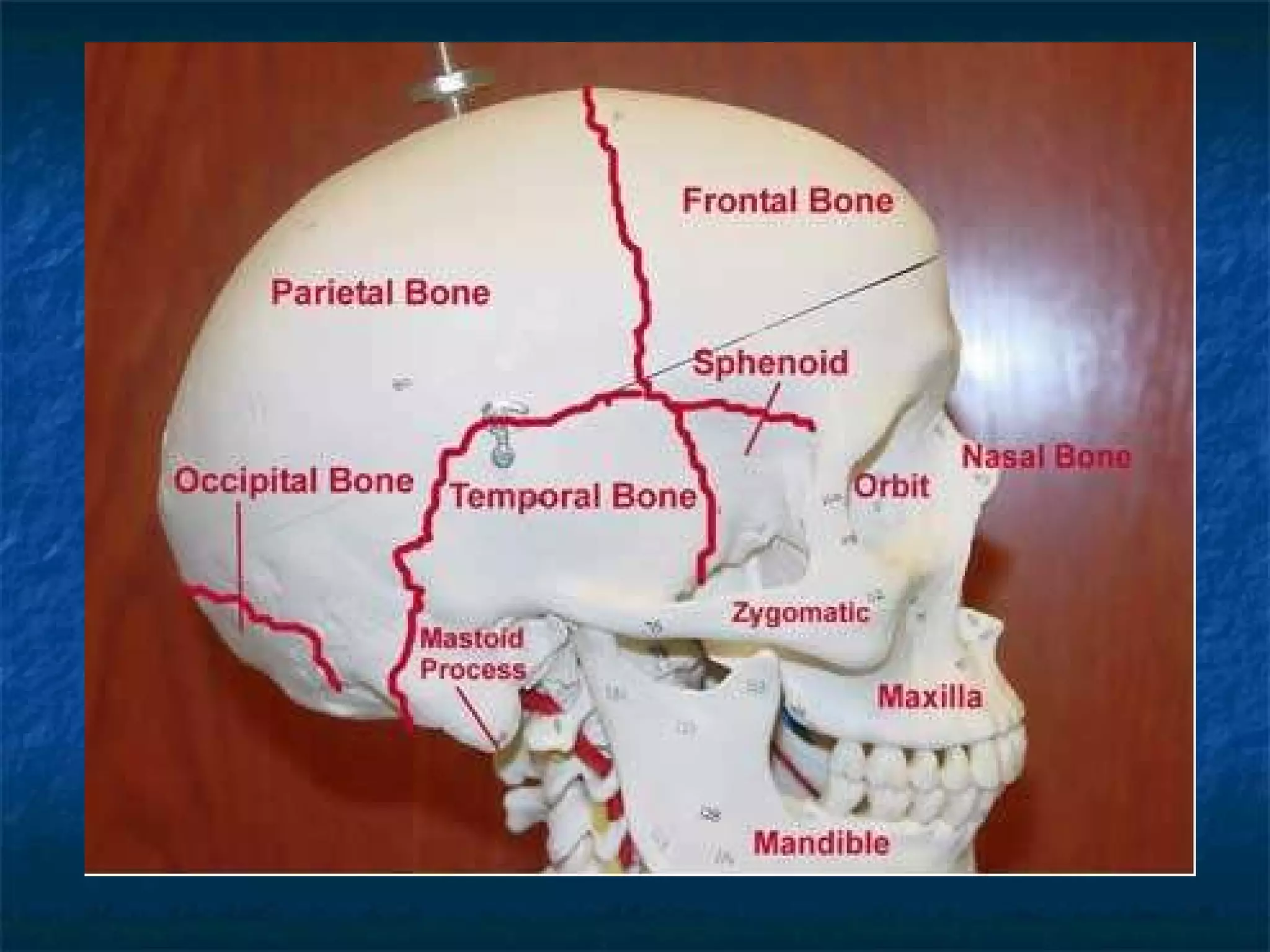
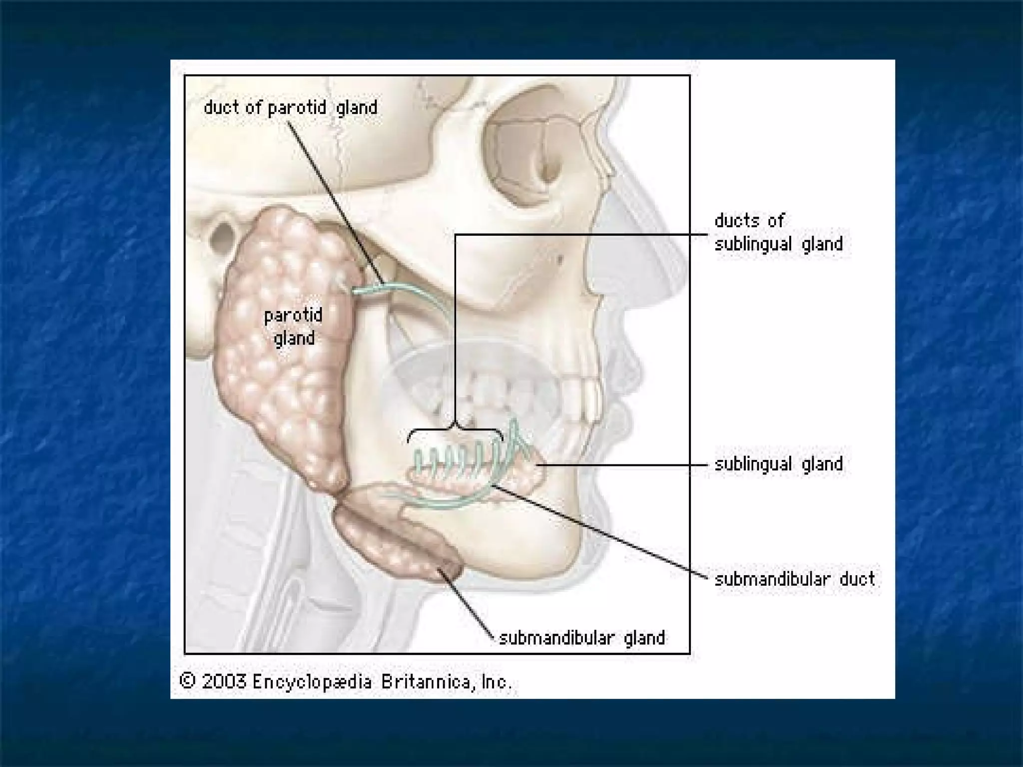
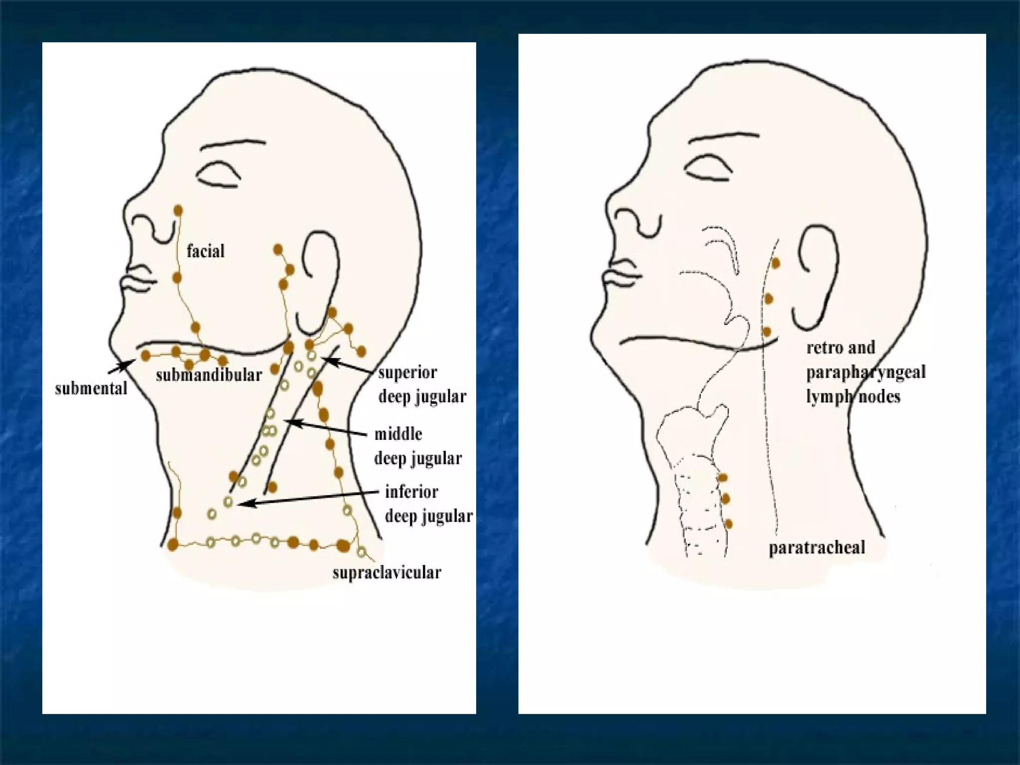
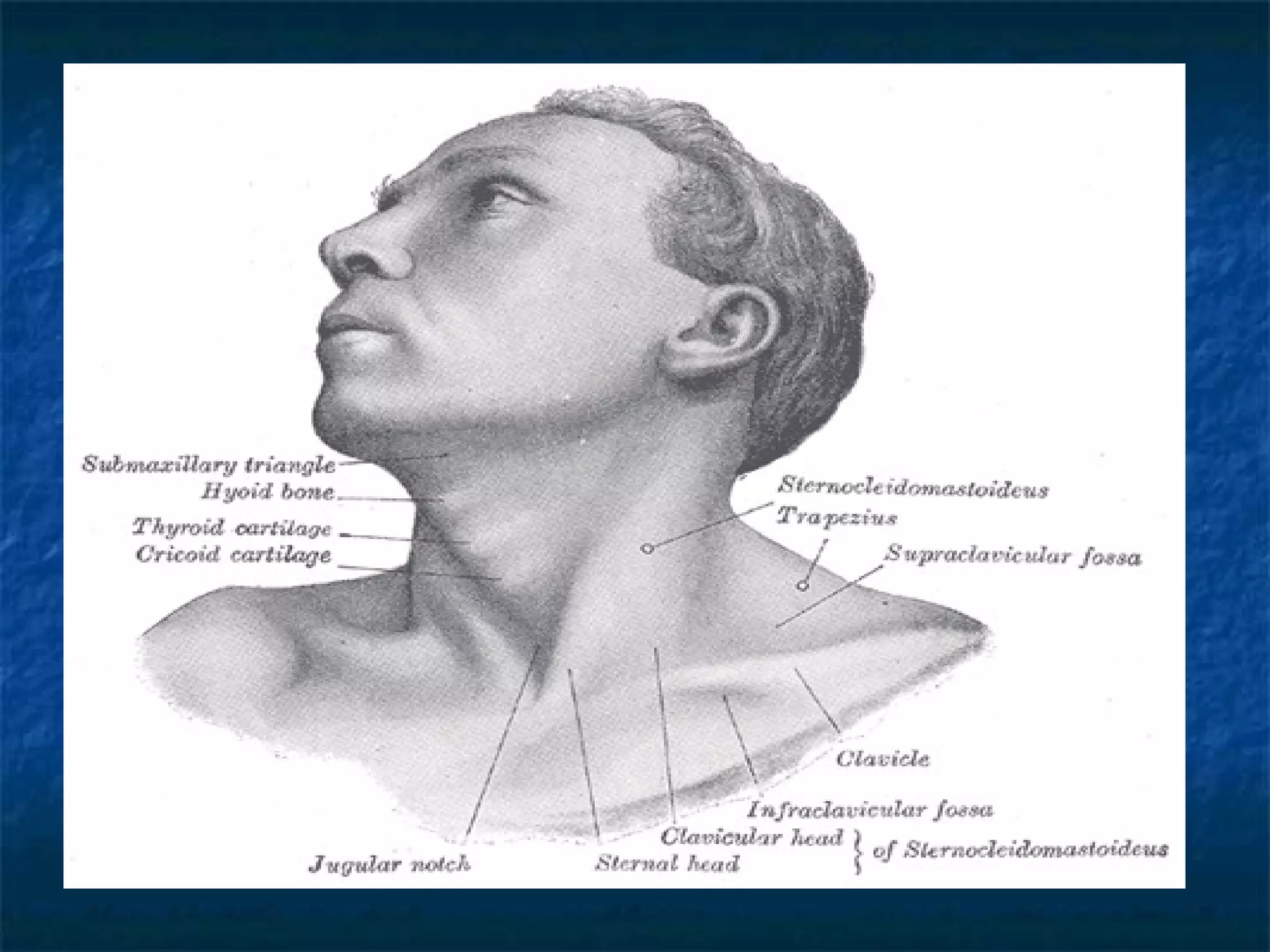
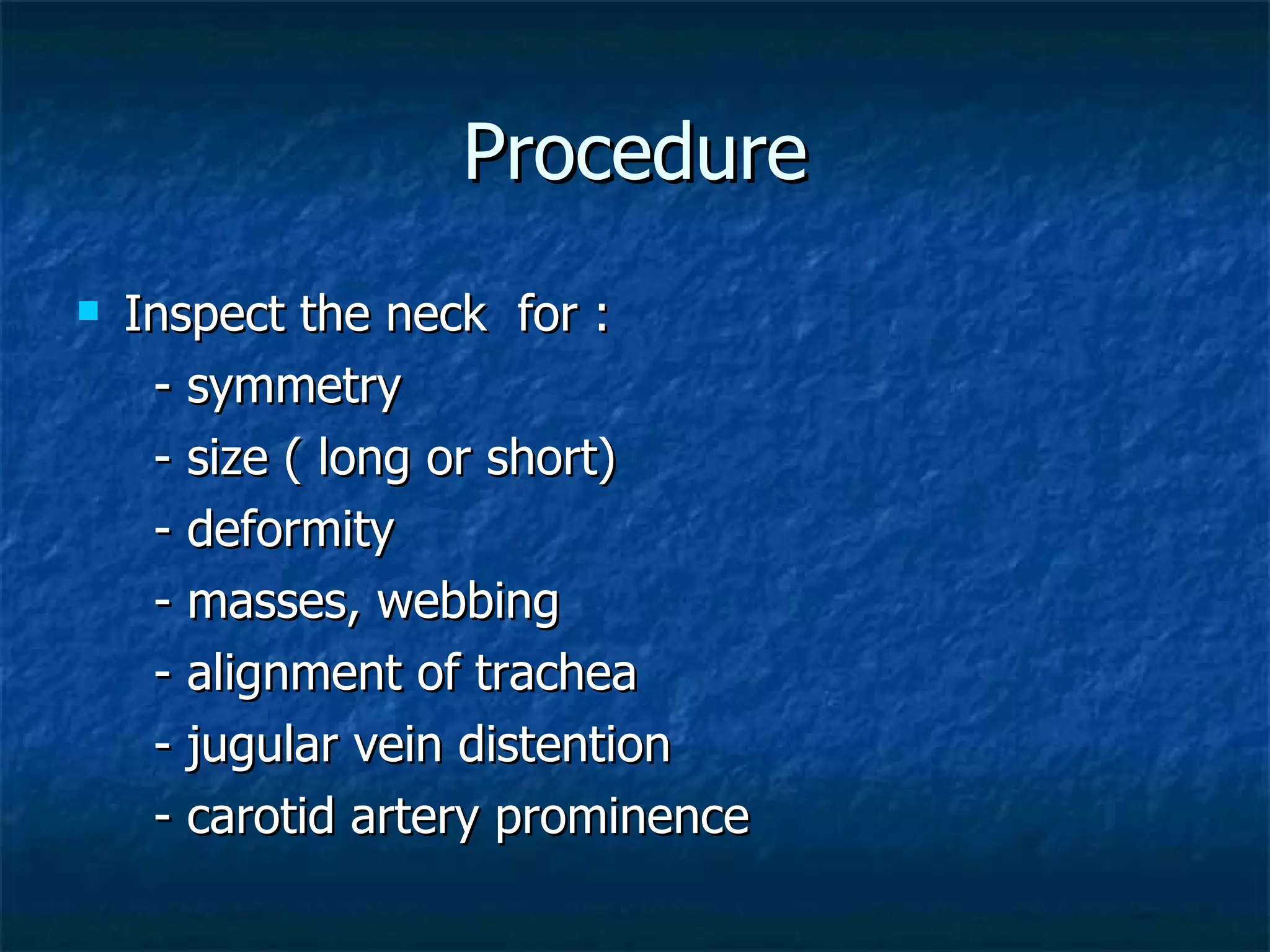
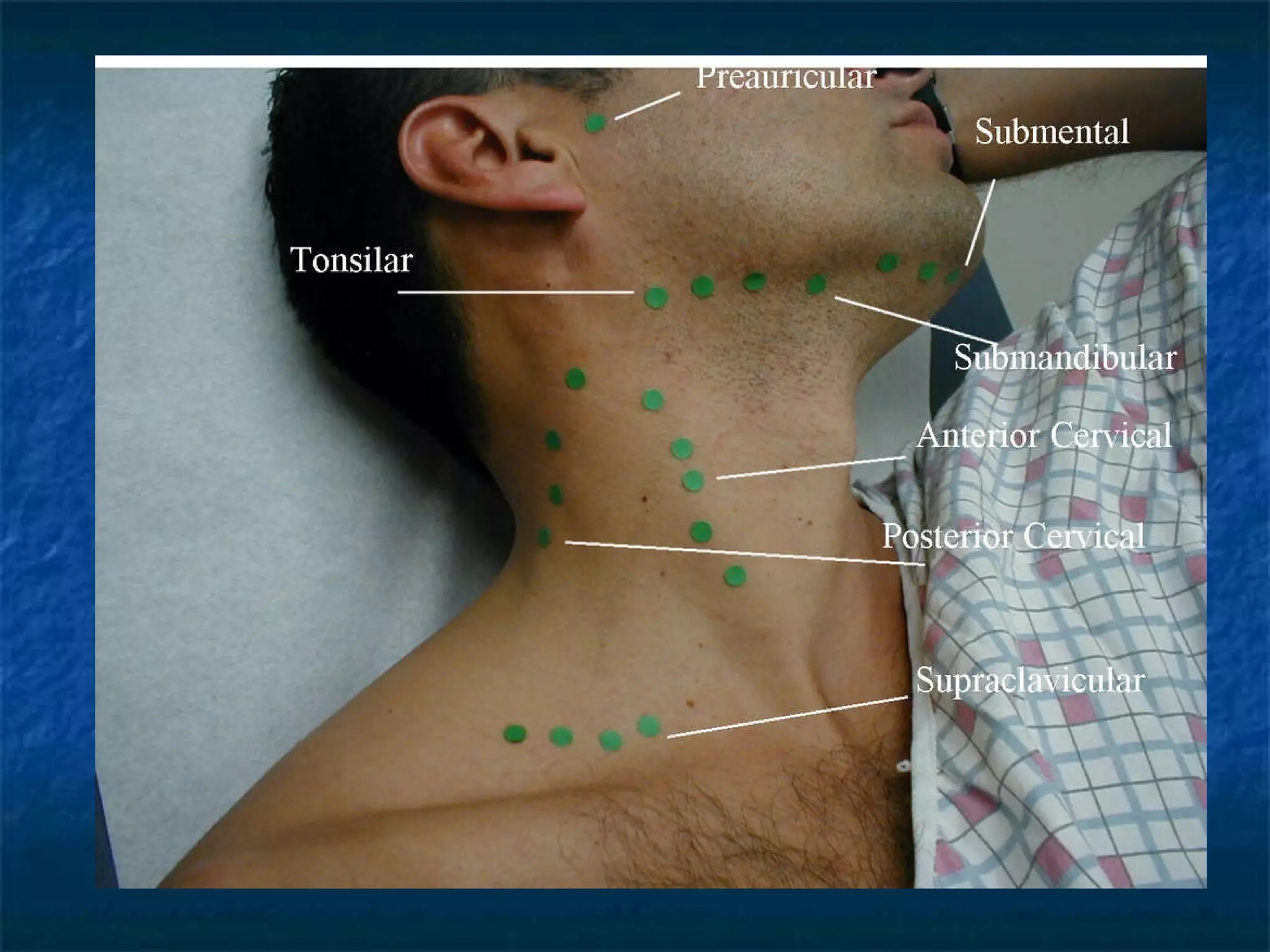
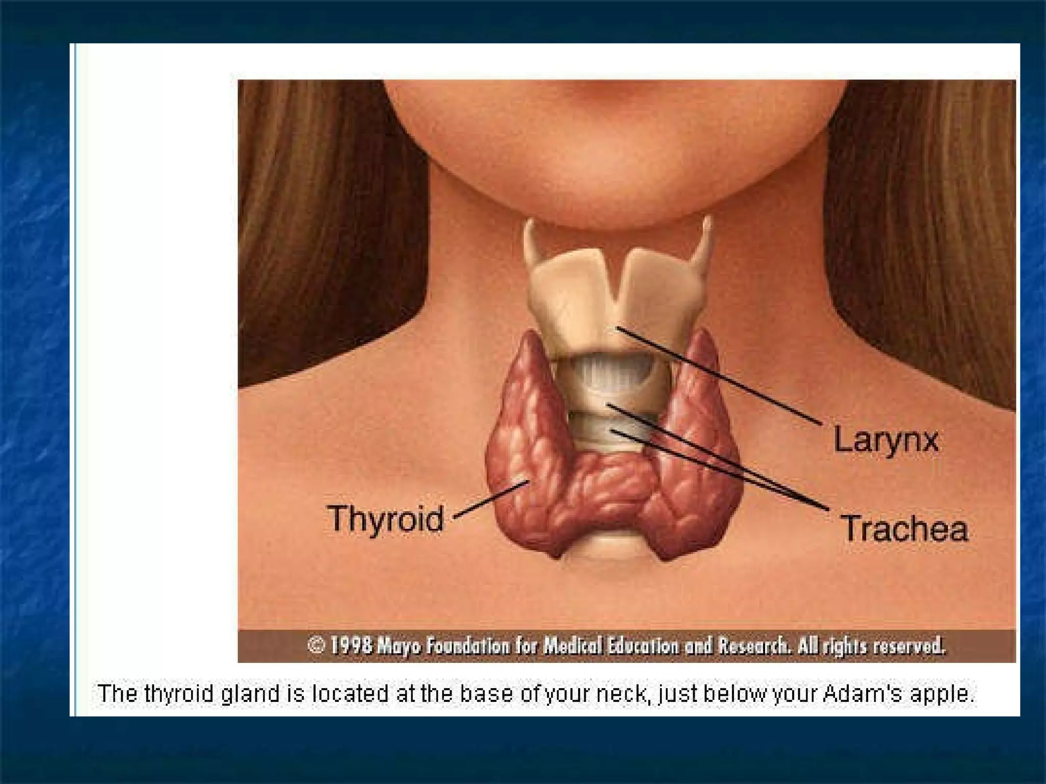
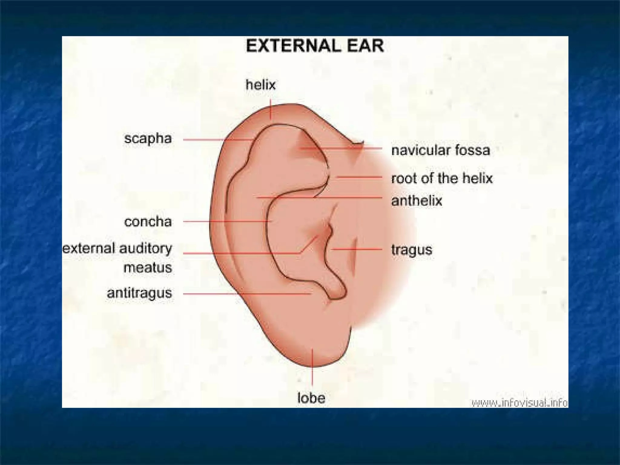
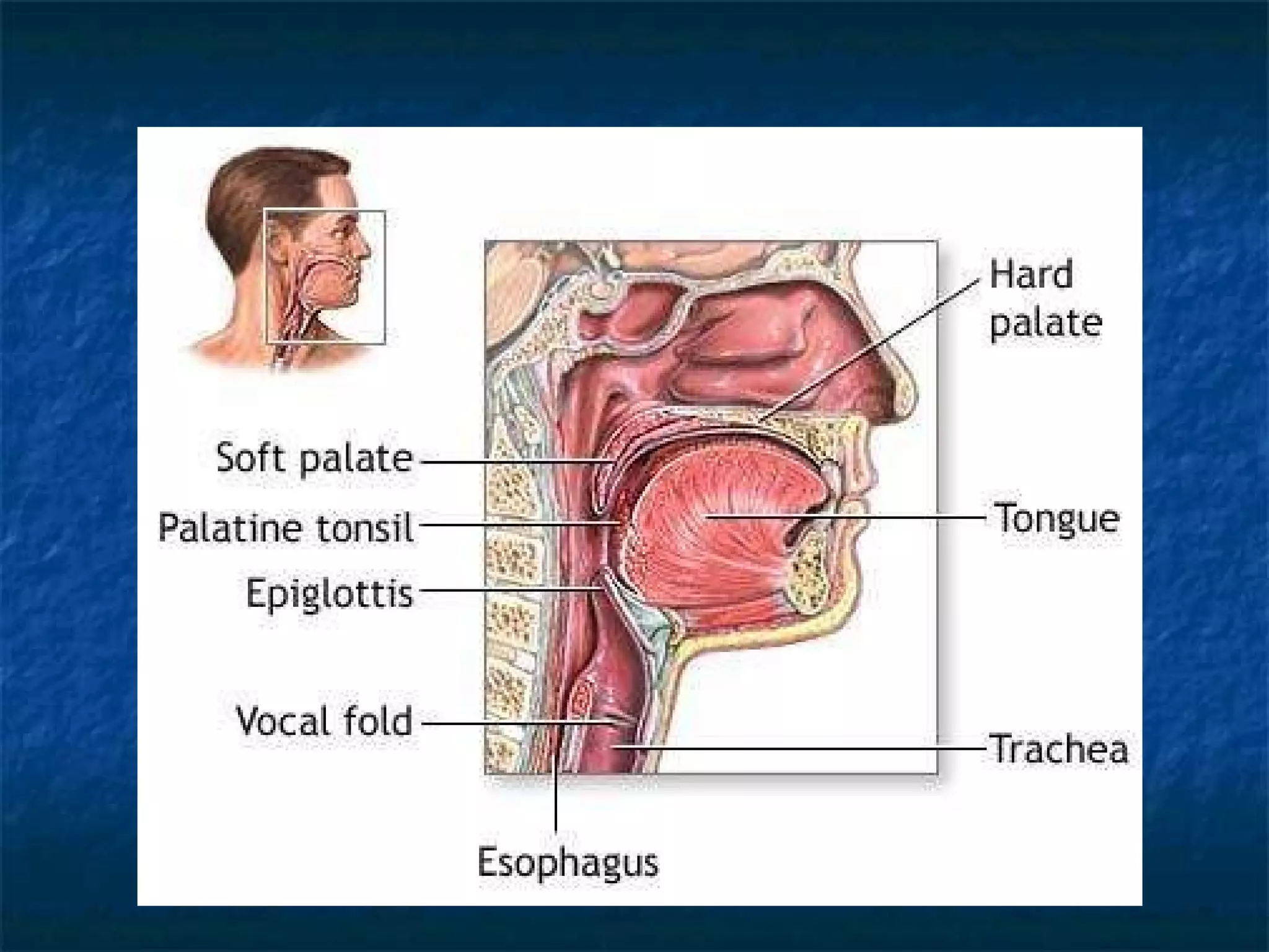
HEENT E. Solis MD, MPH
Learning Objectives The student will be able to identify different components of the head,  eyes, ears, nose, and throat The student will be able to identify anatomic landmarks of the head,  eyes, ears, nose and throat
The student will be able to identify and perform the proper techniques for a basic exam of the head,  eyes, ear, nose and throat The student will be able to describe and record  findings of the head,  eyes, ears, nose and throat examination
Learning Objectives The student will be able to identify different disorders of the head, eyes, ears, nose, and throat The student will be able to identify the signs and symptoms of HEENT disorders
Objective 1: Components of the HEENT Exam HEAD inspection: skull- size ,shape, symmetry, deformity scalp- redness, scaling hair- quantity, distribution ,nits, lice face – symmetry, involuntary movements, skin lesions, color, shape
Palpation: skull ( including temporal artery)- size contour, lumps, deformities,  tenderness scalp- mobility, lesions hair – texture Temporomandibular joint – pain,  decreased ROM
Neck Lymph nodes- enlargement,mobility Trachea – deviation Thyroid gland- size, tenderness, mobility
EYES Inspection: eyebrows- hair loss, scaling eyelids – redness, swelling, lesions
Conjunctiva – paleness, inflammation Sclera – icterus, inflammation Cornea (anterior chamber) – opacities Lens- opacities Pupils – size ,shape, equality, reaction
Tests: -pupillary reactions direct and consensual -accommodation  -Extraocular movement
Funduscopic Examination - red orange  reflex - optic disc  blood vessels  Hemorrhages, exudates
Visual acuity Near Vision Far Vision Peripheral vision
EARS Inspection: Auricle (anterior & posterior)-  deformities, lumps, lesions, position Palpation: Pulls on pinna – tenderness
Otoscope exam  external canal- cerumen, discharge, foreign  bodies, swelling Tympanic membrane- color, landmarks,  bulging ,retraction, perforation light reflex
Tests  Auditory acuity – decreased hearing Weber test- lateralization Rinne test- AC vs BC
Nose and sinuses Inspection: External nares – asymmetry, deformity Internal nares with otoscope – swelling,  turbinates, septal deviation,  perforation, discharge, blood  crusting, ulcers, polyps
Sinuses  Palpation: Frontal – tenderness Maxillary- tenderness
Mouth and pharynx (throat) Inspection: Lips- color, moisture, lumps, ulcers,  cracking Buccal mucosa- color, moisture, lesions Teeth – loose, missing, dental caries Gums- inflammation, swelling, bleeding, discoloration
Tongue (dorsal, ventral, sides, floor) - asymmetry, lesions, salivary  ducts Palate- lumps, lesions Tonsils – presence, size, color, pus  symmetry Pharynx – inflammation, exudates Uvula- inflammation, deviation
Objective 2: Anatomic Landmark To be able to identify common structures in this region which are routinely assessed during physical exam
HEAD Describe anatomy and landmarks of the head
The SKULL
 
 
Temporomandibular Joint


Objective  3: Physical Exam To be able to identify and perform proper techniques for the basic examination of the head, neck, eyes, ears, nose and throat
Procedure HEAD Stand beside or behind the seated patient Observe head position midline, tilted to one side, rotated
Skull Scalp Hair
Inspect the skull for: - size – normocephalic, micro/macrocephalic  - shape  - deformity Palpate the skull for: - symmetry - mass-if present give the exact location, size , shape ,mobility and tenderness -deformity -tenderness- if present localize
The SCALP
 
Inspect the scalp for: - scales - scars -  parasites, nits - mass - pay special attention to the areas behind the  ears, at the hairline and at the crown of the  head. Note for any hair loss pattern
Palpate the scalp for: - tenderness -mass ( sebaceous cyst, lipoma, tumor) - or fluctuant scalp masses like hematoma, abscess, depressed fracture - scalp movement
Hair : inspect and palpate - color - length - distribution- well distributed - pattern of hair loss- receding hair line - quantity –thin, thick or fairly abundant - texture- fine or coarse - moisture – dry or oily - look for  lice and nits
 
Palpation of Temporomandibular joint
Locate the TMJ with your fingertips placed just anterior to the tragus of each ear. Allow your fingertips to slip into the joint space as the patient’s mouth open and gently palpate the joint space
An audible or palpable snapping or clicking in the TMJ is not unusual, but pain, crepitus, locking or popping may indicate TMJ syndrome
 
Objective 4: Record Findings To be able the describe and record findings of the head, neck, eyes, ears and throat
Record the Findings Normocephalic, head held erect and in the midline, thick hair, well-distributed, no focal areas of hair loss, coarse and dry, scalp moves freely under examining fingers, no mass or tenderness, temporal arteries palpable but not thickened
Describe anatomy and landmark of the  FACE
 
Procedure FACE Stand or sit in front of the patient at the same level Inspect the face for: - skin : color, pigmentation and lesions - shape – oval, round, prominent and protruding chin - facial expression and involuntary movements - edema - symmetry – if asymmetric present describe  eg..shallow nasolabial fold.
 
Palpate the temporal arteries, noting the ff: - thickening - hardness - tenderness Auscultate temporal arteries for bruits
 
Record findings Face is oval in shape, symmetrical, fair skinned, with occasional pigmented papules scattered over the face, no masses, nor involuntary movements, temporal artery not visible but palpable with strong pulsation, walls not thickened
NECK Describe the anatomy and landmarks of  the NECK
Thyroid Gland Trachea Lymph Nodes Carotid pulsations
 
 
.


Procedure Inspect the neck  for : - symmetry  - size ( long or short)  - deformity  - masses, webbing - alignment of trachea - jugular vein distention - carotid artery prominence
Evaluate range of motion of the neck - flex, extend ,rotate and lateral turn of the  head and neck - movement should be smooth ,painless and should not cause dizziness
Palpate the neck for: - tracheal position - carotid pulsations - lymph nodes - thyroid gland
The LYMPH NODES  group
 
 
Procedure LYMPH NODES The examiner should stand behind the seated patient Use the pads of both index and middle fingers as you move the skin over the underlying tissues in each area rather than moving your fingers over the skin in a rotatory fashion
Feel in sequence for the following nodes Preauricular –in front of the ear Posterior auricular – superficial to the mastoid process Occipital – at the base of the skull Tonsillar – at the angle of the mandible Submandibular – midway between the angle and the tip of the mandible
6. Submental – in the midline 7. Superficial cervical – superficial to the SCM 8. Posterior cervical chain –along the anterior edge of the trapezius 9. Deep cervical chain –deep into the SCM but often inaccessible to examination 10. Supraclavicular –deep in the angle formed by the clavicle and the SCM
Palpate the lymph nodes for: - size - shape - delimitation ( discrete or matted together) - mobility - consistency - tenderness Small, mobile, discrete, nontender nodes are frequently found in normal persons


Carotid pulsation
Locate for the carotid pulse, in the neck just medial to and below the angle of the jaw ( do not palpate simultaneously) Excessive carotid massage can cause slowing of the pulse or a drop in blood pressure
If you have difficulty feeling the pulse, rotate the patient’s head to the side being examined to relax the SCM muscle Examine the arterial pulse with the distal part of the 2 nd  and 3 rd  fingers
 
The TRACHEA
 
Procedure Inspect the trachea for any deviation from its usual midline position Then feel for any deviation by placing your finger along one side of the trachea and note the space between it and the SCM.
Compare it with the other side. The spaces should be symmetrical. If asymmetrical there is deviation
 
Describe the anatomy and landmarks of  the THYROID GLAND
 
 
Procedure THYROID GLAND Patient should be seated Inspect the lower half of the neck in the anterior triangles Have him swallow or sip a glass of water to note any ascending mass in the midline or behind the SCM
If the patient is obese or has a short neck , tilt the head back to be supported by his hands clasped at the occiput. Ask him to swallow while in this posture The thyroid gland, thyroid cartilage, and cricoid cartilage all normally rise as the person swallows
Palpation  -best done from behind the patient - cricoid cartilage is the basic landmark for examination 2 methods of palpation: Palpation from behind Frontal palpation of the thyroid gland
Palpate the  thyroid gland for : - size - shape - symmetry - consistency of the gland, tenderness - presence of nodules - movement


Record findings NECK Neck is supple with full range of motion, trachea midline, no lymphadenopathy noted. A 1x2 cm nodule is palpated in the right lobe of the thyroid; smooth, soft, nontender, moves freely when patient swallows
Objective 5:Record abnormal Findings To be able to identify and record different disorders of head ,neck, eyes, ears, nose and throat
Facies Expression or appearance of the face and features of the head and neck that when considered together, are characteristics of a clinical condition or syndrome
Acromegaly -large head - forward projection of jaw - protrusion of frontal bone
  Cushing Syndrome   - thin erythematous skin - hirsutism - rounded or moon  shaped face
Mxyedema  - dull, puffy, yellowed  skin - coarse sparse hair - temporal loss of  eyebrows - periorbital edema - prominent tongue
  Hyperthyroid Facies - fine moist skin - fine hair - prominent eyes - lid retraction - startled expression
(R) Facial Palsy - assymmetry of one side of  the face - eyelid not closing completely - loss of nasolabial fold - drooping lower eyelid and  corner of the mouth
SIGNS
Hydrocephalus  Scars  Head tumor
Alopecia areata


SYMPTOMS HEADACHE
HEADACHE - refers to pain perceived more than momentarily in the cranial vault , orbits and the nape. Pain elsewhere in the face is not included
Mechanisms of Headache 1. infection – meningitis, encephalitis 2. arterial dilatation – Malignant Hypertension 3. hemorrhage – intracebral , subdural and SAH 4.Expanding mass lesion – brain tumor
5. Trauma – head trauma , inc. ICP 6. Tissue Ischemia – hypoxia, hypoglycemia
Muscle contraction headache
Muscle Contraction Headache: Tension Headache - Mild or moderate discomfort, a heavy feeling, a sense of pressure, tight band, steady rather than throbbing - related to emotional tension  - not intensified by coughing - improved by shaking the head, massage, mild analgesics, application of hot packs
Migraine Headache
Classic Migraine 4 phases 1. Prodrome- an attack is often triggered  by period of anxiety, tension,  bright light, loud noise,  skipped meals, foods and beverages, strong odors and change in sleep patterns
2. Aura – visual disturbances 3. Headache - frequently present on awakening -severe throbbing, boring, aching  headache over 1 hr. - does not disrupt sleep - increased in the reclining position, shaking the head, coughing or straining at stool
Associated symptoms are N/V, photophobia, annoyance for odors,  maybe normal or cold limbs and pale skin 4. Recovery
Cluster Headache
Cluster headache : Histamine headache or Histamine Cephalgia - due to dilatation of branches of the  internal carotid artery - 5-6x more common in men - onset is typically 3 rd - 4 th  decade of life - commonly episodic and begins w/o aura
Unilateral , severe ,boring,, and throbbing headache that recurs consistently on the same side lasting an average of 40 min Associated symptoms are flushing, rhinorrhea, conjunctivitis, lacrimation,  temporal artery dilatation on the affected side, sweating of the skin
Other causes of headache: Hypertensive headache Brain tumor Hemorrhage  -intracereberal hemorrhage -subarachnoid hemorrhage Bacterial meningitis Lumbar puncture headache
Hypertensive headache - due to segmental dilatation of branches of external carotid artery - headache occurs in half of patients with accelerated HPN without encepalopathy - Headache often occipital, no aura - Diastolic pressure must exceed 120 mm hg to cause headache
Brain tumor Benign and malignant intracranial neoplasms compress and place traction on surrounding structures Headache maybe the first symptom, the onset is recent, a recent change in the customary headache pattern has occured
An apparent migraine aura persists after the headache subsides Headache starts by abrupt change in position, exertion and inc. in recumbent position May interfere with sleep
Subarachnoid hemorrhage Results fr. rupture of a saccular anuerysm of the circle of willis, preceeded often by a leakage Excruciating generalized headache, followed by nuchal rigidity, then coma, often death
Meningitis  Headache , fever and signs of meningeal irritation ( nuchal rigidity) Headache intensified by sudden movement of the head
NECK Stiff neck: 1) Torticollis  ( wryneck)  - the congenital type is due to hematoma or partial rupture of the muscle at birth resulting in unilateral muscle shortening
2) Idiopathic – fibromyalgia 3) inflammatory/immune – osteomyelitis 4) Infectious – pharyngitis, meningitis 5) Metabolic - Tetanus
6) Mechanical /trauma- fracture, dislocation 7) Neoplastic – thyroid cancer, lymphoma 8) Neurologic – parkinson’s disease 9) Psychosocial – malingering
Nongoitrous cervical masses Midline cervical mass Thyroglossal cysts Suprahyoid cysts Subhyoid cysts Pyramidal lobe of thyroid Thyroid cartilage cysts Cricoid cartilage cysts
Lateral cervical cyst Branchial cyst Hygroma Carotid body tumor Cavernous hemangioma Branchial fistula  Zenker’s diverticulum ( pharyngeal pouch)
Thyroid Thyroid enlargement ( GOITER) - results from: a) hyperplasia of the thyroid tissue  b) infection c) neoplastic growth ( primary thyroid cancer, metastatic growth, lymphoma) d) infiltration with foreign substances ( amyloid) -
- patient complains of fullness of mass in the neck , pressure symptoms - Determine the size of the component of the gland, extension the gland within the neck or into the retrosternal space, fixation to surrounding structures
-characterize the enlarge thyroid as diffuse, focal, nodular, or smooth - tenderness
-make an assessment of the state of thyroid function: Hypothyroid, Hyperthyroid, euthyroid Clinical classification is based whether thyroid is diffuse or nodular Level of functional thyroid state:  a) toxic goiter b) nontoxic goiter ( euthyroid or hypothyroid)
Retrosternal Goiter ( substernal, intrathoracic , or submerged goiter) - when the lower border of a goiter can’t be palpated - Goiter may rise only with inc. intrathoracic pressure like coughing. This is also called plunging goiter.
2 physical signs of retrosternal goiter Tracheal displacement Venous engorgement in the neck
Diffuse toxic goiter ( Grave’s Disease) -autoimmune disease char. by goiter, exophthalmos, pretibial edema, hyperthyroidism
Thyroid syndromes - excess or deficit of thyroid hormones alter the physical structure of the body to produce physical signs - examine your patient to determine the size of the TG, to assess thyroid function, to judge the likehood of cancer
Thyroid syndromes a)  Hyperthyroidism  - overproduction of the thyroid  hormone or excessive  thyroid  medication - often with generalized muscle weakness,  energetic, irritable, tachycardic, tremor  frequent defecation, wt. loss , inc appetite
b) Hypothyroidism -due to iodine deficiency, deficit of TH, excessive dose of thiouracil drugs,  lithium, thiocyanates, paraaminosalicylic acid, phenylbutazone - slow metabolism, fatigue, loss of energy, wt gain, constipation, coldness
Lymph Nodes Determine if the lymph node is localized to the neck or generalized in other parts of the body Acute cervical lymphadenopathy 1) Localized lymphadenitis - common infections of the scalp, face,  mouth, teeth, pharynx or ear
Submental lymph nodes - primary lesions from the lower lip, anterior tongue, floor of the mouth Posterior cervical lymph nodes and occipital - primary lesions from the posterior 2/3 of the scalp and nasopharynx
Anterior cervical lymph nodes - primary lesions from anterior 2/3 of the scalp, face including maxillary sinus, oral cavity ( tongue, tonsils, larynx)
2) Generalized lymphadenitis - syphilis - rubella - IM - HIV - Generalized Furunculosis
Chronic localized cervical lymphadenopathy a) TB b) Hodgkin disease c) actinomycosis
d) Virchow node ( sentinel node) -enlargement of a single lymph node usually in the left supraclavicular group - it may be the result of either abdominal or thoracic malignancy
Thank You
 
 
Definitions of Parts Shown Above Helix - The in-curve rim of the external ear Antihelix - A landmark of the outer ear Lobule - A landmark of the outer ear. The very bottom part of the outer ear Crest of Helix - A landmark of the outer ear ExternalAuditory Meatus - or External Auditory Canal. The auditory canal is the channel through which the sounds are led from the ear outside to the middle ear. Eardrum - (tympanic membrane) A thin layer of skin at the end of the external ear canal Auditory Ossicles - The three small bones in the middle ear, know as the hammer (malleus), anvil (incus) and stirrup (stapes) which are connected to one another. Together these ossicles are called the ossicular chain. Their purpose is to lead the sound striking the eardrum further into the inner ear Oval Window - An opening in the bone between the air filled middle ear cavity and the fluid filled inner ear, and is covered by a thin membrane Cochlea - Part of the inner ear that contains part of the hearing organs. Semicircular Canals - Part of the organ of balance that is part of the inner ear Eighth Nerve - Nerve that transmits messages from the inner ear to the brain. Eustachian Tube - A tube connecting the middle ear cavity and the pharynx (back of the throat). It can be opened by coughing or swallowing, though it is normally closed. The occasional opening of the Eustachian tube is necessary to equalize the are in the middle ear cavity


Describe  common changes with age that occur in the head and neck Identify common abnormalities which may be found on examination of hair, scalp, skull
Identify the purpose of testing : visual acuity visual fields by confrontation Define exophthalmos and name 2 possible causes
Identify common abnormalities which may be found in examination of: eyebrows eyelids lacrimal apparatus Identify the potential significance of: yellow schlera pale palpebral conjuctiva
Define or describe the ff: terms or tests related to eyes a) anisocoria b) miosis c) mydriasis d) direct and consensual reactions e) near reaction f) tests for weakness or imbalance of the EOM g) nystagmus h) lid lag
Be able to describe the ff: parts of the normal fundoscopic exam and identify them if given a diagram a) red reflex b) optic disc c) physiologic cup d) arterioles e) veins f)macula fovea
Describe or identify each of the ff: and give its significance a) normal blurring of the disc outline on the nasal side b) differentiate arterioles from veins c)  absent red reflex d) AV nicking
Describe or identify the common abnormalities which may be found in the PE of patients with Otitis media Otitis externa Serous effusions Retracted drum
Describe the Weber and Rinne test. How they are used to distinguished between conductive hearing loss and sensorineural hearing loss Describe or identify the common abnormalities which may be seen in the nasal exam of a) nasal mucosa b) nasal septum c) frontal and maxillary sinus (palpation,  transillumination)
Describe or identify common abnormalities which may be found on examination of the mouth and pharynx List 6 characteristics which can be noted in describing lymph nodes Identify potential significance of tender nodes, hard or fixed nodes
Identify the significance of tracheal deviation Define GOITER Describe physical characteristics of the thyroid in the normal and abnormal states
Define the ff: terms Ptosis Ectropion Entropion Define the ff: terms a) Pinguecula b) Sty( hordeolum) c) Chalazion d) Xanthelasma e) Episcleritis f) Dacryocystitis
Describe the ff: common causes and presentation of red eye Define or identify the ff: Corneal arcus Corneal scars Pterygium  cataract
Define the ff: a) anisocoria  b) Argyll Robertson pupil c) oculomotor nerve paralysis d) strabismus Describe the normal appearance and variation of the optic disc. Be able to describe papilledema  and glaucomatous cupping
Describe the normal retinal arteries and AV crossings. Describe the change that occur with HPN including: a) narrowed light reflex b) copper wire arteries c) silver wire arteries
For each of the ff: identify physical finding  and cause: a) superficial retinal hemorrhage b) deep retinal hemorrhage c) pre retinal hemorrhage d) microaneurysm e) neovascularization
For each of the ff: identify physical finding and cause: a) cotton wool patch b) hard exudates
If a patient has a chief complaint of any one of the head, eyes, ears, nose and throat (HEENT) symptoms, you must generally ask all of the HEENT ROS questions.
The ears, nose, and throat are anatomically connected, hence infection or obstruction in one structure can lead to illness or symptoms in the others
 
 
Facial pain Trigeminal neuralgia (Tic Doulourex) Compression of the 5 th  nerve root by a vessel or a neoplasm 2 nd  maxillary division ,commonly involved Light touch, chewing, sneezing provokes a paroxysm Hot lancinating ,periodic, unilateral pain
Herpes zoster - sharp , burning, unilateral pain along the distribution of a branch of the trigeminal nerve Other causes: acute suppurative sinusitis Orbital cellulitis
Swelling of the face Parotitis Preauricular abscess and ulcer Masseter muscle hypertrophy
Acromegaly
myxedema face
Ptosis

Heent

  • 1.
  • 2.
    Learning Objectives Thestudent will be able to identify different components of the head, eyes, ears, nose, and throat The student will be able to identify anatomic landmarks of the head, eyes, ears, nose and throat
  • 3.
    The student willbe able to identify and perform the proper techniques for a basic exam of the head, eyes, ear, nose and throat The student will be able to describe and record findings of the head, eyes, ears, nose and throat examination
  • 4.
    Learning Objectives Thestudent will be able to identify different disorders of the head, eyes, ears, nose, and throat The student will be able to identify the signs and symptoms of HEENT disorders
  • 5.
    Objective 1: Componentsof the HEENT Exam HEAD inspection: skull- size ,shape, symmetry, deformity scalp- redness, scaling hair- quantity, distribution ,nits, lice face – symmetry, involuntary movements, skin lesions, color, shape
  • 6.
    Palpation: skull (including temporal artery)- size contour, lumps, deformities, tenderness scalp- mobility, lesions hair – texture Temporomandibular joint – pain, decreased ROM
  • 7.
    Neck Lymph nodes-enlargement,mobility Trachea – deviation Thyroid gland- size, tenderness, mobility
  • 8.
    EYES Inspection: eyebrows-hair loss, scaling eyelids – redness, swelling, lesions
  • 9.
    Conjunctiva – paleness,inflammation Sclera – icterus, inflammation Cornea (anterior chamber) – opacities Lens- opacities Pupils – size ,shape, equality, reaction
  • 10.
    Tests: -pupillary reactionsdirect and consensual -accommodation -Extraocular movement
  • 11.
    Funduscopic Examination -red orange reflex - optic disc blood vessels Hemorrhages, exudates
  • 12.
    Visual acuity NearVision Far Vision Peripheral vision
  • 13.
    EARS Inspection: Auricle(anterior & posterior)- deformities, lumps, lesions, position Palpation: Pulls on pinna – tenderness
  • 14.
    Otoscope exam external canal- cerumen, discharge, foreign bodies, swelling Tympanic membrane- color, landmarks, bulging ,retraction, perforation light reflex
  • 15.
    Tests Auditoryacuity – decreased hearing Weber test- lateralization Rinne test- AC vs BC
  • 16.
    Nose and sinusesInspection: External nares – asymmetry, deformity Internal nares with otoscope – swelling, turbinates, septal deviation, perforation, discharge, blood crusting, ulcers, polyps
  • 17.
    Sinuses Palpation:Frontal – tenderness Maxillary- tenderness
  • 18.
    Mouth and pharynx(throat) Inspection: Lips- color, moisture, lumps, ulcers, cracking Buccal mucosa- color, moisture, lesions Teeth – loose, missing, dental caries Gums- inflammation, swelling, bleeding, discoloration
  • 19.
    Tongue (dorsal, ventral,sides, floor) - asymmetry, lesions, salivary ducts Palate- lumps, lesions Tonsils – presence, size, color, pus symmetry Pharynx – inflammation, exudates Uvula- inflammation, deviation
  • 20.
    Objective 2: AnatomicLandmark To be able to identify common structures in this region which are routinely assessed during physical exam
  • 21.
    HEAD Describe anatomyand landmarks of the head
  • 22.
  • 23.
  • 24.
  • 25.
  • 26.
  • 27.
  • 28.
  • 29.
    Objective 3:Physical Exam To be able to identify and perform proper techniques for the basic examination of the head, neck, eyes, ears, nose and throat
  • 30.
    Procedure HEAD Standbeside or behind the seated patient Observe head position midline, tilted to one side, rotated
  • 31.
  • 32.
    Inspect the skullfor: - size – normocephalic, micro/macrocephalic - shape - deformity Palpate the skull for: - symmetry - mass-if present give the exact location, size , shape ,mobility and tenderness -deformity -tenderness- if present localize
  • 33.
  • 34.
  • 35.
    Inspect the scalpfor: - scales - scars - parasites, nits - mass - pay special attention to the areas behind the ears, at the hairline and at the crown of the head. Note for any hair loss pattern
  • 36.
    Palpate the scalpfor: - tenderness -mass ( sebaceous cyst, lipoma, tumor) - or fluctuant scalp masses like hematoma, abscess, depressed fracture - scalp movement
  • 37.
    Hair : inspectand palpate - color - length - distribution- well distributed - pattern of hair loss- receding hair line - quantity –thin, thick or fairly abundant - texture- fine or coarse - moisture – dry or oily - look for lice and nits
  • 38.
  • 39.
  • 40.
    Locate the TMJwith your fingertips placed just anterior to the tragus of each ear. Allow your fingertips to slip into the joint space as the patient’s mouth open and gently palpate the joint space
  • 41.
    An audible orpalpable snapping or clicking in the TMJ is not unusual, but pain, crepitus, locking or popping may indicate TMJ syndrome
  • 42.
  • 43.
    Objective 4: RecordFindings To be able the describe and record findings of the head, neck, eyes, ears and throat
  • 44.
    Record the FindingsNormocephalic, head held erect and in the midline, thick hair, well-distributed, no focal areas of hair loss, coarse and dry, scalp moves freely under examining fingers, no mass or tenderness, temporal arteries palpable but not thickened
  • 45.
    Describe anatomy andlandmark of the FACE
  • 46.
  • 47.
    Procedure FACE Standor sit in front of the patient at the same level Inspect the face for: - skin : color, pigmentation and lesions - shape – oval, round, prominent and protruding chin - facial expression and involuntary movements - edema - symmetry – if asymmetric present describe eg..shallow nasolabial fold.
  • 48.
  • 49.
    Palpate the temporalarteries, noting the ff: - thickening - hardness - tenderness Auscultate temporal arteries for bruits
  • 50.
  • 51.
    Record findings Faceis oval in shape, symmetrical, fair skinned, with occasional pigmented papules scattered over the face, no masses, nor involuntary movements, temporal artery not visible but palpable with strong pulsation, walls not thickened
  • 52.
    NECK Describe theanatomy and landmarks of the NECK
  • 53.
    Thyroid Gland TracheaLymph Nodes Carotid pulsations
  • 54.
  • 55.
  • 56.
  • 57.
  • 58.
  • 59.
  • 60.
    Procedure Inspect theneck for : - symmetry - size ( long or short) - deformity - masses, webbing - alignment of trachea - jugular vein distention - carotid artery prominence
  • 61.
    Evaluate range ofmotion of the neck - flex, extend ,rotate and lateral turn of the head and neck - movement should be smooth ,painless and should not cause dizziness
  • 62.
    Palpate the neckfor: - tracheal position - carotid pulsations - lymph nodes - thyroid gland
  • 63.
  • 64.
  • 65.
  • 66.
    Procedure LYMPH NODESThe examiner should stand behind the seated patient Use the pads of both index and middle fingers as you move the skin over the underlying tissues in each area rather than moving your fingers over the skin in a rotatory fashion
  • 67.
    Feel in sequencefor the following nodes Preauricular –in front of the ear Posterior auricular – superficial to the mastoid process Occipital – at the base of the skull Tonsillar – at the angle of the mandible Submandibular – midway between the angle and the tip of the mandible
  • 68.
    6. Submental –in the midline 7. Superficial cervical – superficial to the SCM 8. Posterior cervical chain –along the anterior edge of the trapezius 9. Deep cervical chain –deep into the SCM but often inaccessible to examination 10. Supraclavicular –deep in the angle formed by the clavicle and the SCM
  • 69.
    Palpate the lymphnodes for: - size - shape - delimitation ( discrete or matted together) - mobility - consistency - tenderness Small, mobile, discrete, nontender nodes are frequently found in normal persons
  • 70.
  • 71.
  • 72.
  • 73.
  • 74.
    Locate for thecarotid pulse, in the neck just medial to and below the angle of the jaw ( do not palpate simultaneously) Excessive carotid massage can cause slowing of the pulse or a drop in blood pressure
  • 75.
    If you havedifficulty feeling the pulse, rotate the patient’s head to the side being examined to relax the SCM muscle Examine the arterial pulse with the distal part of the 2 nd and 3 rd fingers
  • 76.
  • 77.
  • 78.
  • 79.
    Procedure Inspect thetrachea for any deviation from its usual midline position Then feel for any deviation by placing your finger along one side of the trachea and note the space between it and the SCM.
  • 80.
    Compare it withthe other side. The spaces should be symmetrical. If asymmetrical there is deviation
  • 81.
  • 82.
    Describe the anatomyand landmarks of the THYROID GLAND
  • 83.
  • 84.
  • 85.
    Procedure THYROID GLANDPatient should be seated Inspect the lower half of the neck in the anterior triangles Have him swallow or sip a glass of water to note any ascending mass in the midline or behind the SCM
  • 86.
    If the patientis obese or has a short neck , tilt the head back to be supported by his hands clasped at the occiput. Ask him to swallow while in this posture The thyroid gland, thyroid cartilage, and cricoid cartilage all normally rise as the person swallows
  • 87.
    Palpation -bestdone from behind the patient - cricoid cartilage is the basic landmark for examination 2 methods of palpation: Palpation from behind Frontal palpation of the thyroid gland
  • 88.
    Palpate the thyroid gland for : - size - shape - symmetry - consistency of the gland, tenderness - presence of nodules - movement
  • 89.
  • 90.
  • 91.
  • 92.
    Record findings NECKNeck is supple with full range of motion, trachea midline, no lymphadenopathy noted. A 1x2 cm nodule is palpated in the right lobe of the thyroid; smooth, soft, nontender, moves freely when patient swallows
  • 93.
    Objective 5:Record abnormalFindings To be able to identify and record different disorders of head ,neck, eyes, ears, nose and throat
  • 94.
    Facies Expression orappearance of the face and features of the head and neck that when considered together, are characteristics of a clinical condition or syndrome
  • 95.
    Acromegaly -large head- forward projection of jaw - protrusion of frontal bone
  • 96.
    CushingSyndrome - thin erythematous skin - hirsutism - rounded or moon shaped face
  • 97.
    Mxyedema -dull, puffy, yellowed skin - coarse sparse hair - temporal loss of eyebrows - periorbital edema - prominent tongue
  • 98.
    HyperthyroidFacies - fine moist skin - fine hair - prominent eyes - lid retraction - startled expression
  • 99.
    (R) Facial Palsy- assymmetry of one side of the face - eyelid not closing completely - loss of nasolabial fold - drooping lower eyelid and corner of the mouth
  • 100.
  • 101.
  • 102.
  • 103.
  • 104.
  • 105.
  • 106.
  • 107.
  • 108.
  • 109.
    HEADACHE - refersto pain perceived more than momentarily in the cranial vault , orbits and the nape. Pain elsewhere in the face is not included
  • 110.
    Mechanisms of Headache1. infection – meningitis, encephalitis 2. arterial dilatation – Malignant Hypertension 3. hemorrhage – intracebral , subdural and SAH 4.Expanding mass lesion – brain tumor
  • 111.
    5. Trauma –head trauma , inc. ICP 6. Tissue Ischemia – hypoxia, hypoglycemia
  • 112.
  • 113.
    Muscle Contraction Headache:Tension Headache - Mild or moderate discomfort, a heavy feeling, a sense of pressure, tight band, steady rather than throbbing - related to emotional tension - not intensified by coughing - improved by shaking the head, massage, mild analgesics, application of hot packs
  • 114.
  • 115.
    Classic Migraine 4phases 1. Prodrome- an attack is often triggered by period of anxiety, tension, bright light, loud noise, skipped meals, foods and beverages, strong odors and change in sleep patterns
  • 116.
    2. Aura –visual disturbances 3. Headache - frequently present on awakening -severe throbbing, boring, aching headache over 1 hr. - does not disrupt sleep - increased in the reclining position, shaking the head, coughing or straining at stool
  • 117.
    Associated symptoms areN/V, photophobia, annoyance for odors, maybe normal or cold limbs and pale skin 4. Recovery
  • 118.
  • 119.
    Cluster headache :Histamine headache or Histamine Cephalgia - due to dilatation of branches of the internal carotid artery - 5-6x more common in men - onset is typically 3 rd - 4 th decade of life - commonly episodic and begins w/o aura
  • 120.
    Unilateral , severe,boring,, and throbbing headache that recurs consistently on the same side lasting an average of 40 min Associated symptoms are flushing, rhinorrhea, conjunctivitis, lacrimation, temporal artery dilatation on the affected side, sweating of the skin
  • 121.
    Other causes ofheadache: Hypertensive headache Brain tumor Hemorrhage -intracereberal hemorrhage -subarachnoid hemorrhage Bacterial meningitis Lumbar puncture headache
  • 122.
    Hypertensive headache -due to segmental dilatation of branches of external carotid artery - headache occurs in half of patients with accelerated HPN without encepalopathy - Headache often occipital, no aura - Diastolic pressure must exceed 120 mm hg to cause headache
  • 123.
    Brain tumor Benignand malignant intracranial neoplasms compress and place traction on surrounding structures Headache maybe the first symptom, the onset is recent, a recent change in the customary headache pattern has occured
  • 124.
    An apparent migraineaura persists after the headache subsides Headache starts by abrupt change in position, exertion and inc. in recumbent position May interfere with sleep
  • 125.
    Subarachnoid hemorrhage Resultsfr. rupture of a saccular anuerysm of the circle of willis, preceeded often by a leakage Excruciating generalized headache, followed by nuchal rigidity, then coma, often death
  • 126.
    Meningitis Headache, fever and signs of meningeal irritation ( nuchal rigidity) Headache intensified by sudden movement of the head
  • 127.
    NECK Stiff neck:1) Torticollis ( wryneck) - the congenital type is due to hematoma or partial rupture of the muscle at birth resulting in unilateral muscle shortening
  • 128.
    2) Idiopathic –fibromyalgia 3) inflammatory/immune – osteomyelitis 4) Infectious – pharyngitis, meningitis 5) Metabolic - Tetanus
  • 129.
    6) Mechanical /trauma-fracture, dislocation 7) Neoplastic – thyroid cancer, lymphoma 8) Neurologic – parkinson’s disease 9) Psychosocial – malingering
  • 130.
    Nongoitrous cervical massesMidline cervical mass Thyroglossal cysts Suprahyoid cysts Subhyoid cysts Pyramidal lobe of thyroid Thyroid cartilage cysts Cricoid cartilage cysts
  • 131.
    Lateral cervical cystBranchial cyst Hygroma Carotid body tumor Cavernous hemangioma Branchial fistula Zenker’s diverticulum ( pharyngeal pouch)
  • 132.
    Thyroid Thyroid enlargement( GOITER) - results from: a) hyperplasia of the thyroid tissue b) infection c) neoplastic growth ( primary thyroid cancer, metastatic growth, lymphoma) d) infiltration with foreign substances ( amyloid) -
  • 133.
    - patient complainsof fullness of mass in the neck , pressure symptoms - Determine the size of the component of the gland, extension the gland within the neck or into the retrosternal space, fixation to surrounding structures
  • 134.
    -characterize the enlargethyroid as diffuse, focal, nodular, or smooth - tenderness
  • 135.
    -make an assessmentof the state of thyroid function: Hypothyroid, Hyperthyroid, euthyroid Clinical classification is based whether thyroid is diffuse or nodular Level of functional thyroid state: a) toxic goiter b) nontoxic goiter ( euthyroid or hypothyroid)
  • 136.
    Retrosternal Goiter (substernal, intrathoracic , or submerged goiter) - when the lower border of a goiter can’t be palpated - Goiter may rise only with inc. intrathoracic pressure like coughing. This is also called plunging goiter.
  • 137.
    2 physical signsof retrosternal goiter Tracheal displacement Venous engorgement in the neck
  • 138.
    Diffuse toxic goiter( Grave’s Disease) -autoimmune disease char. by goiter, exophthalmos, pretibial edema, hyperthyroidism
  • 139.
    Thyroid syndromes -excess or deficit of thyroid hormones alter the physical structure of the body to produce physical signs - examine your patient to determine the size of the TG, to assess thyroid function, to judge the likehood of cancer
  • 140.
    Thyroid syndromes a) Hyperthyroidism - overproduction of the thyroid hormone or excessive thyroid medication - often with generalized muscle weakness, energetic, irritable, tachycardic, tremor frequent defecation, wt. loss , inc appetite
  • 141.
    b) Hypothyroidism -dueto iodine deficiency, deficit of TH, excessive dose of thiouracil drugs, lithium, thiocyanates, paraaminosalicylic acid, phenylbutazone - slow metabolism, fatigue, loss of energy, wt gain, constipation, coldness
  • 142.
    Lymph Nodes Determineif the lymph node is localized to the neck or generalized in other parts of the body Acute cervical lymphadenopathy 1) Localized lymphadenitis - common infections of the scalp, face, mouth, teeth, pharynx or ear
  • 143.
    Submental lymph nodes- primary lesions from the lower lip, anterior tongue, floor of the mouth Posterior cervical lymph nodes and occipital - primary lesions from the posterior 2/3 of the scalp and nasopharynx
  • 144.
    Anterior cervical lymphnodes - primary lesions from anterior 2/3 of the scalp, face including maxillary sinus, oral cavity ( tongue, tonsils, larynx)
  • 145.
    2) Generalized lymphadenitis- syphilis - rubella - IM - HIV - Generalized Furunculosis
  • 146.
    Chronic localized cervicallymphadenopathy a) TB b) Hodgkin disease c) actinomycosis
  • 147.
    d) Virchow node( sentinel node) -enlargement of a single lymph node usually in the left supraclavicular group - it may be the result of either abdominal or thoracic malignancy
  • 148.
  • 149.
  • 150.
  • 151.
    Definitions of PartsShown Above Helix - The in-curve rim of the external ear Antihelix - A landmark of the outer ear Lobule - A landmark of the outer ear. The very bottom part of the outer ear Crest of Helix - A landmark of the outer ear ExternalAuditory Meatus - or External Auditory Canal. The auditory canal is the channel through which the sounds are led from the ear outside to the middle ear. Eardrum - (tympanic membrane) A thin layer of skin at the end of the external ear canal Auditory Ossicles - The three small bones in the middle ear, know as the hammer (malleus), anvil (incus) and stirrup (stapes) which are connected to one another. Together these ossicles are called the ossicular chain. Their purpose is to lead the sound striking the eardrum further into the inner ear Oval Window - An opening in the bone between the air filled middle ear cavity and the fluid filled inner ear, and is covered by a thin membrane Cochlea - Part of the inner ear that contains part of the hearing organs. Semicircular Canals - Part of the organ of balance that is part of the inner ear Eighth Nerve - Nerve that transmits messages from the inner ear to the brain. Eustachian Tube - A tube connecting the middle ear cavity and the pharynx (back of the throat). It can be opened by coughing or swallowing, though it is normally closed. The occasional opening of the Eustachian tube is necessary to equalize the are in the middle ear cavity
  • 152.
  • 153.
  • 154.
  • 155.
  • 156.
  • 157.
  • 158.
    Describe commonchanges with age that occur in the head and neck Identify common abnormalities which may be found on examination of hair, scalp, skull
  • 159.
    Identify the purposeof testing : visual acuity visual fields by confrontation Define exophthalmos and name 2 possible causes
  • 160.
    Identify common abnormalitieswhich may be found in examination of: eyebrows eyelids lacrimal apparatus Identify the potential significance of: yellow schlera pale palpebral conjuctiva
  • 161.
    Define or describethe ff: terms or tests related to eyes a) anisocoria b) miosis c) mydriasis d) direct and consensual reactions e) near reaction f) tests for weakness or imbalance of the EOM g) nystagmus h) lid lag
  • 162.
    Be able todescribe the ff: parts of the normal fundoscopic exam and identify them if given a diagram a) red reflex b) optic disc c) physiologic cup d) arterioles e) veins f)macula fovea
  • 163.
    Describe or identifyeach of the ff: and give its significance a) normal blurring of the disc outline on the nasal side b) differentiate arterioles from veins c) absent red reflex d) AV nicking
  • 164.
    Describe or identifythe common abnormalities which may be found in the PE of patients with Otitis media Otitis externa Serous effusions Retracted drum
  • 165.
    Describe the Weberand Rinne test. How they are used to distinguished between conductive hearing loss and sensorineural hearing loss Describe or identify the common abnormalities which may be seen in the nasal exam of a) nasal mucosa b) nasal septum c) frontal and maxillary sinus (palpation, transillumination)
  • 166.
    Describe or identifycommon abnormalities which may be found on examination of the mouth and pharynx List 6 characteristics which can be noted in describing lymph nodes Identify potential significance of tender nodes, hard or fixed nodes
  • 167.
    Identify the significanceof tracheal deviation Define GOITER Describe physical characteristics of the thyroid in the normal and abnormal states
  • 168.
    Define the ff:terms Ptosis Ectropion Entropion Define the ff: terms a) Pinguecula b) Sty( hordeolum) c) Chalazion d) Xanthelasma e) Episcleritis f) Dacryocystitis
  • 169.
    Describe the ff:common causes and presentation of red eye Define or identify the ff: Corneal arcus Corneal scars Pterygium cataract
  • 170.
    Define the ff:a) anisocoria b) Argyll Robertson pupil c) oculomotor nerve paralysis d) strabismus Describe the normal appearance and variation of the optic disc. Be able to describe papilledema and glaucomatous cupping
  • 171.
    Describe the normalretinal arteries and AV crossings. Describe the change that occur with HPN including: a) narrowed light reflex b) copper wire arteries c) silver wire arteries
  • 172.
    For each ofthe ff: identify physical finding and cause: a) superficial retinal hemorrhage b) deep retinal hemorrhage c) pre retinal hemorrhage d) microaneurysm e) neovascularization
  • 173.
    For each ofthe ff: identify physical finding and cause: a) cotton wool patch b) hard exudates
  • 174.
    If a patienthas a chief complaint of any one of the head, eyes, ears, nose and throat (HEENT) symptoms, you must generally ask all of the HEENT ROS questions.
  • 175.
    The ears, nose,and throat are anatomically connected, hence infection or obstruction in one structure can lead to illness or symptoms in the others
  • 176.
  • 177.
  • 178.
    Facial pain Trigeminalneuralgia (Tic Doulourex) Compression of the 5 th nerve root by a vessel or a neoplasm 2 nd maxillary division ,commonly involved Light touch, chewing, sneezing provokes a paroxysm Hot lancinating ,periodic, unilateral pain
  • 179.
    Herpes zoster -sharp , burning, unilateral pain along the distribution of a branch of the trigeminal nerve Other causes: acute suppurative sinusitis Orbital cellulitis
  • 180.
    Swelling of theface Parotitis Preauricular abscess and ulcer Masseter muscle hypertrophy
  • 181.
  • 182.
  • 183.