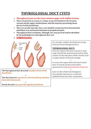
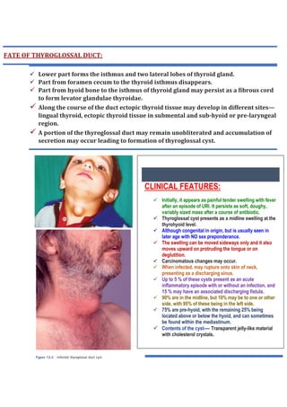
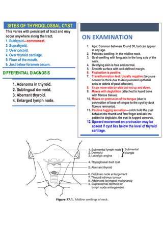
THYROGLOSSAL DUCT CYSTS
 Thyroglossal cysts are the most common upper neck midline lesions.
 They can present as a mass or a lump, at any level between the foramen
caecum and the upper mediastinum, with the majority presenting about
the level of the hyoid bone.
 They are usually sporadic, but a rare familial variant has been documented,
identified as an autosomal dominant in prepubertal girls.
 Thyroglossal duct carcinoma, although rare, may present and be identified
by the pathologist in a thyroglossal duct cyst.
 EMBRYOLOGY:
It is actually a tubular dermoid cyst arising
from persistent thyroglossal duct.
THYROGLOSSAL DUCT:
From the region of foramen caecum in early
stage of foetal life, downward growth of solid
column of cells take place which extends as far
as upper border of thyroid cartilage.
↓
It crosses the region where the future hyoid
bone is located and descends further
downward to form pyramidal lobe of thyroid.
↓
Later on, this whole tract disappears.
Occasionally it persists as a columnar
epithelial lined tract after canalization.
The thyroglossal duct descends usually in front of the
hyoid bone.
↓
The duct however may form a retro-hyoid loop and then
descends downwards.
↓
Rarely the duct may pass through the hyoid bone.
COURSE OF THYROGLOSSAL DUCT:
From the region of foramen cecum, it passes
down in the midline through genioglossus
muscles, geniohyoid muscles and then
between two mylohyoid muscles and comes in
close relation with the central part of body of
the hyoid bone.
↓
At the level of the hyoid bone it may either:
a. Pass in front of central part of hyoid bone.
b. Traverse the hyoid bone.
c. Hook behind the hyoid bone.
↓
From the hyoid bone it comes down to the
upper border of thyroid cartilage and to
continue up to the pyramidal lobe or isthmus
of thyroid gland.
 Thyroid follicles are found in one
third of the specimens of
thyroglossal duct remnant.
A fistula is usually caused by spontaneous drainage of an abscess or more commonly
resulting following an attempted drainage of a misdiagnosed midline neck abscess or
resulting from an inadequate attempt at excision usually associated with leaving an
intact hyoid bone.
DIFFERENT SITES OF THYROGLOSSAL CYST:
 At the base of tongue near the foramen cecum.
 In the floor of mouth.
 Submental region.
 Sub-hyoid region.
 Pre-laryngeal in front of thyroid cartilage.
At the level of cricoid cartilage.
FATE OF THYROGLOSSAL DUCT:
 Lower part forms the isthmus and two lateral lobes of thyroid gland.
 Part from foramen cecum to the thyroid isthmus disappears.
 Part from hyoid bone to the isthmus of thyroid gland may persist as a fibrous cord
to form levator glandulae thyroidae.
 Along the course of the duct ectopic thyroid tissue may develop in different sites—
lingual thyroid, ectopic thyroid tissue in submental and sub-hyoid or pre-laryngeal
region.
 A portion of the thyroglossal duct may remain unobliterated and accumulation of
secretion may occur leading to formation of thyroglossal cyst.
CLINICAL FEATURES:
 Initially, it appears as painful tender swelling with fever
after an episode of URI. It persists as soft, doughy,
variably sized mass after a course of antibiotic.
 Thyroglossal cyst presents as a midline swelling at the
thyrohyoid level.
 Although congenital in origin, but is usually seen in
later age with NO sex preponderance.
 The swelling can be moved sideways only and it also
moves upward on protruding the tongue or on
deglutition.
 Carcinomatous changes may occur.
 When infected, may rupture onto skin of neck,
presenting as a discharging sinus.
 Up to 5 % of these cysts present as an acute
inflammatory episode with or without an infection, and
15 % may have an associated discharging fistula.
 90% are in the midline, but 10% may be to one or other
side, with 95% of these being in the left side.
 75% are pre-hyoid, with the remaining 25% being
located above or below the hyoid, and can sometimes
be found within the mediastinum.
 Contents of the cyst---- Transparent jelly-like material
with cholesterol crystals.
SITES OF THYROGLOSSAL CYST
This varies with persistent of tract and may
occur anywhere along the tract.
1. Subhyoid—commonest.
2. Suprahyoid.
3. Over cricoid.
4. Over thyroid cartilage.
5. Floor of the mouth.
6. Just below foramen cecum.
ON EXAMINATION
1. Age: Common between 15 and 30, but can appear
at any age.
2. Painless swelling: In the midline neck.
3. Oval swelling with long axis in the long axis of the
neck.
4. Overlying skin is free and normal.
5. Smooth surface with well-defined margin.
6. Fluctuation is positive.
7. Transillumination test: Usually negative (because
content is thick due to desquamated epithelial
cells or debris of past infection).
8. It can move side by side but not up and down.
9. Moves with deglutition (attached to hyoid bone
with fibrous tissue).
10. Moves on protrusion of the tongue (due to
connection of base of tongue to the cyst by duct
fibrous remnants).
11. Positive tugging sensation—catch hold the cyst
between the thumb and fore finger and ask the
patient to deglutate, the cyst is tugged upwards.
12.Upward movement on protrusion may be
absent if cyst lies below the level of thyroid
cartilage.
DIFFERENTIAL DIAGNOSIS
1. Adenoma in thyroid.
2. Sublingual dermoid.
3. Aberrant thyroid.
4. Enlarged lymph node.
SISTRUNK OPERATION EXCISED TRACK OF THE SINUS
TREATMENT
SISTRUNK OPERATION:
 This operation involves complete excision of the cyst along with the thyroglossal tract.
 The body of hyoid bone is excised. The excision of the body of the hyoid bone helps in excision of any retro-
hyoid part of the thyroglossal tract and also complete excision of the thyroglossal tract up to the foramen
cecum.
 The operation is done under general anaesthesia → The head is extended by placing a sand bag in between the
shoulder blades and head rest on a head ring → Head end of the bed raised by 15 degree to reduce venous
conges on → An ellip cal incision is made around the cyst and the skin flaps are raised → The cyst is dissected
all around → The thyroglossal duct a ached to the upper part of the cyst is dissected upwards → The body of
the hyoid bone is excised.
VISUALISATION OF THE THYROGLOSSAL TRACT DURING OPERATION:
If methylene blue is injected into the thyroglossal cyst or fistula the thyroglossal tract
may be seen during operation being stained with methylene blue.
This technique has resulted in a significant reduction of the recurrence rates
when compared with a simple excision or removal of the cyst alone
(SHALANG PROCEDURE).
A modification of Sistrunk’s technique is currently being employed as the
standard surgical procedure without the need to excise the tongue base
epithelium. The excision is performed through a transverse midline neck
incision just below the cyst.
CT SCAN NECK
USG NECK
DIAGNOSIS & INVESTIGATIONS
 TSH and T4 will determine the thyroid status of the patient.
 ULTRASOUND-GUIDED FNAC---
o Ultrasound scanning may help with the location and diagnosis of the cyst,
and will also confirm the presence of a normal thyroid gland in its
anatomical position.
o FNAC will demonstrate cystic contents containing colloid.
 Isotope scan either 99
Tc or 123
I may be useful if the cyst is located above the
hyoid or in the posterior tongue or to identify and exclude the possibility of a
lingual thyroid.
 CT scanning or MRI should be considered in the presence of large cysts, when
malignancy is suspected and when the possibility of a lingual thyroid has been
considered.
MRI NECK
(a) Sagittal T2-weighted and
(b) contrast-enhanced T1-weighted MR images
9 yrs male presented with
midline neck swelling (A) that showed thyroglossal duct cyst on USG (B) and
Tc99 Scan showed only functioning thyroid tissue in the thyroglossal duct
cyst (C and D).
PECULIARITIES OF THYROGLOSSAL CYST---
1. There are lymphoid tissue in the wall so infection is common.
2. If hyoid bone is not excised, it can recur.
3. Cyst wall contains thyroid tissue. Sometimes these tissues may be the only thyroid tissue acting as thyroid
gland. Thyroid scan should be done before operation.
TREATMENT OF RECURRENT CYSTS AND FISTULAE:
Up to 8 % of thyroglossal cysts may recur following adequate surgical excision.
If a substandard excision, with no excision of the hyoid bone, has been carried out, a full
Sistrunk’s procedure should be undertaken.
However, if a full Sistrunk’s procedure has been carried out, further surgery may be
difficult. In these cases, the previous incision scar should be excised, with a full central
compartment neck dissection and excision of the scar tissue up to the foramen caecum.
THYROGLOSSAL DUCT CYST WITH CARCINOMA
 The incidence of carcinoma arising in thyroglossal duct cysts is reported to be approximately
1%, with the majority of such lesions being papillary carcinoma.
 Follicular, mixed papillary-follicular, and squamous cell carcinomas are less commonly seen.
 The median age for manifestation of thyroglossal duct carcinoma (TGDC) is 40 years old with
less than 25 cases reported in children.
 Malignancy is typically identified incidentally at the time of the removal of a thyroglossal duct
cyst and usually is not suspected preoperatively.
 The treatment for thyroid carcinoma arising in a thyroglossal duct remnants is an
en bloc resection via the Sistrunk procedure.
 The need to perform a thyroidectomy is debated.
 If the TGDC focus is microscopic without invasion of the cyst wall and if there are no palpable
masses in the thyroid or neck, then an en bloc resection without thyroidectomy should
suffice.
 Advocates of total thyroidectomy point to an associated 25% incidence of papillary carcinoma
in thyroid glands removed in the setting of TGDC.
 However, a review of the glands of 12 children who underwent total thyroidectomy revealed
no evidence of carcinoma.
 As such, in children, a frank discussion concerning limiting to a Sistrunk versus a total
thyroidectomy with possible 131I ablation seems warranted.

Thyroglossal duct cysts

  • 1.
    THYROGLOSSAL DUCT CYSTS Thyroglossal cysts are the most common upper neck midline lesions.  They can present as a mass or a lump, at any level between the foramen caecum and the upper mediastinum, with the majority presenting about the level of the hyoid bone.  They are usually sporadic, but a rare familial variant has been documented, identified as an autosomal dominant in prepubertal girls.  Thyroglossal duct carcinoma, although rare, may present and be identified by the pathologist in a thyroglossal duct cyst.  EMBRYOLOGY: It is actually a tubular dermoid cyst arising from persistent thyroglossal duct. THYROGLOSSAL DUCT: From the region of foramen caecum in early stage of foetal life, downward growth of solid column of cells take place which extends as far as upper border of thyroid cartilage. ↓ It crosses the region where the future hyoid bone is located and descends further downward to form pyramidal lobe of thyroid. ↓ Later on, this whole tract disappears. Occasionally it persists as a columnar epithelial lined tract after canalization. The thyroglossal duct descends usually in front of the hyoid bone. ↓ The duct however may form a retro-hyoid loop and then descends downwards. ↓ Rarely the duct may pass through the hyoid bone.
  • 2.
    COURSE OF THYROGLOSSALDUCT: From the region of foramen cecum, it passes down in the midline through genioglossus muscles, geniohyoid muscles and then between two mylohyoid muscles and comes in close relation with the central part of body of the hyoid bone. ↓ At the level of the hyoid bone it may either: a. Pass in front of central part of hyoid bone. b. Traverse the hyoid bone. c. Hook behind the hyoid bone. ↓ From the hyoid bone it comes down to the upper border of thyroid cartilage and to continue up to the pyramidal lobe or isthmus of thyroid gland.  Thyroid follicles are found in one third of the specimens of thyroglossal duct remnant. A fistula is usually caused by spontaneous drainage of an abscess or more commonly resulting following an attempted drainage of a misdiagnosed midline neck abscess or resulting from an inadequate attempt at excision usually associated with leaving an intact hyoid bone. DIFFERENT SITES OF THYROGLOSSAL CYST:  At the base of tongue near the foramen cecum.  In the floor of mouth.  Submental region.  Sub-hyoid region.  Pre-laryngeal in front of thyroid cartilage. At the level of cricoid cartilage.
  • 3.
    FATE OF THYROGLOSSALDUCT:  Lower part forms the isthmus and two lateral lobes of thyroid gland.  Part from foramen cecum to the thyroid isthmus disappears.  Part from hyoid bone to the isthmus of thyroid gland may persist as a fibrous cord to form levator glandulae thyroidae.  Along the course of the duct ectopic thyroid tissue may develop in different sites— lingual thyroid, ectopic thyroid tissue in submental and sub-hyoid or pre-laryngeal region.  A portion of the thyroglossal duct may remain unobliterated and accumulation of secretion may occur leading to formation of thyroglossal cyst. CLINICAL FEATURES:  Initially, it appears as painful tender swelling with fever after an episode of URI. It persists as soft, doughy, variably sized mass after a course of antibiotic.  Thyroglossal cyst presents as a midline swelling at the thyrohyoid level.  Although congenital in origin, but is usually seen in later age with NO sex preponderance.  The swelling can be moved sideways only and it also moves upward on protruding the tongue or on deglutition.  Carcinomatous changes may occur.  When infected, may rupture onto skin of neck, presenting as a discharging sinus.  Up to 5 % of these cysts present as an acute inflammatory episode with or without an infection, and 15 % may have an associated discharging fistula.  90% are in the midline, but 10% may be to one or other side, with 95% of these being in the left side.  75% are pre-hyoid, with the remaining 25% being located above or below the hyoid, and can sometimes be found within the mediastinum.  Contents of the cyst---- Transparent jelly-like material with cholesterol crystals.
  • 4.
    SITES OF THYROGLOSSALCYST This varies with persistent of tract and may occur anywhere along the tract. 1. Subhyoid—commonest. 2. Suprahyoid. 3. Over cricoid. 4. Over thyroid cartilage. 5. Floor of the mouth. 6. Just below foramen cecum. ON EXAMINATION 1. Age: Common between 15 and 30, but can appear at any age. 2. Painless swelling: In the midline neck. 3. Oval swelling with long axis in the long axis of the neck. 4. Overlying skin is free and normal. 5. Smooth surface with well-defined margin. 6. Fluctuation is positive. 7. Transillumination test: Usually negative (because content is thick due to desquamated epithelial cells or debris of past infection). 8. It can move side by side but not up and down. 9. Moves with deglutition (attached to hyoid bone with fibrous tissue). 10. Moves on protrusion of the tongue (due to connection of base of tongue to the cyst by duct fibrous remnants). 11. Positive tugging sensation—catch hold the cyst between the thumb and fore finger and ask the patient to deglutate, the cyst is tugged upwards. 12.Upward movement on protrusion may be absent if cyst lies below the level of thyroid cartilage. DIFFERENTIAL DIAGNOSIS 1. Adenoma in thyroid. 2. Sublingual dermoid. 3. Aberrant thyroid. 4. Enlarged lymph node.
  • 5.
    SISTRUNK OPERATION EXCISEDTRACK OF THE SINUS TREATMENT SISTRUNK OPERATION:  This operation involves complete excision of the cyst along with the thyroglossal tract.  The body of hyoid bone is excised. The excision of the body of the hyoid bone helps in excision of any retro- hyoid part of the thyroglossal tract and also complete excision of the thyroglossal tract up to the foramen cecum.  The operation is done under general anaesthesia → The head is extended by placing a sand bag in between the shoulder blades and head rest on a head ring → Head end of the bed raised by 15 degree to reduce venous conges on → An ellip cal incision is made around the cyst and the skin flaps are raised → The cyst is dissected all around → The thyroglossal duct a ached to the upper part of the cyst is dissected upwards → The body of the hyoid bone is excised. VISUALISATION OF THE THYROGLOSSAL TRACT DURING OPERATION: If methylene blue is injected into the thyroglossal cyst or fistula the thyroglossal tract may be seen during operation being stained with methylene blue. This technique has resulted in a significant reduction of the recurrence rates when compared with a simple excision or removal of the cyst alone (SHALANG PROCEDURE). A modification of Sistrunk’s technique is currently being employed as the standard surgical procedure without the need to excise the tongue base epithelium. The excision is performed through a transverse midline neck incision just below the cyst.
  • 6.
    CT SCAN NECK USGNECK DIAGNOSIS & INVESTIGATIONS  TSH and T4 will determine the thyroid status of the patient.  ULTRASOUND-GUIDED FNAC--- o Ultrasound scanning may help with the location and diagnosis of the cyst, and will also confirm the presence of a normal thyroid gland in its anatomical position. o FNAC will demonstrate cystic contents containing colloid.  Isotope scan either 99 Tc or 123 I may be useful if the cyst is located above the hyoid or in the posterior tongue or to identify and exclude the possibility of a lingual thyroid.  CT scanning or MRI should be considered in the presence of large cysts, when malignancy is suspected and when the possibility of a lingual thyroid has been considered.
  • 7.
    MRI NECK (a) SagittalT2-weighted and (b) contrast-enhanced T1-weighted MR images
  • 10.
    9 yrs malepresented with midline neck swelling (A) that showed thyroglossal duct cyst on USG (B) and Tc99 Scan showed only functioning thyroid tissue in the thyroglossal duct cyst (C and D).
  • 12.
    PECULIARITIES OF THYROGLOSSALCYST--- 1. There are lymphoid tissue in the wall so infection is common. 2. If hyoid bone is not excised, it can recur. 3. Cyst wall contains thyroid tissue. Sometimes these tissues may be the only thyroid tissue acting as thyroid gland. Thyroid scan should be done before operation. TREATMENT OF RECURRENT CYSTS AND FISTULAE: Up to 8 % of thyroglossal cysts may recur following adequate surgical excision. If a substandard excision, with no excision of the hyoid bone, has been carried out, a full Sistrunk’s procedure should be undertaken. However, if a full Sistrunk’s procedure has been carried out, further surgery may be difficult. In these cases, the previous incision scar should be excised, with a full central compartment neck dissection and excision of the scar tissue up to the foramen caecum. THYROGLOSSAL DUCT CYST WITH CARCINOMA  The incidence of carcinoma arising in thyroglossal duct cysts is reported to be approximately 1%, with the majority of such lesions being papillary carcinoma.  Follicular, mixed papillary-follicular, and squamous cell carcinomas are less commonly seen.  The median age for manifestation of thyroglossal duct carcinoma (TGDC) is 40 years old with less than 25 cases reported in children.  Malignancy is typically identified incidentally at the time of the removal of a thyroglossal duct cyst and usually is not suspected preoperatively.  The treatment for thyroid carcinoma arising in a thyroglossal duct remnants is an en bloc resection via the Sistrunk procedure.  The need to perform a thyroidectomy is debated.  If the TGDC focus is microscopic without invasion of the cyst wall and if there are no palpable masses in the thyroid or neck, then an en bloc resection without thyroidectomy should suffice.  Advocates of total thyroidectomy point to an associated 25% incidence of papillary carcinoma in thyroid glands removed in the setting of TGDC.  However, a review of the glands of 12 children who underwent total thyroidectomy revealed no evidence of carcinoma.  As such, in children, a frank discussion concerning limiting to a Sistrunk versus a total thyroidectomy with possible 131I ablation seems warranted.