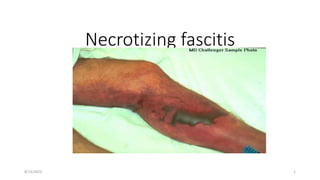
2. Necrotizing fasciitis
• Necrotizing fasciitis is an infection of the deeper tissues that results
in progressive destruction of the muscle fascia and overlying
subcutaneous fat.
• Infection spreads very quickly along the fascial planes and unless
rapidly treated, may prove fatal.
• Muscle tissue is frequently spared because of its generous blood
supply
• Initially the overlying tissue can appear unaffected. It is this feature
that makes necrotizing fasciitis difficult to diagnose without surgical
intervention.
8/13/2023 2
3. Pathophysiology
• Common pathologic features are extensive tissue destruction,
thrombosis of blood vessels, abundant bacteria spreading along
fascial planes, and an unimpressive infiltration of acute inflammatory
cells
8/13/2023 3
4. • Two clinical types exist
Type 1 necrotiscing fascites
Caused by mixed infection caused by aerobic and anaerobic bacteria
and occurs most commonly after surgical procedures and in patients
with diabetes and peripheral vascular disease other risk factors
including malnutrition, obesity, immune compromising states.
Patients with diabetes & /or pvd frequently have lower extremity
involvement
The predominant organisms isolated were Staphylococcus aureus,
streptococci, enterococci, Escherichia coli, Peptostreptococcus species,
Prevotella and Porphyromonas species, Bacteroides fragilis group, and
Clostridium species
8/13/2023 4
5. • Cervical necrotizing fasciitis — Cervical necrotizing fasciitis can result
from a breach of the integrity of mucous membranes after surgery or
instrumentation or from an odontogenic infection.
-GAS peritonsillar abscess, which can extend into the deep structures
of the neck can also cause cervical necrotizing fasciitis.
8/13/2023 5
6. • Fournier's gangrene — In the perennial area, penetration of the
gastrointestinal or urethral mucosa by enteric organisms can cause
Fournier's gangrene. Begin abruptly with severe pain and may spread
rapidly onto the anterior abdominal wall, into the gluteal muscles
and, in males, onto the scrotum and penis.
8/13/2023 6
7. • Necrotizing fasciitis can also occur in the setting of surgical wound
infection. It is characterized by a copious dishwater-like drainage,
dusky and friable subcutaneous tissue, and pale, devitalized fascia.
8/13/2023 7
8. Type II necrotizing fasciitis
• caused by GAS or other B hemolytic streptococci either alone or in
combination with other pathogens most commonly S.aureus and was
previously called "streptococcal gangrene”.
• Can occurs in young healthy adults
• Predisposing factors include a history of blunt trauma, varicella
(chickenpox), injection drug use, a penetrating injury such as
laceration, surgical procedures, childbirth, exposure to a "case,"
burns, and perhaps NSAIDs.
8/13/2023 8
9. Clinical manifestations
• Is an acute process but rarely may follow a subacute progressive
course
The affected area may be erythematous (without sharp margins),
swollen, warm, shiny, and exquisitely tender
Pain out of proportion to physical exam findings
Lymphangitis and lymphadenitis are infrequent.
The process progresses rapidly over several days, with changes in
skin color from red-purple to patches of blue-gray
8/13/2023 9
10. Within three to five days after onset, skin breakdown with bullae
(containing thick pink or purple fluid) and frank cutaneous gangrene
can be seen.
• By this time the involved area is no longer tender but has become
anesthetic secondary to thrombosis of small blood vessels and
destruction of superficial nerves in the subcutaneous tissue.
• The development of anesthesia may precede the appearance of skin
necrosis and provide a clue to the presence of necrotizing fasciitis
8/13/2023 10
11. Marked swelling and edema may produce a compartment syndrome
with complicating myonecrosis requiring fasciotomy
Subcutaneous gas is often present in the polymicrobial form of
necrotizing fasciitis, particularly in patients with diabetes
• Most cases of necrotizing fasciitis involve a single site of soft tissue
infection; multifocal necrotizing fasciitis has also been described .
Most cases were caused by gram-positive cocci; Vibrio vulnificus may
also be associated with multifocal infection.
8/13/2023 11
12. • In advanced infection, fever, tachycardia, and systemic toxicity are
generally observed, with temperature elevation in the range of 38.9°
to 40.5°C (102° to 105°F).
• Other symptoms include malaise, myalgias, diarrhea, and anorexia.
Hypotension may be present initially or develop with progressive
infection.
8/13/2023 12
13. Diagnosis
Laboratory
leukocytosis with a marked left shift,
Coagulopathy
elevations in the serum lactate,
creatine kinase
creatinine concentrations
8/13/2023 13
14. Risk score
• Serum C-reactive protein ≥150 mg/L (4 points)
• White blood cell count 15,000 to 25,000/microL (1 point) or >25,000/microL (2
points)
• Hemoglobin 11.0 to 13.5 g/dL (1 point) or ≤11 g/dL (2 points)
• Serum sodium less than 135 meq/L (2 points)
• Serum creatinine greater than 1.6 mg/dL (141 micromol/L) (2 points)
• Serum glucose greater than 180 mg/dL (10 mmol/L) (1 point)
Score ≥6 - the suspicion for necrotizing fasciitis
Score ≥8 - highly predictive (>75 percent)
The score is only useful when severe soft tissue infection is strongly
suspected.
8/13/2023 14
15. Culture
• Blood Cx positive in 60% with type II, 20% with type I
• Surgical wound cultures almost always positive
Histopathology
• Characteristic pathologic features include extensive tissue destruction,
thrombosis of blood vessels, abundant bacteria spreading along fascial
planes, and infiltration of acute inflammatory cells.
Imaging
• soft tissue x-rays, CT scan and MRI- if there is gas in the tissue, seen
in type I necrotizing fasciitis
8/13/2023 15
16. • Surgical exploration is the only way to definitively establish the
diagnosis of necrotizing infection and distinguish it from other entities
8/13/2023 16
17. Treatment
• Treatment of necrotizing infection consists of early and aggressive
surgical exploration and debridement of necrotic tissue, together with
broad spectrum empiric antibiotic therapy and hemodynamic
support.
8/13/2023 17
18. Surgery
• Is surgical emergency
• Perform aggressive debridement of all necrotic tissue until healthy,
viable (bleeding) tissue is reached. Tissue obtained in the operating
room should be sent for Gram stain and culture.
• The wound is covered with a sterile dressing, re-evaluated in the
operating room approximately 24 hours later, and aggressively
debrided again if necrotic tissue is present.
• The wound is closed after all necrotic tissue is completely debrided.
8/13/2023 18
19. • In some cases, allografting or myocutaneous tissue reconstruction is
required to cover the defect. For severe necrotizing infections
involving the digits or extremities, amputation may be required to
control the infection.
8/13/2023 19
20. Antibiotic therapy
• Empiric treatment consist of broad-spectrum antimicrobial therapy,
including activity against gram-positive, gram-negative, and anaerobic
organisms( special consideration for group A streptococcus and
Clostridium species should be taken).
include ampicillin-sulbactam OR ampicillin with either clindamycin or
metronidazole. had prior hospitalization or if antibiotics have been
used recently broader gram-Negative( can be accomplished by
substituting ticarcillin-clavulanate or piperacillin-tazobactam for
ampicillin-sulbactam or by adding a fluoroquinolone, an
aminoglycoside, an extended spectrum cephalosporin, or a
carbapenem.)
8/13/2023 20
21. • Anaerobic coverage - Clindamycin or metronidazole should be added
to the antibiotic regimen unless a beta-lactam-beta-lactamase
inhibitor or carbapenem was selected
• Antibiotic treatment should be tailored to Gram stain, culture, and
sensitivity results when available
8/13/2023 21
22. • Antibiotics should be continued until no further debridements are
needed and the patient’s hemodynamic status has normalized; this
duration must be tailored to individual patient circumstances.
8/13/2023 22
24. Outcome
• Overall mortality 17%
• Type I 21%
• Type II 14-34%
• Predictors of mortality
WBC >30 000
Cr >2.0
Clostridial infection
Presence of heart disease at admission
8/13/2023 24