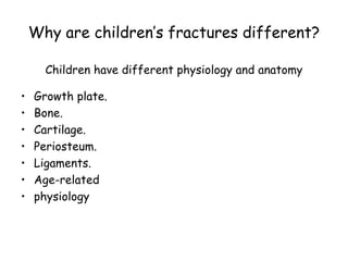
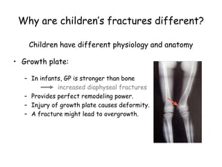
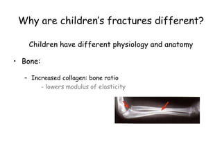
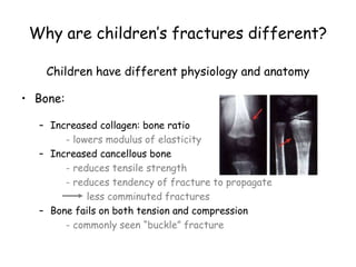
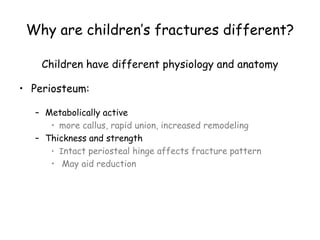
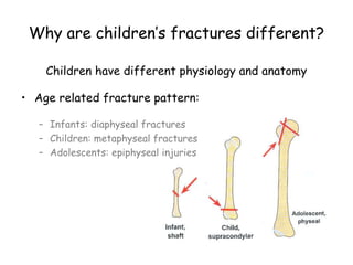
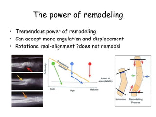
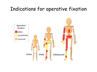
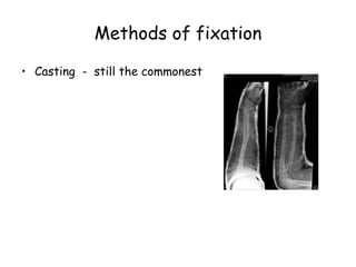
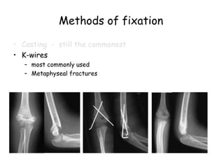
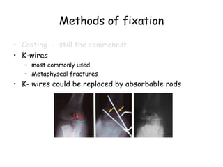
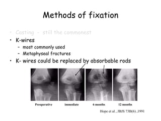
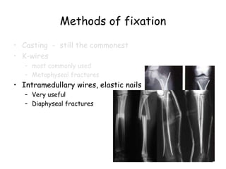
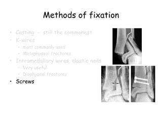
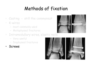
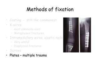
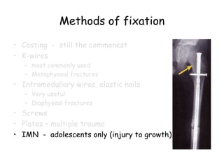
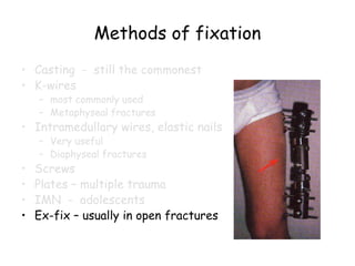
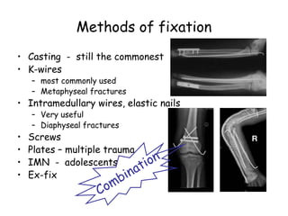
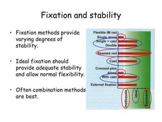
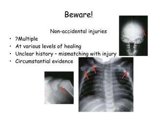
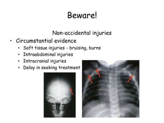
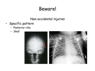
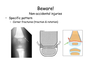
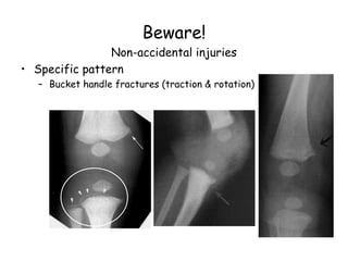
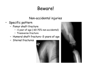
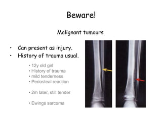
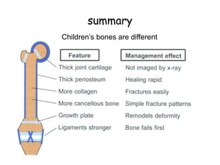
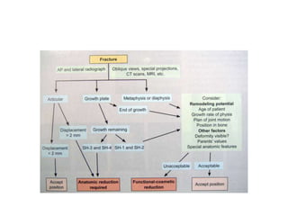
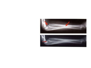
Principles of management
Pediatric Fractures
Objectives
• Statistics about fractures in children
• How children’s bones are different
• Outline principles of management
• Point out specific precautions
Acknowledgement and recommendation
Lynn T Staheli
introduction
• In Middle East ~60% of population are < 20 yrs.
• Fractures account for ~15% of all injuries in children.
• Different from adult fractures
• Vary in various age groups
( Infants, children, adolescents )
Statistics
• ~ 50% of boys and 25% of girls, expected to have a
fracture during childhood.
• Boys > girls
• Rate increases with age.
Mizulta, 1987
Statistics
• ~ 50% of boys and 25% of girls, expected to have a
fracture during childhood.
• Boys > girls
• Rate increases with age.
• Physeal injuries with age.
Mizulta, 1987
Statistics
Most frequent sites
(sample of 923 children, Mizulta, 1987)
Why are children’s fractures different?
Children have different physiology and anatomy
• Growth plate.
• Bone.
• Cartilage.
• Periosteum.
• Ligaments.
• Age-related
• physiology
Why are children’s fractures different?
Children have different physiology and anatomy
• Growth plate:
– In infants, GP is stronger than bone
increased diaphyseal fractures
– Provides perfect remodeling power.
– Injury of growth plate causes deformity.
– A fracture might lead to overgrowth.
Why are children’s fractures different?
Children have different physiology and anatomy
• Bone:
– Increased collagen: bone ratio
- lowers modulus of elasticity
Why are children’s fractures different?
Children have different physiology and anatomy
• Bone:
– Increased collagen: bone ratio
- lowers modulus of elasticity
– Increased cancellous bone
- reduces tensile strength
- reduces tendency of fracture to propagate
less comminuted fractures
– Bone fails on both tension and compression
- commonly seen “buckle” fracture
Why are children’s fractures different?
Children have different physiology and anatomy
• Cartilage:
– Increased ratio of cartilage to bone
- better resilience
- difficult x-ray evaluation
- size of articular fragment often under-estimated
Why are children’s fractures different?
Children have different physiology and anatomy
• Periosteum:
– Metabolically active
• more callus, rapid union, increased remodeling
– Thickness and strength
• Intact periosteal hinge affects fracture pattern
• May aid reduction
Why are children’s fractures different?
Children have different physiology and anatomy
• Age related fracture pattern:
– Infants: diaphyseal fractures
– Children: metaphyseal fractures
– Adolescents: epiphyseal injuries
Why are children’s fractures different?
Children have different physiology and anatomy
• Physiology
– Better blood supply
rare incidence of delayed and non-union
Physeal injuries
• Account for ~25% of all children’s fractures.
• More in boys.
• More in upper limb.
• Most heal well rapidly with good remodeling.
• Growth may be affected.
Physeal injuries
Classification: Salter-Harris, Peterson, Ogden
Physeal injuries
• Less than 1% cause physeal bridging affecting growth.
– Small bridges (<10%) may lyse spontaneously.
– Central bridges more likely to lyse.
– Peripheral bridges more likely to cause deformity
– Avoid injury to physis during fixation.
– Monitor growth over a long period.
– Image suspected physeal bar (CT, MRI)
The power of remodeling
• Tremendous power of remodeling
• Can accept more angulation and displacement
• Rotational mal-alignment ?does not remodel
The power of remodeling
Factors affecting remodeling potential
• Years of remaining growth – most important factor
• Position in the bone – the nearer to physis the better
• Plane of motion –
greatest in sagittal, the frontal, and least for transverse plane
• Physeal status – if damaged, less potential for correction
• Growth potential of adjacent physis
e.g. upper humerus better than lower humerus
The power of remodeling
Factors affecting remodeling potential
• Growth potential of adjacent physis
e.g. upper humerus better than lower humerus
Indications for operative fixation
• Open fractures
• Displaced intra articular fractures
( Salter-Harris III-IV )
• fractures with vascular injury
• ? Compartment syndrome
• Fractures not reduced by closed reduction
( soft tissue interposition, button-holing of periosteum )
• If reduction could be only maintained in an abnormal
position
Indications for operative fixation
Methods of fixation
• Casting - still the commonest
Methods of fixation
• Casting - still the commonest
• K-wires
– most commonly used
– Metaphyseal fractures
Methods of fixation
• Casting - still the commonest
• K-wires
– most commonly used
– Metaphyseal fractures
• K- wires could be replaced by absorbable rods
Methods of fixation
• Casting - still the commonest
• K-wires
– most commonly used
– Metaphyseal fractures
• K- wires could be replaced by absorbable rods
Preoperative immediate 6 months 12 months
Hope et al , JBJS 73B(6) ,1991
Methods of fixation
• Casting - still the commonest
• K-wires
– most commonly used
– Metaphyseal fractures
• Intramedullary wires, elastic nails
– Very useful
– Diaphyseal fractures
Methods of fixation
• Casting - still the commonest
• K-wires
– most commonly used
– Metaphyseal fractures
• Intramedullary wires, elastic nails
– Very useful
– Diaphyseal fractures
• Screws
Methods of fixation
• Casting - still the commonest
• K-wires
– most commonly used
– Metaphyseal fractures
• Intramedullary wires, elastic nails
– Very useful
– Diaphyseal fractures
• Screws
Methods of fixation
• Casting - still the commonest
• K-wires
– most commonly used
– Metaphyseal fractures
• Intramedullary wires, elastic nails
– Very useful
– Diaphyseal fractures
• Screws
• Plates – multiple trauma
Methods of fixation
• Casting - still the commonest
• K-wires
– most commonly used
– Metaphyseal fractures
• Intramedullary wires, elastic nails
– Very useful
– Diaphyseal fractures
• Screws
• Plates – multiple trauma
• IMN - adolescents only (injury to growth)
Methods of fixation
• Casting - still the commonest
• K-wires
– most commonly used
– Metaphyseal fractures
• Intramedullary wires, elastic nails
– Very useful
– Diaphyseal fractures
• Screws
• Plates – multiple trauma
• IMN - adolescents
• Ex-fix – usually in open fractures
Methods of fixation
• Casting - still the commonest
• K-wires
– most commonly used
– Metaphyseal fractures
• Intramedullary wires, elastic nails
– Very useful
– Diaphyseal fractures
• Screws
• Plates – multiple trauma
• IMN - adolescents
• Ex-fix
Fixation and stability
• Fixation methods provide
varying degrees of
stability.
• Ideal fixation should
provide adequate stability
and allow normal flexibility.
• Often combination methods
are best.
Complications
• Ma-lunion is not usually a problem
( except cubitus varus )
• Non-union is hardly seen
( except in the lateral condyle )
• Growth disturbance – epiphyseal damage
• Vascular – volkmann’s ischemia
• Infection - rare
Beware!
Non-accidental injuries
Beware!
Non-accidental injuries
• ?Multiple
• At various levels of healing
• Unclear history – mismatching with injury
• Circumstantial evidence
Beware!
Non-accidental injuries
• Circumstantial evidence
• Soft tissue injuries - bruising, burns
• Intraabdominal injuries
• Intracranial injuries
• Delay in seeking treatment
Beware!
Non-accidental injuries
• Specific pattern
– Posterior ribs
– Skull
Beware!
Non-accidental injuries
• Specific pattern
– Corner fractures (traction & rotation)
Beware!
Non-accidental injuries
• Specific pattern
– Bucket handle fractures (traction & rotation)
Beware!
Non-accidental injuries
• Specific pattern
– Femur shaft fracture
• <1 year of age ( 60-70% non accidental)
• Transverse fracture
– Humeral shaft fracture <3 years of age
– Sternal fractures
Beware!
Malignant tumours
• Can present as injury.
• History of trauma usual.
• 12y old girl
• History of trauma
• mild tenderness
• Periosteal reaction
• 2m later, still tender
• Ewings sarcoma
Special considerations
During resuscitation
summary
Children’s bones are different
summary
• About 60% of population in ME are children!
• Fractures in children are common.
• Children’s bones are different
• Outline principles of management.
• Specific treatment plans (combinations possible)
• Specific precautions.
• Beware
– Non-accidental trauma
– Malignant tumors
Principles_of_Pediatric_Fractures.ppt

Principles_of_Pediatric_Fractures.ppt

  • 1.
  • 2.
    Objectives • Statistics aboutfractures in children • How children’s bones are different • Outline principles of management • Point out specific precautions Acknowledgement and recommendation Lynn T Staheli
  • 3.
    introduction • In MiddleEast ~60% of population are < 20 yrs. • Fractures account for ~15% of all injuries in children. • Different from adult fractures • Vary in various age groups ( Infants, children, adolescents )
  • 4.
    Statistics • ~ 50%of boys and 25% of girls, expected to have a fracture during childhood. • Boys > girls • Rate increases with age. Mizulta, 1987
  • 5.
    Statistics • ~ 50%of boys and 25% of girls, expected to have a fracture during childhood. • Boys > girls • Rate increases with age. • Physeal injuries with age. Mizulta, 1987
  • 6.
    Statistics Most frequent sites (sampleof 923 children, Mizulta, 1987)
  • 7.
    Why are children’sfractures different? Children have different physiology and anatomy • Growth plate. • Bone. • Cartilage. • Periosteum. • Ligaments. • Age-related • physiology
  • 8.
    Why are children’sfractures different? Children have different physiology and anatomy • Growth plate: – In infants, GP is stronger than bone increased diaphyseal fractures – Provides perfect remodeling power. – Injury of growth plate causes deformity. – A fracture might lead to overgrowth.
  • 9.
    Why are children’sfractures different? Children have different physiology and anatomy • Bone: – Increased collagen: bone ratio - lowers modulus of elasticity
  • 10.
    Why are children’sfractures different? Children have different physiology and anatomy • Bone: – Increased collagen: bone ratio - lowers modulus of elasticity – Increased cancellous bone - reduces tensile strength - reduces tendency of fracture to propagate less comminuted fractures – Bone fails on both tension and compression - commonly seen “buckle” fracture
  • 11.
    Why are children’sfractures different? Children have different physiology and anatomy • Cartilage: – Increased ratio of cartilage to bone - better resilience - difficult x-ray evaluation - size of articular fragment often under-estimated
  • 12.
    Why are children’sfractures different? Children have different physiology and anatomy • Periosteum: – Metabolically active • more callus, rapid union, increased remodeling – Thickness and strength • Intact periosteal hinge affects fracture pattern • May aid reduction
  • 13.
    Why are children’sfractures different? Children have different physiology and anatomy • Age related fracture pattern: – Infants: diaphyseal fractures – Children: metaphyseal fractures – Adolescents: epiphyseal injuries
  • 14.
    Why are children’sfractures different? Children have different physiology and anatomy • Physiology – Better blood supply rare incidence of delayed and non-union
  • 15.
    Physeal injuries • Accountfor ~25% of all children’s fractures. • More in boys. • More in upper limb. • Most heal well rapidly with good remodeling. • Growth may be affected.
  • 16.
  • 17.
    Physeal injuries • Lessthan 1% cause physeal bridging affecting growth. – Small bridges (<10%) may lyse spontaneously. – Central bridges more likely to lyse. – Peripheral bridges more likely to cause deformity – Avoid injury to physis during fixation. – Monitor growth over a long period. – Image suspected physeal bar (CT, MRI)
  • 18.
    The power ofremodeling • Tremendous power of remodeling • Can accept more angulation and displacement • Rotational mal-alignment ?does not remodel
  • 19.
    The power ofremodeling Factors affecting remodeling potential • Years of remaining growth – most important factor • Position in the bone – the nearer to physis the better • Plane of motion – greatest in sagittal, the frontal, and least for transverse plane • Physeal status – if damaged, less potential for correction • Growth potential of adjacent physis e.g. upper humerus better than lower humerus
  • 20.
    The power ofremodeling Factors affecting remodeling potential • Growth potential of adjacent physis e.g. upper humerus better than lower humerus
  • 21.
    Indications for operativefixation • Open fractures • Displaced intra articular fractures ( Salter-Harris III-IV ) • fractures with vascular injury • ? Compartment syndrome • Fractures not reduced by closed reduction ( soft tissue interposition, button-holing of periosteum ) • If reduction could be only maintained in an abnormal position
  • 22.
  • 23.
    Methods of fixation •Casting - still the commonest
  • 24.
    Methods of fixation •Casting - still the commonest • K-wires – most commonly used – Metaphyseal fractures
  • 25.
    Methods of fixation •Casting - still the commonest • K-wires – most commonly used – Metaphyseal fractures • K- wires could be replaced by absorbable rods
  • 26.
    Methods of fixation •Casting - still the commonest • K-wires – most commonly used – Metaphyseal fractures • K- wires could be replaced by absorbable rods Preoperative immediate 6 months 12 months Hope et al , JBJS 73B(6) ,1991
  • 27.
    Methods of fixation •Casting - still the commonest • K-wires – most commonly used – Metaphyseal fractures • Intramedullary wires, elastic nails – Very useful – Diaphyseal fractures
  • 28.
    Methods of fixation •Casting - still the commonest • K-wires – most commonly used – Metaphyseal fractures • Intramedullary wires, elastic nails – Very useful – Diaphyseal fractures • Screws
  • 29.
    Methods of fixation •Casting - still the commonest • K-wires – most commonly used – Metaphyseal fractures • Intramedullary wires, elastic nails – Very useful – Diaphyseal fractures • Screws
  • 30.
    Methods of fixation •Casting - still the commonest • K-wires – most commonly used – Metaphyseal fractures • Intramedullary wires, elastic nails – Very useful – Diaphyseal fractures • Screws • Plates – multiple trauma
  • 31.
    Methods of fixation •Casting - still the commonest • K-wires – most commonly used – Metaphyseal fractures • Intramedullary wires, elastic nails – Very useful – Diaphyseal fractures • Screws • Plates – multiple trauma • IMN - adolescents only (injury to growth)
  • 32.
    Methods of fixation •Casting - still the commonest • K-wires – most commonly used – Metaphyseal fractures • Intramedullary wires, elastic nails – Very useful – Diaphyseal fractures • Screws • Plates – multiple trauma • IMN - adolescents • Ex-fix – usually in open fractures
  • 33.
    Methods of fixation •Casting - still the commonest • K-wires – most commonly used – Metaphyseal fractures • Intramedullary wires, elastic nails – Very useful – Diaphyseal fractures • Screws • Plates – multiple trauma • IMN - adolescents • Ex-fix
  • 34.
    Fixation and stability •Fixation methods provide varying degrees of stability. • Ideal fixation should provide adequate stability and allow normal flexibility. • Often combination methods are best.
  • 35.
    Complications • Ma-lunion isnot usually a problem ( except cubitus varus ) • Non-union is hardly seen ( except in the lateral condyle ) • Growth disturbance – epiphyseal damage • Vascular – volkmann’s ischemia • Infection - rare
  • 36.
  • 37.
    Beware! Non-accidental injuries • ?Multiple •At various levels of healing • Unclear history – mismatching with injury • Circumstantial evidence
  • 38.
    Beware! Non-accidental injuries • Circumstantialevidence • Soft tissue injuries - bruising, burns • Intraabdominal injuries • Intracranial injuries • Delay in seeking treatment
  • 39.
    Beware! Non-accidental injuries • Specificpattern – Posterior ribs – Skull
  • 40.
    Beware! Non-accidental injuries • Specificpattern – Corner fractures (traction & rotation)
  • 41.
    Beware! Non-accidental injuries • Specificpattern – Bucket handle fractures (traction & rotation)
  • 42.
    Beware! Non-accidental injuries • Specificpattern – Femur shaft fracture • <1 year of age ( 60-70% non accidental) • Transverse fracture – Humeral shaft fracture <3 years of age – Sternal fractures
  • 43.
    Beware! Malignant tumours • Canpresent as injury. • History of trauma usual. • 12y old girl • History of trauma • mild tenderness • Periosteal reaction • 2m later, still tender • Ewings sarcoma
  • 44.
  • 45.
  • 46.
    summary • About 60%of population in ME are children! • Fractures in children are common. • Children’s bones are different • Outline principles of management. • Specific treatment plans (combinations possible) • Specific precautions. • Beware – Non-accidental trauma – Malignant tumors