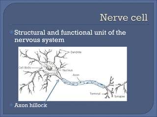
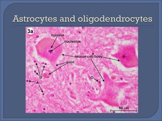
Overlapping functions: Sensory input Integration Motor output
Collect information from the environment Internal and external Also called the sensory input Give the specific type of nerves found in the five basic senses
Processing of input to be interpreted and associated with the correct response Processed in the CNS Give an example of a correct response from a stimulus
Conduction of signals from integration to effector cells Signals are conducted by nerves Nerve= tissue (composed of nerve cells) Neuron= nerve cells
Through a combination of chemical and electrical signals
Structural and functional unit of the nervous system Axon hillock
Dendrites Axons Axon hillock Myelin sheath Synaptic terminals Synapse Presynaptic cell Postsynaptic cell Neurotransmitter
Simplest- reflex arc Reflex – an automatic response Simplest reflex arc= two kinds of nerve cells Motor and sensory Effector cells – muscle or gland Knee-jerk= more complex  Front thigh contracts/inhibition of back thigh Involves second circuit (three parts) Sensory neurons from quadriceps= synapse with interneuron and motor neuron
 
Always in the CNS Also called association neuron, local circuit neuron Multi-branched Always communicate with each other “ memory”
Both are clusters of cell bodies Nuclei- found inside the CNS Ganglion- found outside CNS Take note the difference in structures of sensory neuron, motor neuron and interneuron Relate it to their function
Single presynaptic neuron to several postsynaptic neurons Convergent of several presynaptic neuron to a single postsynaptic neuron Circular path (memory)
Outnumber neurons 10:1 Then: glial cells do not participate in nerve signalling Now: presence of some synaptic interactions Important in structural integrity of the Nervous system Normal functioning of neurons
Embryo: radial glia- form tracks where neurons migrate Mature CNS: astrocytes – structural and metabolic support; stimulate formation of tight junctions between cells lining the capillaries of the brain (blood-brain barrier
Glia that form myelin sheath around axons of neurons Oligodendrocytes – CNS Schwann cells- PNS Myelin sheath- provides electrical insulation


Change in the voltage across the plasma membrane of neurons Caused by movement ions across the plasma membrane Ion channels
All cells have voltage across theri plasma membrane This membrane potential exists because of difference in ion concentration Electrically polarized Anion- inside Cation-outside
Animal cell: normally -50V to -100V Resting state of neuron: -70V Resting potential- membrane potential of an unstimulated neuron
Presence of special ion channels  Selective permeability of the membrane Anions do not diffuse readily outside the membrane
Plasma membrane- lipid bilayer Lipid- not electrically charged Ions cannot dissolved readily (do not diffuse easily) Presence of pumps or channels
Presence of more K +  channels than Na +  channels due to high permeability to K +
Ion channels do not determine the rate and direction of ion movement Electrochemical concentration is responsible
 
-85mV- amount that can counterbalance concentration gradient of K Called the equilibrium potential of potassium
http://images.google.com.ph/imgres?imgurl=http://bp3.blogger.com/_v2GFIISzHOU/SAfTN-3b72I/AAAAAAAAAM0/gxoCzSqXK5o/s400/Action%2BPotential.jpg&imgrefurl=http://katie-humanbio.blogspot.com/2008/04/nerve-impulses.html&usg=__vMf6_WjdaeIWniQU8SyB-39dsQE=&h=324&w=383&sz=25&hl=en&start=1&um=1&tbnid=Co_p3fdsxS3QEM:&tbnh=104&tbnw=123&prev=/images%3Fq%3Daction%2Bpotential%2Bnerve%2Bcell%2Bsodium%2Bpotassium%2Bpump%26um%3D1%26hl%3Den%26rlz%3D1T4PBEA_en-GBPH281PH282%26sa%3DN
All cells have membrane potential but only neurons and muscle cells can generate large amount of membrane potential Excitable cells Resting potential- membrane potential of resting cell
Nerve impulses are the action potential generated in a cell that travel through pulse-like wave of voltage though membranes Due to stimulus
Ungated ion channels- open all the time Gated ion channels Chemically-gated ion channel- stimulus: chemical (e.g. Neurotransmitter) Voltage-gated ion channel- stimulus: change in membrane potential
Graded potential: change in membrane potential due to strength of stimulus Hyperpolarization- membrane potential is more negative Depolarization- membrane potential is more positive
Recall: muscle contraction at the cellular level Threshold potential Action potential- response when threshold potential is met (generated only in axons) Threshold potential usually -50mV to -55 mV Hyperpolarization do not produce action potentials
Voltage-gated ion channels are stimulated Potassium- single voltage-sensitive  gate Closed- resting state Opens slowly in response depolarization Sodium- two voltage sensitive gates Activation- closed (resting state); opens rapidly during depolarization Inactivation- open (resting state); closes slowly in response to depolarization
Responsible in the restoration of the internal membrane potential  Due to low permeability to sodium  Inc permeability to potassium Hyperpolarization- responsible for refractory period
 
A domino effect always in a forward direction due to refractory period
Diameter of axon- faster if higher diameter Current is inversely proportional to the cross section of a wire Presence of Schwann cells Node of Ranvier- gaps between Schwann cells Ion channels are concentrated, extracellular fluid is in contact with the axon membrane at the node Saltatory conduction
Unique cell junction that control communication Between: 2 neurons, sensory receptors and sensory neurons, motor neurons and muscle cell, neurons and gland cell Two types Electrical synapse Chemical synapse
Less common than chemical synapse Cells are connected by gap junctions Allows action potential to spread directly from presynaptic terminals to postsynaptic terminals No loss of signal strength and delay Responsible for rapid movement
Presence of synaptic cleft (gap) Electrical signals not directly transmitted Signal: electrical-chemical-electrical Presence of synaptic vesicles containing neurotransmitter in the presynaptic axon Presence of chemical-gated ions
Recall: structure of a neuron Presence of inhibitory and excitatory synapses
Depolarization of the plasma membrane Results from influx of positive ions (e.g. Na ions) Depolarization may lead to an action potential if the threshold potential is met This is called EXCITATORY POSTSYNAPTIC POTENTIAL or EPSP
Inc permeability of Cl and K ion channels Hyperpolarization INHIBITORY POSTSYNAPTIC POTENTIAL or IPSP
Depends on the type of receptor and ion channel at the receiveing end
Region where voltage-gated sodium channels open Production of action potential:  Several synaptic terminals acting simultaneously on one postsynaptic terminal Few synaptic terminal discharging neurotransmitter at a greater frequency Summation is produced
Temporal: frequency of chemical transmission is great; no time to return to resting potential Spatial: presence of many presynaptic neurons stimulating one postsynaptic neuron Both are present in IPSP and EPSP
Effects vary Can be within a few millisecond Can be longer due to signal transduction pathways it enters Can remain active over a long period of time
One of the most common among vertebrate and invertebrate Can be inhibitory or excitatory depending on the receptor Excitatory in muscle cell In heart muscle: inhibitory Dec ability to create action potential through hyperpolarization
Derived from amino acids Function as transmitters in the CNS Imbalances result in different disorders Parkinson’s disease- low dopamine Schizophrenia-high dopamine Psychoactive drugs like LSD and mescaline- produce hallucinatory effects by binding to receptors of serotonin and dopamine
Catecholamines- from tyrosine Dopamine  Epinephrine and norepinephrine Also functions as hormones  Serotonin- from tryptophan Affects signal transduction pathway Affects biochemical processes
GABA- gamma aminobutyric acid Transmitter of most inhibitory synapses Produces IPSPs by inc Cl permeability Present in Valium  Glycine Glutamate Aspartate
Short chain amino acids Operate via signal transduction pathways Substance P- mediates pain Endorphins- feel good hormone Analgesic; counters pain Dec urine output Also acts as a hormone
NO and CO as regulators Release of NO in penis Viagra inhibits enzyme that masks the effect of NO Gaseous messengers are not store rather are synthesized
 

The Nervous System2

  • 1.
  • 2.
    Overlapping functions: Sensoryinput Integration Motor output
  • 3.
    Collect information fromthe environment Internal and external Also called the sensory input Give the specific type of nerves found in the five basic senses
  • 4.
    Processing of inputto be interpreted and associated with the correct response Processed in the CNS Give an example of a correct response from a stimulus
  • 5.
    Conduction of signalsfrom integration to effector cells Signals are conducted by nerves Nerve= tissue (composed of nerve cells) Neuron= nerve cells
  • 6.
    Through a combinationof chemical and electrical signals
  • 7.
    Structural and functionalunit of the nervous system Axon hillock
  • 8.
    Dendrites Axons Axonhillock Myelin sheath Synaptic terminals Synapse Presynaptic cell Postsynaptic cell Neurotransmitter
  • 9.
    Simplest- reflex arcReflex – an automatic response Simplest reflex arc= two kinds of nerve cells Motor and sensory Effector cells – muscle or gland Knee-jerk= more complex Front thigh contracts/inhibition of back thigh Involves second circuit (three parts) Sensory neurons from quadriceps= synapse with interneuron and motor neuron
  • 10.
  • 11.
    Always in theCNS Also called association neuron, local circuit neuron Multi-branched Always communicate with each other “ memory”
  • 12.
    Both are clustersof cell bodies Nuclei- found inside the CNS Ganglion- found outside CNS Take note the difference in structures of sensory neuron, motor neuron and interneuron Relate it to their function
  • 13.
    Single presynaptic neuronto several postsynaptic neurons Convergent of several presynaptic neuron to a single postsynaptic neuron Circular path (memory)
  • 14.
    Outnumber neurons 10:1Then: glial cells do not participate in nerve signalling Now: presence of some synaptic interactions Important in structural integrity of the Nervous system Normal functioning of neurons
  • 15.
    Embryo: radial glia-form tracks where neurons migrate Mature CNS: astrocytes – structural and metabolic support; stimulate formation of tight junctions between cells lining the capillaries of the brain (blood-brain barrier
  • 16.
    Glia that formmyelin sheath around axons of neurons Oligodendrocytes – CNS Schwann cells- PNS Myelin sheath- provides electrical insulation
  • 17.
  • 18.
  • 19.
  • 20.
  • 21.
    Change in thevoltage across the plasma membrane of neurons Caused by movement ions across the plasma membrane Ion channels
  • 22.
    All cells havevoltage across theri plasma membrane This membrane potential exists because of difference in ion concentration Electrically polarized Anion- inside Cation-outside
  • 23.
    Animal cell: normally-50V to -100V Resting state of neuron: -70V Resting potential- membrane potential of an unstimulated neuron
  • 24.
    Presence of specialion channels Selective permeability of the membrane Anions do not diffuse readily outside the membrane
  • 25.
    Plasma membrane- lipidbilayer Lipid- not electrically charged Ions cannot dissolved readily (do not diffuse easily) Presence of pumps or channels
  • 26.
    Presence of moreK + channels than Na + channels due to high permeability to K +
  • 27.
    Ion channels donot determine the rate and direction of ion movement Electrochemical concentration is responsible
  • 28.
  • 29.
    -85mV- amount thatcan counterbalance concentration gradient of K Called the equilibrium potential of potassium
  • 30.
  • 31.
    All cells havemembrane potential but only neurons and muscle cells can generate large amount of membrane potential Excitable cells Resting potential- membrane potential of resting cell
  • 32.
    Nerve impulses arethe action potential generated in a cell that travel through pulse-like wave of voltage though membranes Due to stimulus
  • 33.
    Ungated ion channels-open all the time Gated ion channels Chemically-gated ion channel- stimulus: chemical (e.g. Neurotransmitter) Voltage-gated ion channel- stimulus: change in membrane potential
  • 34.
    Graded potential: changein membrane potential due to strength of stimulus Hyperpolarization- membrane potential is more negative Depolarization- membrane potential is more positive
  • 35.
    Recall: muscle contractionat the cellular level Threshold potential Action potential- response when threshold potential is met (generated only in axons) Threshold potential usually -50mV to -55 mV Hyperpolarization do not produce action potentials
  • 36.
    Voltage-gated ion channelsare stimulated Potassium- single voltage-sensitive gate Closed- resting state Opens slowly in response depolarization Sodium- two voltage sensitive gates Activation- closed (resting state); opens rapidly during depolarization Inactivation- open (resting state); closes slowly in response to depolarization
  • 37.
    Responsible in therestoration of the internal membrane potential Due to low permeability to sodium Inc permeability to potassium Hyperpolarization- responsible for refractory period
  • 38.
  • 39.
    A domino effectalways in a forward direction due to refractory period
  • 40.
    Diameter of axon-faster if higher diameter Current is inversely proportional to the cross section of a wire Presence of Schwann cells Node of Ranvier- gaps between Schwann cells Ion channels are concentrated, extracellular fluid is in contact with the axon membrane at the node Saltatory conduction
  • 41.
    Unique cell junctionthat control communication Between: 2 neurons, sensory receptors and sensory neurons, motor neurons and muscle cell, neurons and gland cell Two types Electrical synapse Chemical synapse
  • 42.
    Less common thanchemical synapse Cells are connected by gap junctions Allows action potential to spread directly from presynaptic terminals to postsynaptic terminals No loss of signal strength and delay Responsible for rapid movement
  • 43.
    Presence of synapticcleft (gap) Electrical signals not directly transmitted Signal: electrical-chemical-electrical Presence of synaptic vesicles containing neurotransmitter in the presynaptic axon Presence of chemical-gated ions
  • 44.
    Recall: structure ofa neuron Presence of inhibitory and excitatory synapses
  • 45.
    Depolarization of theplasma membrane Results from influx of positive ions (e.g. Na ions) Depolarization may lead to an action potential if the threshold potential is met This is called EXCITATORY POSTSYNAPTIC POTENTIAL or EPSP
  • 46.
    Inc permeability ofCl and K ion channels Hyperpolarization INHIBITORY POSTSYNAPTIC POTENTIAL or IPSP
  • 47.
    Depends on thetype of receptor and ion channel at the receiveing end
  • 48.
    Region where voltage-gatedsodium channels open Production of action potential: Several synaptic terminals acting simultaneously on one postsynaptic terminal Few synaptic terminal discharging neurotransmitter at a greater frequency Summation is produced
  • 49.
    Temporal: frequency ofchemical transmission is great; no time to return to resting potential Spatial: presence of many presynaptic neurons stimulating one postsynaptic neuron Both are present in IPSP and EPSP
  • 50.
    Effects vary Canbe within a few millisecond Can be longer due to signal transduction pathways it enters Can remain active over a long period of time
  • 51.
    One of themost common among vertebrate and invertebrate Can be inhibitory or excitatory depending on the receptor Excitatory in muscle cell In heart muscle: inhibitory Dec ability to create action potential through hyperpolarization
  • 52.
    Derived from aminoacids Function as transmitters in the CNS Imbalances result in different disorders Parkinson’s disease- low dopamine Schizophrenia-high dopamine Psychoactive drugs like LSD and mescaline- produce hallucinatory effects by binding to receptors of serotonin and dopamine
  • 53.
    Catecholamines- from tyrosineDopamine Epinephrine and norepinephrine Also functions as hormones Serotonin- from tryptophan Affects signal transduction pathway Affects biochemical processes
  • 54.
    GABA- gamma aminobutyricacid Transmitter of most inhibitory synapses Produces IPSPs by inc Cl permeability Present in Valium Glycine Glutamate Aspartate
  • 55.
    Short chain aminoacids Operate via signal transduction pathways Substance P- mediates pain Endorphins- feel good hormone Analgesic; counters pain Dec urine output Also acts as a hormone
  • 56.
    NO and COas regulators Release of NO in penis Viagra inhibits enzyme that masks the effect of NO Gaseous messengers are not store rather are synthesized
  • 57.