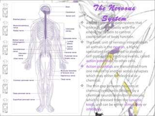
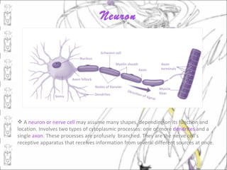
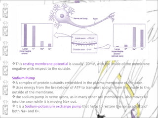
The document discusses the nervous system and sense organs. It begins by describing the basic functions and components of the nervous system, including neurons, action potentials, and synapses. It then provides details on the types of neurons, glial cells, and how the resting membrane potential and action potentials work. The document also discusses the evolution of nervous systems in invertebrates and vertebrates. It concludes by describing the peripheral nervous system and different types of sense organs.
Invertebrates are not ‘simple animals’, but they are indeed
masters of economy: their small nervous systems contain
many fewer nerve cells than those of even the tiniest
vertebrates, yet these animals solve all of the same survival
problems, can live in highly organized societies and can
communicate complex messages. The goal of this article is
to outline general features of the nervous systems of
invertebrates, and to begin to ask how these tiny
information-processing systems drive such diverse behaviour.
Central nervous system: The central nervous system consists of the brain and spinal cord. The brain plays a central role in the control of most bodily functions, including awareness, movements, sensations, thoughts, speech, and memory. Some reflex movements can occur via spinal cord pathways without the participation of brain structures. The spinal cord is connected to a section of the brain called the brainstem and runs through the spinal canal.
Peripheral Nervous System: Nerve fibers that exit the brainstem and spinal cord become part of the peripheral nervous system. Cranial nerves exit the brainstem and function as peripheral nervous system mediators of many functions, including eye movements, facial strength and sensation, hearing, and taste.
The autonomic nervous system: The autonomic nervous system is a control system that acts largely unconsciously and regulates bodily functions, such as the heart rate, digestion, respiratory rate, pupillary response, urination, and sexual arousal. This system is the primary mechanism in control of the fight-or-flight response.
The autonomic nervous system comprises two antagonistic sets of nerves, the sympathetic and parasympathetic nervous systems. The hypothalamus is the key brain site for central control of the autonomic nervous system, and the paraventricular nucleus is the key hypothalamic site for this control.
Divisions of Nervous System:
The vertebrate nervous system has three divisions:
(i) A central nervous system comprising the brain and spinal cord. Its function is to receive the stimulus from the receptors and transmit its response to the effectors. Thus, it coordinates all the functions of the body.
(ii) A peripheral nervous system consisting of cranial and spinal nerves arising from the brain and spinal cord respectively. It forms a connecting link between the receptors, central nervous system (CNS) and effectors.
(iii) An autonomic nervous system made of two ganglionated sympathetic nerves, ganglia in the head and viscera, and their connecting nerves. The autonomic nervous system is often regarded as a part of the peripheral nervous system because the two are connected. But all the three divisions of the nervous system are connected intimately both structurally and functionally.
The following power point presentation talks about neural control and coordination in humans. In this, we study about neurons, the conduction of nerve impulse, about Central Nervous System and also about sense organs
The chordates are named for the notochord: a flexible, rod-shaped structure that is found in the embryonic stage of all chordates and also in the adult stage of some chordate species.
It is located between the digestive tube and the nerve cord, providing skeletal support through the length of the body.
In some chordates, the notochord acts as the primary axial support of the body throughout the animal's lifetime.
Chordata is the last phylum of kingdom Animalia.
Which is further subdivided into subphylums, divisions and classes.
The Slides shows the classification of the phylum along with the basis on which it is classified.
(includes examples along with pictures for easy understanding and memorizing)
Origin of the Lateral Line System
Lateral line is a canal along the side of a fish containing pores that open into tubes supplied with sense organs sensitive to low vibrations.
Robert H. Denison explained the origin of the lateral line system. He explained that early vertebrates had a pore-canal system in the dermis which functioned as a primitive sensory system in detecting water movement.
Through the evidences from fossils, embryology and comparative anatomy, Denison (1966) established that the inner ear is closely related to the lateral line system. He found a distinct relationship between the pore canal system and the lateral line in Osteotraci.
The inner ear and the lateral line are developed from ectodermal thickenings, called dorso-lateral placodes. These have a number of similarities, including receptors with sensory hairs, and are both innervated by fibers in the acoustico-lateral area of the brain.
The pore canal system is present and developed in Osteostraci (ostracoderm).
It is also present in Heterostraci which is another group of ostracoderms and includes early vertebrates such as lungfishes and crossopterygians.
As its presence is extensive, it is reasonable to suggest that the pore canal system was a primitive character in early vertebrates .
In transverse sections also , it is very difficult to differentiate the pore canal system from a lateral line canal.
Structure of the Lateral Line System
Epidermal structures called neuromasts form the peripheral area of the lateral line.
Neuromasts consist of two types of cells, hair cells and supporting cells.
Hair cells have an epidermal origin and each hair cell has one high kynocyle (5-10 μm) and 30 to 150 short stereocilia (2-3 μm).
The number of hair cells in each neuromast depends on its size, and they can range from dozens to thousands.
Hair cells can be oriented in two opposite directions with each hair cell surrounded by supporting cells.
At the basal part of each hair cell, there are synaptic contacts with afferent and efferent nerve fibers. Afferent fibers, transmit signals to the neural centres of the lateral line and expand at the neuromast base. The regulation of hair cells is achieved by the action of efferent fibers.
Stereocilia and kinocilium of hair cells are immersed into a cupula and are located above the surface of the sensory epithelium.
The cupula is created by a gel-like media, which is secreted by non-receptor cells of the neuromast.
Sense organs are the specialized organs composed of sensory neurons, which help us to perceive and respond to our surroundings. There are five sense organs – eyes, ears, nose, tongue, and skin.
External receptors (exteroceptors): sense organs for touch, smell, taste, sight and hearing.
Internal receptors (interocepyors): these sense organs found in the body which detect the temperature, pain, hunger, thirst, fatigue and muscle position.
Here I would like to inform you on physiology of impulse transmission in insects. I hope this would increase your understanding -------------------------------------------------
The sensory system is the part of the nervous system that detects ,transfers and processes stimuli from the environment
http://www.asktheneurologist.com/Sensory-System.html
The Indian Dental Academy is the Leader in continuing dental education , training dentists in all aspects of dentistry and
offering a wide range of dental certified courses in different formats.for more details please visit
www.indiandentalacademy.com
Nerve Impulse is defined as a wave of electrical chemical changes across the neuron that helps in the generation of the action potential in response to the stimulus. This transmission of a nerve impulse across the neuron membrane as a result of a change in membrane potential is known as Nerve impulse conduction.
Mechanism of Nerve Impulse Conduction
Nerve impulse conduction is a major process occurring in the body responsible for organized functions of the body. So, for conduction of nerve impulse there are two mechanisms:
Continuous conduction
Saltatory conduction
types of neurons, structure and functions, types of glia cells, their structure and function, functioning of a neuron - resting potential, action potential, graded potential, absolute and relative refractory period.
Nervous system intro, neurons Branches of nervous system (generally .pdfarishmarketing21
Nervous system intro, neurons Branches of nervous system (generally speaking) Neuron
abilities Neuron organization-know whats special about all these components and how they fit
together. See details of AP and Synaptic communication Dendrites Soma Hillock Axon In
MN: myelination and nodes of ranvier Terminals EPSPs and IPSPs Receptors, ions involved,
change in membrane potential Compare properties with APs! How summed, initiate AP AP
Know different phases, ion channels involved, direction of current, effect on Vm, and stimulus
for onset/off of different phases AP continued and Start Synapse Be fluent in concepts and
mechanisms regarding Self propagation Unidirectional movement All or nothing amplitude
Saltatory conduction - why and benefits Refractory period Absolute - inactivation 2 semi-
independently regulated parts of Na channel Relative -hyperpol
Solution
Please find the answer below.
Lec 10:
1. The nervous system is divided into the central nervous system and peripheral nervous system.
2. The basic function of a neuron is to receive the information and send a signal to other neurons,
muscles, or glands. First, a neuron receives information from the external environment or from
other neurons. Then, the neuron integrates, or processes the information from all of its inputs and
determines whether or not to send an output signal.
3. The dendrites and axon are thin cytoplasmic extensions of the neuron. The dendrites, which
branch out in treelike fashion from the cell body, are specialized to receive signals and transmit
them toward the cell body. The single long axon carries signals away from the cell body.
The cell body (soma) is the enlarged portion of a neuron that most closely resembles other cells.
Nodes of Ranvier are a gap in the myelin sheath of a nerve, between adjacent Schwann cells.
4. Ion channels and direction:
As an action potential travels down the axon, there is a change in polarity across the membrane.
The Na+ and K+ gated ion channels open and close as the membrane reaches the threshold
potential, in response to a signal from another neuron. At the beginning of the action potential,
the Na+ channels open and Na+ moves into the axon, causing depolarization. Repolarization
occurs when the K+ channels open and K+ moves out of the axon. This creates a change in
polarity between the outside of the cell and the inside. The impulse travels down the axon in one
direction only, to the axon terminal where it signals other neurons..
These slides contain the basic information and principle of nervous transduction, It also includes the information about the type of the neurons, structure of the neuron, resting and active membrane potential, synapes and events occurring in it, and introduction to the neurotransmitters.
Software Delivery At the Speed of AI: Inflectra Invests In AI-Powered QualityInflectra
In this insightful webinar, Inflectra explores how artificial intelligence (AI) is transforming software development and testing. Discover how AI-powered tools are revolutionizing every stage of the software development lifecycle (SDLC), from design and prototyping to testing, deployment, and monitoring.
Learn about:
• The Future of Testing: How AI is shifting testing towards verification, analysis, and higher-level skills, while reducing repetitive tasks.
• Test Automation: How AI-powered test case generation, optimization, and self-healing tests are making testing more efficient and effective.
• Visual Testing: Explore the emerging capabilities of AI in visual testing and how it's set to revolutionize UI verification.
• Inflectra's AI Solutions: See demonstrations of Inflectra's cutting-edge AI tools like the ChatGPT plugin and Azure Open AI platform, designed to streamline your testing process.
Whether you're a developer, tester, or QA professional, this webinar will give you valuable insights into how AI is shaping the future of software delivery.
UiPath Test Automation using UiPath Test Suite series, part 4DianaGray10
Welcome to UiPath Test Automation using UiPath Test Suite series part 4. In this session, we will cover Test Manager overview along with SAP heatmap.
The UiPath Test Manager overview with SAP heatmap webinar offers a concise yet comprehensive exploration of the role of a Test Manager within SAP environments, coupled with the utilization of heatmaps for effective testing strategies.
Participants will gain insights into the responsibilities, challenges, and best practices associated with test management in SAP projects. Additionally, the webinar delves into the significance of heatmaps as a visual aid for identifying testing priorities, areas of risk, and resource allocation within SAP landscapes. Through this session, attendees can expect to enhance their understanding of test management principles while learning practical approaches to optimize testing processes in SAP environments using heatmap visualization techniques
What will you get from this session?
1. Insights into SAP testing best practices
2. Heatmap utilization for testing
3. Optimization of testing processes
4. Demo
Topics covered:
Execution from the test manager
Orchestrator execution result
Defect reporting
SAP heatmap example with demo
Speaker:
Deepak Rai, Automation Practice Lead, Boundaryless Group and UiPath MVP
Connector Corner: Automate dynamic content and events by pushing a buttonDianaGray10
Here is something new! In our next Connector Corner webinar, we will demonstrate how you can use a single workflow to:
Create a campaign using Mailchimp with merge tags/fields
Send an interactive Slack channel message (using buttons)
Have the message received by managers and peers along with a test email for review
But there’s more:
In a second workflow supporting the same use case, you’ll see:
Your campaign sent to target colleagues for approval
If the “Approve” button is clicked, a Jira/Zendesk ticket is created for the marketing design team
But—if the “Reject” button is pushed, colleagues will be alerted via Slack message
Join us to learn more about this new, human-in-the-loop capability, brought to you by Integration Service connectors.
And...
Speakers:
Akshay Agnihotri, Product Manager
Charlie Greenberg, Host
Slack (or Teams) Automation for Bonterra Impact Management (fka Social Soluti...Jeffrey Haguewood
Sidekick Solutions uses Bonterra Impact Management (fka Social Solutions Apricot) and automation solutions to integrate data for business workflows.
We believe integration and automation are essential to user experience and the promise of efficient work through technology. Automation is the critical ingredient to realizing that full vision. We develop integration products and services for Bonterra Case Management software to support the deployment of automations for a variety of use cases.
This video focuses on the notifications, alerts, and approval requests using Slack for Bonterra Impact Management. The solutions covered in this webinar can also be deployed for Microsoft Teams.
Interested in deploying notification automations for Bonterra Impact Management? Contact us at sales@sidekicksolutionsllc.com to discuss next steps.
The Art of the Pitch: WordPress Relationships and SalesLaura Byrne
Clients don’t know what they don’t know. What web solutions are right for them? How does WordPress come into the picture? How do you make sure you understand scope and timeline? What do you do if sometime changes?
All these questions and more will be explored as we talk about matching clients’ needs with what your agency offers without pulling teeth or pulling your hair out. Practical tips, and strategies for successful relationship building that leads to closing the deal.
PHP Frameworks: I want to break free (IPC Berlin 2024)Ralf Eggert
In this presentation, we examine the challenges and limitations of relying too heavily on PHP frameworks in web development. We discuss the history of PHP and its frameworks to understand how this dependence has evolved. The focus will be on providing concrete tips and strategies to reduce reliance on these frameworks, based on real-world examples and practical considerations. The goal is to equip developers with the skills and knowledge to create more flexible and future-proof web applications. We'll explore the importance of maintaining autonomy in a rapidly changing tech landscape and how to make informed decisions in PHP development.
This talk is aimed at encouraging a more independent approach to using PHP frameworks, moving towards a more flexible and future-proof approach to PHP development.
Accelerate your Kubernetes clusters with Varnish CachingThijs Feryn
A presentation about the usage and availability of Varnish on Kubernetes. This talk explores the capabilities of Varnish caching and shows how to use the Varnish Helm chart to deploy it to Kubernetes.
This presentation was delivered at K8SUG Singapore. See https://feryn.eu/presentations/accelerate-your-kubernetes-clusters-with-varnish-caching-k8sug-singapore-28-2024 for more details.
"Impact of front-end architecture on development cost", Viktor TurskyiFwdays
I have heard many times that architecture is not important for the front-end. Also, many times I have seen how developers implement features on the front-end just following the standard rules for a framework and think that this is enough to successfully launch the project, and then the project fails. How to prevent this and what approach to choose? I have launched dozens of complex projects and during the talk we will analyze which approaches have worked for me and which have not.
UiPath Test Automation using UiPath Test Suite series, part 3DianaGray10
Welcome to UiPath Test Automation using UiPath Test Suite series part 3. In this session, we will cover desktop automation along with UI automation.
Topics covered:
UI automation Introduction,
UI automation Sample
Desktop automation flow
Pradeep Chinnala, Senior Consultant Automation Developer @WonderBotz and UiPath MVP
Deepak Rai, Automation Practice Lead, Boundaryless Group and UiPath MVP
2. The Nervous
System that
a rapid communication system
interacts continuously with the
endocrine system to control
coordination of body function.
The basic unit of nervous integration in
all animals is the neuron, a highly
specialized cell designed to conduct
self-propagating electrical events, called
action potentials, to other cells.
Action potentials are transmitted from
one neuron to another across synapses
which may either be electrical or
chemical.
The thin gap between neurons at
chemical synapses is bridged by a
chemical neurotransmitter molecule,
which is released from the synaptic
knob, and can be either stimulatory or
inhibitory.
4. Neuron
Schwann cell
Nucleus
Myelin sheath Axon
Axon terminals
Axon hillock
Nodes of Ranvier Muscle
Direc
Dendrites t ion o fiber
Soma f sign
al
A neuron or nerve cell may assume many shapes, depending on its function and
location. Involves two types of cytoplasmic processes: one or more dendrites and a
single axon. These processes are profusely branched. They are the nerve cell’s
receptive apparatus that receives information from several different sources at once.
5. The axon is often covered with an insulating sheath of myelin, which speeds up signal
propagation.
Neurons are commonly classified as afferent(sensory), efferent(motor) and
interneurons, which are neither sensory nor motor but connect neurons with other
neurons.
Afferent and efferent neurons lie mostly in the peripheral nervous system, while
interneurons lie entirely within the central nervous system.
Afferent neurons are connected to receptors. Receptors function to convert external
and internal environmental stimuli into nerve signals, which are carried by afferent
neurons into the central nervous system. Nerve signals also move to efferent
neurons, which carry them via the peripheral nervous system to effectors, such as
muscles or glands.
Cell bodies of the nerve processes are located either in the:
*Central nervous system
*Ganglia-discrete bundles of nerve cell bodies located outside the CNS
Neuroglial cells (glial cells)
Extremely numerous in the vertebrate brain and may form almost half the volume of
the brain.
6. Vertebrate nerves are often enclosed by concentric rings of myelin, produced by
special glial cells called Schwann cells in the PNS and oligodendrocytes in the CNS.
Astrocytes are radiating and star like glial cells that serve as nutrients and ion
reservoirs for neurons, as well as scaffold during brain development.
Astrocytes, and microglial cells, are essential for the regenerative process that
follows brain injury.
Astrocytes also participate in several diseases of the nervous system, including
Parkinson’s disease, multiple sclerosis and brain tumor development.
7. Nature of a Nerve Action Potential
A nerve signal or action potential is an electrochemical message of neurons, the
common functional denominator of all nervous system activity.
An action potential is an “all-or-none” phenomenon; either the fiber is conducting an
action potential, or it is not.
Action potentials are alike, the only way a nerve fiber can vary its signal is by
changing the frequency of signal conduction.
Frequency change is the language of a nerve fiber.
The higher the frequency (or rate) of conduction, the greater is the level of
excitation.
Resting Membrane Potential
The membrane of a neuron is selectively permeable to K+, which can transverse the
membrane through special potassium channels. The permeability to Na+ is nearly
zero because Na+ channels are closed in a resting membrane. Potassium ions tend to
diffuse outward through the membrane, following the gradient of potassium
concentration. Very quickly the positive charge outside reaches a level that prevents
anymore K+ from diffusing out of the axon, and because large ions cannot pass
through the membrane, positively charged potassium ions are drawn back into the
cell. Now the resting membrane is at equilibrium, with an electrical gradient that
exactly balances the concentration gradient.
8. This resting membrane potential is usually -70mV, with the inside of the membrane
negative with respect to the outside.
Sodium Pump
A complex of protein subunits embedded in the plasma membrane of the axon.
Uses energy from the breakdown of ATP to transport sodium form the inside to the
outside of the membrane.
the sodium pump in nerve axons, as in many other cell membranes, also moves K+
into the axon while it is moving Na+ out.
It is a Sodium-potassium exchange pump that helps to restore the ion gradients of
both Na+ and K+.
9. Action Potential
A very rapid and brief depolarization of the membrane of the nerve fiber. This means
that the membrane potential changes from rest in a positive direction and
overshoots 0 mV to about +35 mV.
The membrane potential reverses for an instant so that the outside becomes
negative compared with the inside.
As the action potential moves ahead, the membranes returns to its normal resting
membrane potential, ready to conduct another signal.
The entire event occupies approximately a millisecond.
10. What causes the reversal of polarity in the
cell membrane during passage of an action
potential?
When an action potential arrives at a given point in a neuron membrane, the change in
membrane potential causes voltage-gated Na+ channels to suddenly open,
permitting a flood of Na+ to diffuse into the axon from the outside, moving down
the concentration gradient for Na+. Only a very minute amount of Na+ moves
across the membrane but this sudden rush of positive ions cancels the local resting
membrane potential and the membrane is depolarized. Then, as the Na+ channels
close, the membrane quickly regains its resting properties as K+ ions quickly diffuse
out of voltage-gated K+ channels that open briefly in response to the membrane
depolarization. The membrane once again become practically impermeable to Na+
the outward movement of K+ is checked as the voltage-gated K+ channels close, and
the membrane again becomes leaky to movement of K+ as the resting membrane
potential is reestablished.
Increased potassium permeability causes the action potential to drop rapidly toward
the resting membrane level, during the repolarization phase. The membrane is now
ready to transmit another action potential.
12. Synapse is a small gap that separates another neuron or effector organ from an axon
terminal when an action potential passes down.
Two distinct types of synapses:
*electrical synapses-points at which ionic currents flow directly across a narrow gap
junction from one neuron to another.
*chemical synapses-much more complex than electrical synapses which contain
packets or vesicles or specialized chemicals called neurotransmitters.
+presynaptic neurons-neurons bringing action potentials toward chemical
synapses.
+postsynaptic neurons-neurons carrying action potentials away from
chemical synapses.
Synaptic cleft-a narrow gap, having a with of approximately 20nm, that separates
membranes at a synapse.
Synaptic vesicles-found inside the synaptic knobs, each containing several thousand
molecules of acetylcholine.
Acetylcholine-most common neurotransmitter of the PNS, which illustrates typical
synaptic transmission.
Whether the postsynaptic excitatory potential is large enough to trigger an action
potential depends on how many acetylcholine molecules are released and how many
channels are opened.
13. Acetylcholinesterase-enzyme that rapidly destroys acetylcholine, which converts
acetylcholine into acetate and choline.
The final step in the sequence is reabsorption of choline into the presynaptic
terminal, resynthesis of acetylcholine and its storage in synaptic vesicles, ready to
respond to another action potential.
Excitatory synapses-releases chemical neurotransmitters that depolarize
postsynaptic membranes .
Inhibitory synapses-releases chemical neurotransmitters that move the resting
membrane potential in a more negative direction (hyperpolarization).
Neurotransmitters that are :
both excitatory and inhibitory-acetylcholine, norepinephrine, dopamine, serotonin
always excitatory-glutamate
always inhibitory-glycine, GABA(gamma amino butyric acid)
The synapse is a crucial part of the decision making equipment of the central nervous
system, modulating flow of information from one neuron to the next.
15. Invertebrates: Development of
Centralized Nervous Systems
The simplest pattern of invertebrate nervous system is the nerve net of radiate
animals, such as sea anemones, jellyfishes, hydras, and comb jellies.
There are no differentiated sensory, motor or connector components in the strict
meaning of those terms. However, there is evidence of organization into reflex arcs
with branches of a nerve net connecting to sensory receptors in the epidermis and to
epithelial cells that have contractile properties.
This type of nervous system is found among vertebrates in nerve plexuses located,
for example, in the intestinal wall.
Bilateral nervous system represent a distinct increase in complexity over the nerve
net of radiate animals.
The flatworm’s nervous system is the simplest nervous system showing
differentiation into a PNS and a CNS which coordinates everything.
More complex invertebrates exhibit a more centralized nervous system (brain), with
two longitudinal fused nerve cords and many ganglia.
16.
17. Vertebrates: Fruition of
Encephalonbrain.
The basic plan of the vertebrate nervous system is a hollow, dorsal nerve cord
terminating anteriorly in a large ganglionic mass, or
The most important trend in evolution of vertebrate nervous system is the great
elaboration of size, configuration, and functional capacity of the brain, a process
called encephalization.
Spinal Cord Spinal cord
The brain and spinal cord compose
the CNS. They begin as an ectodermal
Ventral root
neural groove, which by folding and
Dorsal root
enlarging becomes a long, hollow neural
Dorsal ganglion
tube. Spinal nerve
Segmental nerves of the spinal cords
Meninges
of the vertebrates are separated into
dorsal sensory roots and ventral motor
roots. Sympathetic
ganglion
Sensory nerve cell bodies are
gathered together into dorsal root
(spinal) ganglia. Both dorsal (sensory) Vertebra
and ventral (motor) roots meet beyond
the spinal cord to form a mixed spinal
nerve.
18. A reflex act is a response to a stimulus acting over a reflex arc. It is involuntary. Some reflex
acts are innate; others are acquired through learning.
Brain
A primitive linear brain, as seen in fishes and amphibians, expanded to form a deeply
fissured and enormously Intricate brain in the lineage leading to mammals.
The spinal cord encloses a central spinal canal and is additionally wrapped in three
layers of membranes called meninges.
An inner zone of gray matter contains the cell bodies of motor neurons and
interconnecting interneurons. An outer zone of white matter contains bundles of axon
and dendrites linking different levels of the spinal cord with each other and with the
brain.
Reflex Arc
Reflex arcs appear to be the fundamental unit of neural operation.
Parts of a typical reflex arc
Receptor-a sense organ in skin, muscle, or another organ.
Afferent-sensory neuron, which carries impulses toward the CNS.
CNS-where synaptic connections are made between sensory and interneurons.
19. Efferent-motor neuron, which makes the synaptic connection with the interneuron
and carries impulses from the CNS.
Effector-by which an animal responds to environmental changes.
A reflex arc in vertebrates in its simplest form contains only two neurons:
sensory(afferent) neuron and a motor(efferent) neuron.
Interneurons are interposed between sensory and motor neurons.
Brains of early vertebrates had three principal divisions:
*prosencephalon (forebrain)
*mesencephalon (midbrain)
*rhombencephalon (hindbrain)
Hindbrain
The medulla oblongata, the most posterior division of the brain, is really a conical
continuation of the spinal cord. The medulla, together with the more anterior
midbrain, constitutes the “brainstem”, an area that controls numerous vital and
largely subconscious activities such as heartbeat, respiration, etc. The pons contains a
thick bundle of fibers that carry impulses from one side of the cerebellum to the
other.
20. The cerebellum, lying dorsally to the medulla, controls equilibrium, posture and
movement. It does not initiate movement but operates as a precision error-control
center, or servomechanism, that programs a movement initiated somewhere else,
such as motor cortex of the cerebrum.
Midbrain
The midbrain consists mainly of the tectum, which contains nuclei that serve as centers
for visual and auditory reflexes. It mediates the most complex behavior of fishes and
amphibians, integrating visual, tactile, and auditory information. In mammals, the
midbrain is mainly a relay center for information on its way to higher brain centers.
Forebrain
Just anterior to the midbrain lie the thalamus and hypothalamus, the most posterior
element of the forebrain. The thalamus is a major relay station the analyzes and
passes sensory information to higher brain centers. In the hypothalamus are several
“house keeping” centers that regulate all functions concerned with maintenance of
internal consistency (homeostasis).
The anterior portion of the forebrain, or cerebrum, can be divide into two anatomical
distinct areas, the paleocortex and neocortex. In mammals and especially in primates
the paleocortex is a deep-lying area called rhinencephalon, because many of its
functions depend on olfaction. Better known as the limbic system, it mediates
several species-specific behaviors that relate to fulfilling needs such as feeding and
sex.
21. The neocortex (cerecral cortex)completely
overshadows the paleocortex and has
become so expanded that it envelops
much of the forebrain and all of the
midbrain.
The cortex contains discrete motor and
sensory ares. The motor ares control
voluntary muscle movements, while the
sensory cortex is the center of
conscious perception of touch, pain,
pressure, temperature, and taste.
The right and left hemispheres of the
cerebral cortex are bridged through the
corpus callosum, a neural connection
through which the two hemispheres are
able to transfer information and
coordinate mental activities. In humans,
the left hemisphere is for language
development, mathematical and
learning capabilities, and sequential
thought processes; the right
hemisphere is for spatial, musical,
artistic, intuitive, and perceptual
activities. Each hemisphere also
controls the opposite side of the body.
22.
23. Peripheral Nervous System
The peripheral nervous system includes all nervous tissue outside the CNS.
Two functional divisions:
*sensory or afferent division, which brings sensory information to the CNS
*motor or efferent division, which conveys motor commands to muscles and
glands
Efferent divisions:
+somatic nervous system-innervates skeletal muscles
+autonomic nervous system-innervates smooth muscle, cardiac muscle, and
glands
Autonomic NS subdivisions:
->parasympathetic NS-associated with non stressful activities
->sympathetic NS-active under conditions of physical or emotional
stress
25. Sense organs are specialized receptors designed for detecting environmental status
and change. Sense organs are its first level of environmental perception; they are
channels for bringing information to the CNS.
A stimulus is some form of energy-electrical, chemical, mechanical, or radiant. A
sense organs transforms energy from a stimulus into nerve action potentials. Sense
organs are biological inducers.
Sense organs are specific for one kind of stimulus
*eyes respond to light, ears to sound, pressure receptors to pressure, and
chemoreceptors to chemicals
Classification of Receptors
By location: Exteroceptors-near the external surface that keep an animal
informed about its external environment.
Interoceptor-internal parts of the body which receive stimuli from
internal organs.
Proprioceptors-in muscles, tendons, and joints which are sensitive to
changes in tension of muscles and provide an organism with a sense
of body position.
By the form of energy to which it responds:
Chemical, Mechanical, Light, or Thermal
26. Chemoreception
Chemoreception is the oldest and most universal sense in the animal kingdom.
*Contact chemical receptors-to locate food and adequately oxygenated water and to
avoid harmful substances. Chemotaxis, orientation behavior toward or away from the
chemical source.
*Distance chemical receptors-often developed to a remarkable degree of sensitivity.
Distance chemoreception is usually called smell or olfaction that guides feeding behavior,
location and selection of sexual mates, etc.
In vertebrates, taste receptors are found in the mouth cavity and especially on the
tongue, where they provide a means for judging foods before they are swallowed. A taste
bud consists of a cluster of receptor cells surrounded by supporting cells; it is provided
with a small external pore through which slender tips of sensory cells project.
Taste sensations are categorized as sweet, salty, acid, bitter, and possibly umami (Jap. For
“meaty” or “savory”)
Taste discrimination depends on assessment by the brain of the relative activity of many
different taste receptors.
Taste buds have short life (5-10 days in mammals) and are continually being replaced.
Olfactory sense is a primal sense for many animals, used for identification of food, sexual
mates, and predators.
Olfactory endings are located in a special epithelium covered by a thin film of mucus,
positioned deep in the nasal cavity.
27. Social insects and many other animals produce species-specific compounds called
pheromones that constitute a highly developed chemical language.
Pheromones are a diverse group of organic compounds that an animal releases to
affect the physiology or behavior of another individual of the same species.
Mechanoreception
Mechanoreceptors are sensitive to quantitative forces such as touch, pressure, or in
short, in motion.
Touch: Pacinian corpuscles, relatively large mechanoreceptors that register deep
touch and pressure in mammalian skin, illustrate the general properties of
mechanoreceptors.
Pain: Pain receptors are relatively unspecialized nerve fiber endings that respond to a
variety of stimuli signaling possible or real damage tissues.
*Slow pain-Pain fibers respond to small peptides which are released by the injured
cell.
*Fast pain-more direct response of the nerve endings to mechanical or thermal
stimuli.
Lateral-line System of Fish and Amphibians: a lateral line is a distant touch receptor
system for detecting wave vibrations and currents in water.
28.
29. Receptors called neuromasts are located on the
body surface in aquatic amphibians and some
fishes. Each neuromast is a collection of hair cells
with sensory endings or cilia, embedded in a
gelatinous, wedge-shape mass, the cupula.
Hearing: An ear is a specialized receptor for
detecting sound waves in the surrounding
environment.
Equilibrium: The vertebrate organ of equilibrium is
the labyrinth, or vestibular organ. Specialized sense
organs for monitoring gravity and low- frequency
vibrations often appear as statocysts, a simple sac
lined with hair cells and containing a heavy
calcareous structure, the statolith.
Photoreception: Vision
Light –sensitive receptors are called photoreceptors.
these receptors range from simple light-sensitive
cells scattered randomly on the body surface of
many invertebrates to the exquisitely developed
camera-type eye of vertebrates and cephalopods.
30. A dinoflagellate bears a lens, a light-gathering
chamber, and a photoreceptive pigment cup-all
developed within a single-celled oragnism.
Vertebrates have a camera eye with focusing
optics. Photoreceptor cells of the retina are
two of kinds:
*Rods-designed for high sensitivity with
dim light
*Cones-designed for color vision in
daylight.
Cones predominate in fovea centralis of
human eyes, the area of keenest vision. Rods
are more abundant in peripheral areas of the
retina.