Downloaded 356 times
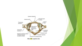
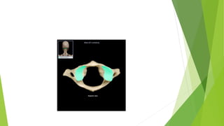

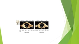


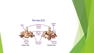
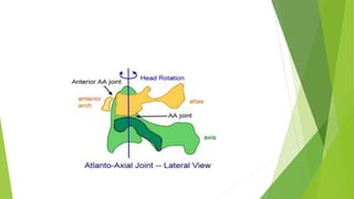
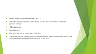

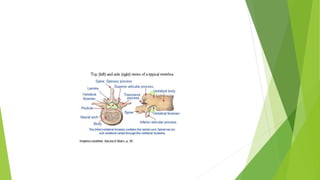
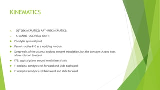
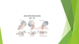


The document details the anatomy and function of the cervical spine, which consists of 7 vertebrae divided into the cranio-vertebral and lower cervical regions. It discusses the specific features, articulations, and ligaments associated with the atlas (C1) and axis (C2), emphasizing their roles in motion and stability. The lower cervical spine characteristics and kinematics during various motions like flexion and extension are also covered.


![Biomechanics_of_spine[1].pptx](https://cdn.slidesharecdn.com/ss_thumbnails/biomechanicsofspine1-230804185208-4b0b1a1a-thumbnail.jpg?width=640&height=640&fit=bounds)


![MT-01 Anatomy and biomechanics of cervical spine-102024 [Autosaved].pptx](https://cdn.slidesharecdn.com/ss_thumbnails/mt-01anatomyandbiomechanicsofcervicalspine-102024autosaved-250827121120-fd4fe0b6-thumbnail.jpg?width=640&height=640&fit=bounds)


