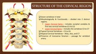
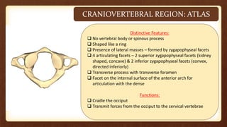
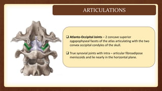
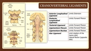
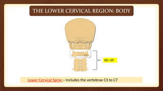
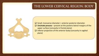
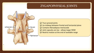
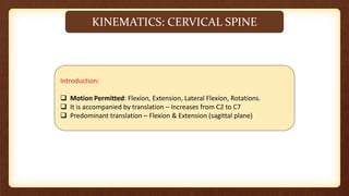
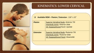
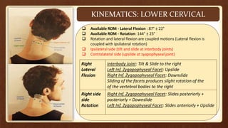
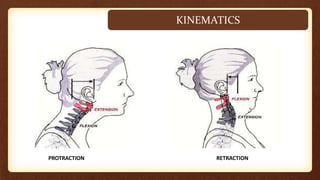
CERVICAL BIOMECHANICS
DR. NITHIN NAIR (PT)
ASST. PROFESSOR
DEPARTMENT OF COMMUNITY PHYSIOTHERAPY
MGM COLLEGE OF PHYSIOTHERAPY
STRUCTURE OF THE CERVICAL REGION
C1
C2
C4
C7
C6
C5
C3
 Seven vertebrae in total
 Morphologically & Functionally – divided into 2 distinct
regions.
 Upper Cervical Spine – includes occipital condyles &
first two cervical vertebrae (C1 & C2)
 Lower Cervical Spine – includes the vertebrae C3 to C7
 Typical Cervical Vertebrae – C3 to C6
 Atypical Cervical Vertebrae – Atlas, Axis, and C7
 Presence of transverse foramen – passage for vertebral
artery.
CRANIOVERTEBRAL REGION: ATLAS
Distinctive Features:
 No vertebral body or spinous process
 Shaped like a ring
 Presence of lateral masses – formed by zygapophyseal facets
 4 articulating facets – 2 superior zygapophyseal facets (kidney
shaped, concave) & 2 inferior zygapophyseal facets (convex,
directed inferiorly)
 Transverse process with transverse foramen
 Facet on the internal surface of the anterior arch for
articulation with the dense
Functions:
 Cradle the occiput
 Transmit forces from the occiput to the cervical vertebrae
CRANIOVERTEBRAL REGION: AXIS
Distinctive Features:
 Body: Anterior portion extends inferiorly, superior surface of
the body has a vertical projection (Dens)
 Dens: Anterior facet for articulation with anterior arch of
atlas & Posterior groove for articulation with the transverse
ligament.
 Arch: Inferior (face anteriorly) & Superior zygapophyseal
facets (face upward & laterally)
 Spinous process: Long and elongated with a bifid.
Functions:
 Transmit the combined load of the head and the atlas to the
remainder of the cervical spine.
 Provide motion into axial rotation of head and the atlas.
TEST YOURSELF
TEST YOURSELF
TEST YOURSELF
ARTICULATIONS
 Atlanto-Occipital Joints – 2 concave superior
zygapophyseal facets of the atlas articulating with the two
convex occipital condyles of the skull.
 True synovial joints with intra – articular fibroadipose
meniscoids and lie nearly in the horizontal plane.
ARTICULATIONS
 Atlantoaxial Joints: 3 synovial joints (1 median and 2
lateral atlantoaxial joints)
Median Atlantoaxial Joint:
 Between Dens & Atlas.
 It’s a synovial trochoid (pivot) joint.
Lateral Atlantoaxial Joints:
 Between the superior zygapophyseal facets of the axis
and the inferior zygapophyseal facets of the atlas.
 It’s a plain synovial joints – however, the articular
cartilages of both the atlantal & axial facets are convex
(thus – biconvex and filled with meniscoids)
CRANIOVERTEBRAL LIGAMENTS
Anterior Longitudinal
Ligament
Limits Extension
Posterior
Longitudinal
Ligament
Limits Forward Flexion
Tectorial Ligament Limits Forward Flexion
Ligamentum Flavum Limits Forward Flexion
Ligamentum Nuchae Limits Forward Flexion
Alar Ligament Limit rotation of the
(same side)
Lateral flexion (opposite
side)
THE LOWER CERVICAL REGION: BODY
Lower Cervical Spine – Includes the vertebrae C3 to C7
C3 – C7
THE LOWER CERVICAL REGION: BODY
 Small, transverse diameter > anterior posterior diameter.
 Uncinate process – present at the postero-lateral margins of the
upper surface (concavity in frontal plane)
 Inferior projection of the anterior body (concavity in sagittal
plane)
THE LOWER CERVICAL REGION: ARCHES
 Pedicles – Projects posterolaterally
 Laminae – Projects posteromedially
 Zygapophyseal articular process – Superior facets are flat, oval
and face superiorly & posteriorly; inferior facets face inferiorly
and anteriorly.
 Transverse process – Presence of transverse foramen, Presence
of groove for spinal nerves.
 Spinous process – Short, extend horizontally, bifid tip. C2 to C3
(Length decreases slightly), C3 to C5 (Length remains constant),
C7 (Length increases)
 Vertebral Foramen – Large, Triangular
TEST YOURSELF
INTERVERTEBRAL DISK
 Annulus Fibrosus – Crescent shape
 Cervical discs – Thick anteriorly and taper laterally
 Fissures in the disc develops to become cleft by 9 years of age –
becomes joint cavity (Uncovertebral Cleft or Joints of Luschka)
INTERBODY JOINTS OF THE LOWER CERVICAL REGION
 Saddle joints
 In sagittal plane, inferior surface of cranial vertebra (concave) and
superior surface of caudal vertebra (convex)
 In frontal plane, inferior surface of cranial vertebra (convex) and
superior surface of caudal vertebra (concave)
 Predominant motions - Tilting
ZYGAPOPHYSEAL JOINTS
 True synovial joints
 Lie midway between frontal and horizontal plane
 Contains fibroadipose meniscoids
 Joint capsules are lax – allows larger ROM
 Restrict motion at the end of available range
KINEMATICS: CERVICAL SPINE
Introduction:
 Motion Permitted: Flexion, Extension, Lateral Flexion, Rotations.
 It is accompanied by translation – Increases from C2 to C7
 Predominant translation – Flexion & Extension (sagittal plane)
KINEMATICS: ATLANTOOCCIPITAL JT.
Atlanto-occipital Joint
 Motion Permitted: Nodding (Flexion / Extension) + Small
amount of Lat. Flexion & Rotation
 Combined ROM - 15° (Flexion / Extension) with approx. 3° lat.
flexion and axial rotation
Flexion Occipital condyles slide backward and role
role forward.
Extension Occipital condyles slide forward and role
role backward.
Lateral Flexion &
& Axial Rotation
Rotation
Extremely limited by tension in the joint
joint capsules
KINEMATICS: ATLANTOAXIAL JOINT
Atlantoaxial Joint:
 Motion Permitted: Rotations, lateral flexion, flexion, and
extension.
 50% approx. – Total rotations of the cervical region occurs at
atlantoaxial joint.
 Remaining 40% - Total rotations of the cervical region evenly
distributed in the lower cervical joints.
 Atlas pivots approx. 45° to either side.
KINEMATICS: LOWER CERVICAL
 Available ROM – Flexion / Extension : 126° ± 22°
Flexion Superior Vertebral body: Anterior Tilt
Interbody Joints: Anterior Slide
Inf. Zygapophyseal Facet: Upslide
Extension Superior Vertebral body: Posterior Tilt
Interbody Joints: Posterior Slide
Inf. Zygapophyseal Facet: Downslide
KINEMATICS: LOWER CERVICAL
 Available ROM - Lateral Flexion : 87° ± 22°
 Available ROM - Rotation: 144° ± 23°
 Rotation and lateral flexion are coupled motions (Lateral flexion is
coupled with ipsilateral rotation)
 Ipsilateral side (tilt and slide at interbody joints)
 Contralateral side (upslide at zyapophyseal joint)
Right
Lateral
Flexion
Interbody Joint: Tilt & Slide to the right
Left Inf. Zygapophyseal Facet: Upslide
Right Inf. Zygapophyseal Facet: Downslide
Sliding of the facets produces slight rotation of the
of the vertebral bodies to the right
Right side
side
Rotation
Right Inf. Zygapophyseal Facet: Slides posteriorly +
posteriorly + Downslide
Left Inf. Zygapophyseal Facet: Slides anteriorly + Upslide
PROTRACTION RETRACTION
KINEMATICS
TEST YOURSELF
KINEMATICS: ATLANTOOCCIPITAL JT.
KINEMATICS: ATLANTOAXIAL JOINT
KINEMATICS: LOWER CERVICAL
KINETICS: MUSCLE GROUPS
4 functional groups – Superficial Posterior, Deep
Posterior, Superficial Anterior, Deep Anterior.
Superficial
Posterior
Trapezius, Levator Scapulae, Splenius
Capitis, Splenius Cervicis
Deep Posterior Multifidi, Suboccipital
Superficial
Anterior
Sternocleidomastoid, Scalenus
Deep Anterior Longus Colli & Longus Capitis
KINETICS
 Cervical region bears less weight and are more mobile
 Cervical spine is lordotic – anterior shear force
 No discs present at atlantooccipital and atlantoaxial articulations – compressive load
transferred directly.
 Laminae of axis and C7 are large to withstand compressive load
 In cervical region from C3 TO C7 – compressive forces are transmitted by 3 columns –
central (major), 2 posterolateral column (one-third)
SUMMARY
REFERENCE
 JOINT STRUCTURE AND FUNCTION – CYNTHIA NORKIN (5TH & 6TH ED.)
 BIOMECHANICS OF JOINT – SHYAM D. GANVIR (1ST ED.)
THANK
YOU!!!

CERVICAL BIOMECHANICS.pptx

  • 1.
    CERVICAL BIOMECHANICS DR. NITHINNAIR (PT) ASST. PROFESSOR DEPARTMENT OF COMMUNITY PHYSIOTHERAPY MGM COLLEGE OF PHYSIOTHERAPY
  • 2.
    STRUCTURE OF THECERVICAL REGION C1 C2 C4 C7 C6 C5 C3  Seven vertebrae in total  Morphologically & Functionally – divided into 2 distinct regions.  Upper Cervical Spine – includes occipital condyles & first two cervical vertebrae (C1 & C2)  Lower Cervical Spine – includes the vertebrae C3 to C7  Typical Cervical Vertebrae – C3 to C6  Atypical Cervical Vertebrae – Atlas, Axis, and C7  Presence of transverse foramen – passage for vertebral artery.
  • 3.
    CRANIOVERTEBRAL REGION: ATLAS DistinctiveFeatures:  No vertebral body or spinous process  Shaped like a ring  Presence of lateral masses – formed by zygapophyseal facets  4 articulating facets – 2 superior zygapophyseal facets (kidney shaped, concave) & 2 inferior zygapophyseal facets (convex, directed inferiorly)  Transverse process with transverse foramen  Facet on the internal surface of the anterior arch for articulation with the dense Functions:  Cradle the occiput  Transmit forces from the occiput to the cervical vertebrae
  • 4.
    CRANIOVERTEBRAL REGION: AXIS DistinctiveFeatures:  Body: Anterior portion extends inferiorly, superior surface of the body has a vertical projection (Dens)  Dens: Anterior facet for articulation with anterior arch of atlas & Posterior groove for articulation with the transverse ligament.  Arch: Inferior (face anteriorly) & Superior zygapophyseal facets (face upward & laterally)  Spinous process: Long and elongated with a bifid. Functions:  Transmit the combined load of the head and the atlas to the remainder of the cervical spine.  Provide motion into axial rotation of head and the atlas.
  • 5.
  • 6.
  • 7.
  • 8.
    ARTICULATIONS  Atlanto-Occipital Joints– 2 concave superior zygapophyseal facets of the atlas articulating with the two convex occipital condyles of the skull.  True synovial joints with intra – articular fibroadipose meniscoids and lie nearly in the horizontal plane.
  • 9.
    ARTICULATIONS  Atlantoaxial Joints:3 synovial joints (1 median and 2 lateral atlantoaxial joints) Median Atlantoaxial Joint:  Between Dens & Atlas.  It’s a synovial trochoid (pivot) joint. Lateral Atlantoaxial Joints:  Between the superior zygapophyseal facets of the axis and the inferior zygapophyseal facets of the atlas.  It’s a plain synovial joints – however, the articular cartilages of both the atlantal & axial facets are convex (thus – biconvex and filled with meniscoids)
  • 10.
    CRANIOVERTEBRAL LIGAMENTS Anterior Longitudinal Ligament LimitsExtension Posterior Longitudinal Ligament Limits Forward Flexion Tectorial Ligament Limits Forward Flexion Ligamentum Flavum Limits Forward Flexion Ligamentum Nuchae Limits Forward Flexion Alar Ligament Limit rotation of the (same side) Lateral flexion (opposite side)
  • 11.
    THE LOWER CERVICALREGION: BODY Lower Cervical Spine – Includes the vertebrae C3 to C7 C3 – C7
  • 12.
    THE LOWER CERVICALREGION: BODY  Small, transverse diameter > anterior posterior diameter.  Uncinate process – present at the postero-lateral margins of the upper surface (concavity in frontal plane)  Inferior projection of the anterior body (concavity in sagittal plane)
  • 13.
    THE LOWER CERVICALREGION: ARCHES  Pedicles – Projects posterolaterally  Laminae – Projects posteromedially  Zygapophyseal articular process – Superior facets are flat, oval and face superiorly & posteriorly; inferior facets face inferiorly and anteriorly.  Transverse process – Presence of transverse foramen, Presence of groove for spinal nerves.  Spinous process – Short, extend horizontally, bifid tip. C2 to C3 (Length decreases slightly), C3 to C5 (Length remains constant), C7 (Length increases)  Vertebral Foramen – Large, Triangular
  • 14.
  • 15.
    INTERVERTEBRAL DISK  AnnulusFibrosus – Crescent shape  Cervical discs – Thick anteriorly and taper laterally  Fissures in the disc develops to become cleft by 9 years of age – becomes joint cavity (Uncovertebral Cleft or Joints of Luschka)
  • 16.
    INTERBODY JOINTS OFTHE LOWER CERVICAL REGION  Saddle joints  In sagittal plane, inferior surface of cranial vertebra (concave) and superior surface of caudal vertebra (convex)  In frontal plane, inferior surface of cranial vertebra (convex) and superior surface of caudal vertebra (concave)  Predominant motions - Tilting
  • 17.
    ZYGAPOPHYSEAL JOINTS  Truesynovial joints  Lie midway between frontal and horizontal plane  Contains fibroadipose meniscoids  Joint capsules are lax – allows larger ROM  Restrict motion at the end of available range
  • 18.
    KINEMATICS: CERVICAL SPINE Introduction: Motion Permitted: Flexion, Extension, Lateral Flexion, Rotations.  It is accompanied by translation – Increases from C2 to C7  Predominant translation – Flexion & Extension (sagittal plane)
  • 19.
    KINEMATICS: ATLANTOOCCIPITAL JT. Atlanto-occipitalJoint  Motion Permitted: Nodding (Flexion / Extension) + Small amount of Lat. Flexion & Rotation  Combined ROM - 15° (Flexion / Extension) with approx. 3° lat. flexion and axial rotation Flexion Occipital condyles slide backward and role role forward. Extension Occipital condyles slide forward and role role backward. Lateral Flexion & & Axial Rotation Rotation Extremely limited by tension in the joint joint capsules
  • 20.
    KINEMATICS: ATLANTOAXIAL JOINT AtlantoaxialJoint:  Motion Permitted: Rotations, lateral flexion, flexion, and extension.  50% approx. – Total rotations of the cervical region occurs at atlantoaxial joint.  Remaining 40% - Total rotations of the cervical region evenly distributed in the lower cervical joints.  Atlas pivots approx. 45° to either side.
  • 21.
    KINEMATICS: LOWER CERVICAL Available ROM – Flexion / Extension : 126° ± 22° Flexion Superior Vertebral body: Anterior Tilt Interbody Joints: Anterior Slide Inf. Zygapophyseal Facet: Upslide Extension Superior Vertebral body: Posterior Tilt Interbody Joints: Posterior Slide Inf. Zygapophyseal Facet: Downslide
  • 22.
    KINEMATICS: LOWER CERVICAL Available ROM - Lateral Flexion : 87° ± 22°  Available ROM - Rotation: 144° ± 23°  Rotation and lateral flexion are coupled motions (Lateral flexion is coupled with ipsilateral rotation)  Ipsilateral side (tilt and slide at interbody joints)  Contralateral side (upslide at zyapophyseal joint) Right Lateral Flexion Interbody Joint: Tilt & Slide to the right Left Inf. Zygapophyseal Facet: Upslide Right Inf. Zygapophyseal Facet: Downslide Sliding of the facets produces slight rotation of the of the vertebral bodies to the right Right side side Rotation Right Inf. Zygapophyseal Facet: Slides posteriorly + posteriorly + Downslide Left Inf. Zygapophyseal Facet: Slides anteriorly + Upslide
  • 23.
  • 24.
    TEST YOURSELF KINEMATICS: ATLANTOOCCIPITALJT. KINEMATICS: ATLANTOAXIAL JOINT KINEMATICS: LOWER CERVICAL
  • 25.
    KINETICS: MUSCLE GROUPS 4functional groups – Superficial Posterior, Deep Posterior, Superficial Anterior, Deep Anterior. Superficial Posterior Trapezius, Levator Scapulae, Splenius Capitis, Splenius Cervicis Deep Posterior Multifidi, Suboccipital Superficial Anterior Sternocleidomastoid, Scalenus Deep Anterior Longus Colli & Longus Capitis
  • 26.
    KINETICS  Cervical regionbears less weight and are more mobile  Cervical spine is lordotic – anterior shear force  No discs present at atlantooccipital and atlantoaxial articulations – compressive load transferred directly.  Laminae of axis and C7 are large to withstand compressive load  In cervical region from C3 TO C7 – compressive forces are transmitted by 3 columns – central (major), 2 posterolateral column (one-third)
  • 27.
  • 28.
    REFERENCE  JOINT STRUCTUREAND FUNCTION – CYNTHIA NORKIN (5TH & 6TH ED.)  BIOMECHANICS OF JOINT – SHYAM D. GANVIR (1ST ED.)
  • 29.