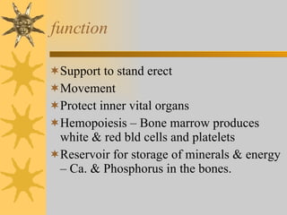
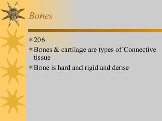
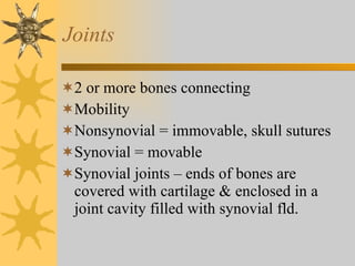
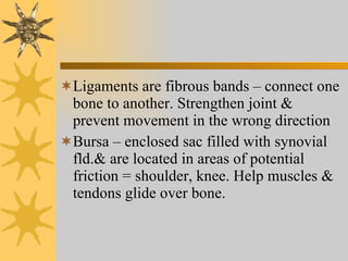
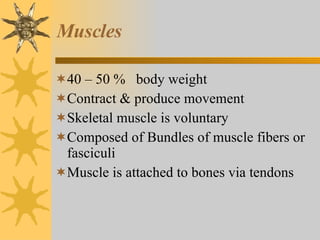
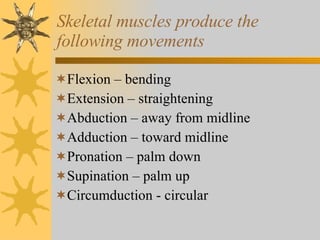
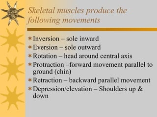
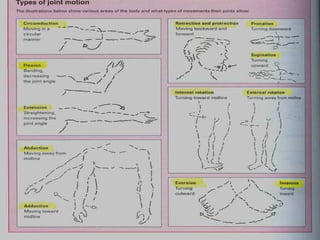
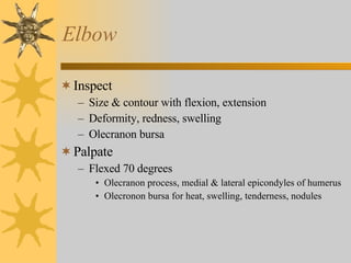
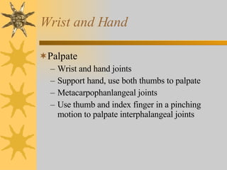
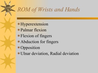
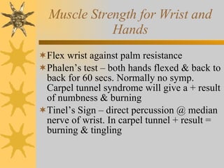
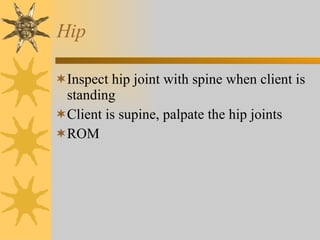
Muscoloskeletal System
Musculoskeletal System Consists of: Bones Muscles Joints cartilage
function Support to stand erect Movement Protect inner vital organs Hemopoiesis – Bone marrow produces white & red bld cells and platelets Reservoir for storage of minerals & energy – Ca. & Phosphorus in the bones.
Bones 206 Bones & cartilage are types of Connective tissue Bone is hard and rigid and dense
Joints 2 or more bones connecting Mobility  Nonsynovial = immovable, skull sutures Synovial = movable Synovial joints – ends of bones are covered with cartilage & enclosed in a joint cavity filled with synovial fld.
Ligaments are fibrous bands – connect one bone to another. Strengthen joint & prevent movement in the wrong direction Bursa – enclosed sac filled with synovial fld.& are located in areas of potential friction = shoulder, knee. Help muscles & tendons glide over bone.
Muscles 40 – 50 %  body weight Contract & produce movement Skeletal muscle is voluntary Composed of Bundles of muscle fibers or fasciculi Muscle is attached to bones via tendons
Skeletal muscles produce the following movements Flexion – bending Extension – straightening Abduction – away from midline Adduction – toward midline Pronation – palm down Supination – palm up Circumduction - circular
Skeletal muscles produce the following movements Inversion – sole inward Eversion – sole outward Rotation – head around central axis Protraction –forward movement parallel to ground (chin) Retraction – backward parallel movement Depression/elevation – Shoulders up & down
 
Temporomandibular Joint (TMJ) Articulation of temporal & mandible Depression anterior to tragus of ear Jaw function for chewing & speaking Movements Hinge – open/close Gliding – protrusion/retraction Gliding- side to side
Spine 33 Vertebrae Spinous process posterior midline 7 Cervical 12 Thoracic 5 Lumbar 5 Sacral 3 – 4 Coccygeal
C7 & T1 prominent base of neck Inferior angle of scapula in line with T7 & T8 Highest point iliac crest at L4 Curves Double S – lateral view cervical & lumbar are concave;(inward)  Thoracic & sacrococcygeal are convex Intervertebral discs cushion the spine = shock absorber
Shoulder Articulation of humerus & glenoid fossa of scapula Ball & socket – enclosed by rotator cuff (4 muscles and tendons) Acromion process – bump at top of shoulder
Elbow Articulation humerus, radius, & ulna  Landmarks are the Medial & lateral epicondyles of the humerus & large olecranon process of the ulna in between Sensitive ulnar nerve
Wrists and Carpals Wrist –articulation of radius & carpal bones Permits flexion, extension & side to side deviation Metacarpophalangeal & interphalangeal joints – permit finger flexion and extension
Hip Acetabulum & femur Ball & socket joint  Weight bearing function Landmarks ( IM injections) Anterior, superior iliac crest Ischial tuberosity ( ↓ gluteus maximus, flex hip) Greater trochanter of femur
Knee Femur, tibia & patella Largest joint Hinged joint & largest synovial membrane 2 cartilages – medial & lateral menisci cushion the tibia & femur
Ankle & Foot Ankle joint is the articulation of Tibia, fibula & talus Hinged joint Dorsiflexion Plantar flexion Landmarks Medial & lateral malleolus
Aging adult Loss of bone density = osteoporosis Postural changes ↓  height due to shortening of the vertebral column
Subjective Data Joints Pain Stiffness Swelling, heat, redness Muscles Pain, cramps weakness
Subjective Data Bones Pain Deformity Trauma Functional Assessment ( ADL’s ) Self – care behaviors
Objective Assessment Physical Exam Musculoskeletal Purpose  To assess function for ADL’s  Screen for abnormalities
Screening Exams Inspection Palpation ROM with movement active or passive if apparent limitations  Age Specific
Important to : Client comfort Systemic approach Support joints Bilateral exam
Equipment Tape measure Goniometer Skin marking pen
Inspection Size & contour of joint Color, swelling, masses, deformity
Palpation Each joint Temperature  Muscles Bony articulations joint capsule Tenderness, swelling, masses
ROM Active ROM Limitation – try passive motion or  in ROM, use a goniometer to measure angles
Muscle Testing Repeat movements for Active ROM Client flexes & holds against opposing force = bilaterally, resists opposing force
Grade muscle strength (pg. 616) Values 0- 5 Grade 5= Normal –Full ROM against gravity, full resistance
 
TMJ Swelling, tenderness, crepitation Crepitation = audible & palpable crunching or grating with movement
Cervical Spine Inspection Head & neck alignment Spine  Palpation Spinous processes, Trapezius, Paravertebral muscles ROM, flexion, extension, hyperextension, lateral flexion, rotation, circumduction Repeat applying opposing force
Shoulders Inspect Bilateral comparison Palpate Bilaterally for muscle spasms, atrophy, swelling, heat, tenderness Clavicle to acromioclavicular joint, scapula, greater tubercle of humerus, subacromal bursa, biceps groove & anterior aspect glenohumeral joint
Test for Shoulder ROM Flexion Extension Internal rotation  External rotation Abduction Adduction Circumduction  Test for strength; shrug shoulders, flex forward, up & abduct against resistance
Elbow Inspect Size & contour with flexion, extension Deformity, redness, swelling Olecranon bursa Palpate Flexed 70 degrees Olecranon process, medial & lateral epicondyles of humerus Olecronon bursa for heat, swelling, tenderness, nodules
ROM of Elbow Flexion Extension pronation supination
Muscle Strength of Elbow Flex elbow – then extend against resistance applied just proximal to the wrist
Wrist and Hand Inspect Palmar & dorsal surface Position, contour and shape Swelling, redness, deformity or nodules
Wrist and Hand Palpate Wrist and hand joints Support hand, use both thumbs to palpate Metacarpophanlangeal joints Use thumb and index finger in a pinching motion to palpate interphalangeal joints
ROM of Wrists and Hands Hyperextension Palmar flexion Flexion of fingers Abduction for fingers  Opposition  Ulnar deviation, Radial deviation
Muscle Strength for Wrist and Hands Flex wrist against palm resistance Phalen’s test – both hands flexed & back to back for 60 secs. Normally no symp. Carpel tunnel syndrome will give a + result of numbness & burning Tinel’s Sign – direct percussion @ median nerve of wrist. In carpel tunnel + result = burning & tingling
Hip Inspect hip joint with spine when client is standing Client is supine, palpate the hip joints ROM
Knee Supine with legs extended ( knees can be flexed or dangling for inspection) Swelling = ? Soft tissue or  ↑ fld in the joint Bulge Sign – stroke up medial aspect 2-3x. Tap lateral aspect. Watch for a bulge in the medial hollow. Ballottement of the Patella – lger amt of flds
Ankle & foot Inspect while nonweight- bearing, then standing & walking ROM Muscle strength
Spine Standing Inspect  Palpate spinous processes ROM of spine is checked by asking to touch toes
Leg measurement True leg length = measure b/t fixed points, the anterior iliac spine cross the medial side of the knee to the medial malleolus

NurseReview.Org - Muscoloskeletal System

  • 1.
  • 2.
    Musculoskeletal System Consistsof: Bones Muscles Joints cartilage
  • 3.
    function Support tostand erect Movement Protect inner vital organs Hemopoiesis – Bone marrow produces white & red bld cells and platelets Reservoir for storage of minerals & energy – Ca. & Phosphorus in the bones.
  • 4.
    Bones 206 Bones& cartilage are types of Connective tissue Bone is hard and rigid and dense
  • 5.
    Joints 2 ormore bones connecting Mobility Nonsynovial = immovable, skull sutures Synovial = movable Synovial joints – ends of bones are covered with cartilage & enclosed in a joint cavity filled with synovial fld.
  • 6.
    Ligaments are fibrousbands – connect one bone to another. Strengthen joint & prevent movement in the wrong direction Bursa – enclosed sac filled with synovial fld.& are located in areas of potential friction = shoulder, knee. Help muscles & tendons glide over bone.
  • 7.
    Muscles 40 –50 % body weight Contract & produce movement Skeletal muscle is voluntary Composed of Bundles of muscle fibers or fasciculi Muscle is attached to bones via tendons
  • 8.
    Skeletal muscles producethe following movements Flexion – bending Extension – straightening Abduction – away from midline Adduction – toward midline Pronation – palm down Supination – palm up Circumduction - circular
  • 9.
    Skeletal muscles producethe following movements Inversion – sole inward Eversion – sole outward Rotation – head around central axis Protraction –forward movement parallel to ground (chin) Retraction – backward parallel movement Depression/elevation – Shoulders up & down
  • 10.
  • 11.
    Temporomandibular Joint (TMJ)Articulation of temporal & mandible Depression anterior to tragus of ear Jaw function for chewing & speaking Movements Hinge – open/close Gliding – protrusion/retraction Gliding- side to side
  • 12.
    Spine 33 VertebraeSpinous process posterior midline 7 Cervical 12 Thoracic 5 Lumbar 5 Sacral 3 – 4 Coccygeal
  • 13.
    C7 & T1prominent base of neck Inferior angle of scapula in line with T7 & T8 Highest point iliac crest at L4 Curves Double S – lateral view cervical & lumbar are concave;(inward) Thoracic & sacrococcygeal are convex Intervertebral discs cushion the spine = shock absorber
  • 14.
    Shoulder Articulation ofhumerus & glenoid fossa of scapula Ball & socket – enclosed by rotator cuff (4 muscles and tendons) Acromion process – bump at top of shoulder
  • 15.
    Elbow Articulation humerus,radius, & ulna Landmarks are the Medial & lateral epicondyles of the humerus & large olecranon process of the ulna in between Sensitive ulnar nerve
  • 16.
    Wrists and CarpalsWrist –articulation of radius & carpal bones Permits flexion, extension & side to side deviation Metacarpophalangeal & interphalangeal joints – permit finger flexion and extension
  • 17.
    Hip Acetabulum &femur Ball & socket joint Weight bearing function Landmarks ( IM injections) Anterior, superior iliac crest Ischial tuberosity ( ↓ gluteus maximus, flex hip) Greater trochanter of femur
  • 18.
    Knee Femur, tibia& patella Largest joint Hinged joint & largest synovial membrane 2 cartilages – medial & lateral menisci cushion the tibia & femur
  • 19.
    Ankle & FootAnkle joint is the articulation of Tibia, fibula & talus Hinged joint Dorsiflexion Plantar flexion Landmarks Medial & lateral malleolus
  • 20.
    Aging adult Lossof bone density = osteoporosis Postural changes ↓ height due to shortening of the vertebral column
  • 21.
    Subjective Data JointsPain Stiffness Swelling, heat, redness Muscles Pain, cramps weakness
  • 22.
    Subjective Data BonesPain Deformity Trauma Functional Assessment ( ADL’s ) Self – care behaviors
  • 23.
    Objective Assessment PhysicalExam Musculoskeletal Purpose To assess function for ADL’s Screen for abnormalities
  • 24.
    Screening Exams InspectionPalpation ROM with movement active or passive if apparent limitations Age Specific
  • 25.
    Important to :Client comfort Systemic approach Support joints Bilateral exam
  • 26.
    Equipment Tape measureGoniometer Skin marking pen
  • 27.
    Inspection Size &contour of joint Color, swelling, masses, deformity
  • 28.
    Palpation Each jointTemperature Muscles Bony articulations joint capsule Tenderness, swelling, masses
  • 29.
    ROM Active ROMLimitation – try passive motion or in ROM, use a goniometer to measure angles
  • 30.
    Muscle Testing Repeatmovements for Active ROM Client flexes & holds against opposing force = bilaterally, resists opposing force
  • 31.
    Grade muscle strength(pg. 616) Values 0- 5 Grade 5= Normal –Full ROM against gravity, full resistance
  • 32.
  • 33.
    TMJ Swelling, tenderness,crepitation Crepitation = audible & palpable crunching or grating with movement
  • 34.
    Cervical Spine InspectionHead & neck alignment Spine Palpation Spinous processes, Trapezius, Paravertebral muscles ROM, flexion, extension, hyperextension, lateral flexion, rotation, circumduction Repeat applying opposing force
  • 35.
    Shoulders Inspect Bilateralcomparison Palpate Bilaterally for muscle spasms, atrophy, swelling, heat, tenderness Clavicle to acromioclavicular joint, scapula, greater tubercle of humerus, subacromal bursa, biceps groove & anterior aspect glenohumeral joint
  • 36.
    Test for ShoulderROM Flexion Extension Internal rotation External rotation Abduction Adduction Circumduction Test for strength; shrug shoulders, flex forward, up & abduct against resistance
  • 37.
    Elbow Inspect Size& contour with flexion, extension Deformity, redness, swelling Olecranon bursa Palpate Flexed 70 degrees Olecranon process, medial & lateral epicondyles of humerus Olecronon bursa for heat, swelling, tenderness, nodules
  • 38.
    ROM of ElbowFlexion Extension pronation supination
  • 39.
    Muscle Strength ofElbow Flex elbow – then extend against resistance applied just proximal to the wrist
  • 40.
    Wrist and HandInspect Palmar & dorsal surface Position, contour and shape Swelling, redness, deformity or nodules
  • 41.
    Wrist and HandPalpate Wrist and hand joints Support hand, use both thumbs to palpate Metacarpophanlangeal joints Use thumb and index finger in a pinching motion to palpate interphalangeal joints
  • 42.
    ROM of Wristsand Hands Hyperextension Palmar flexion Flexion of fingers Abduction for fingers Opposition Ulnar deviation, Radial deviation
  • 43.
    Muscle Strength forWrist and Hands Flex wrist against palm resistance Phalen’s test – both hands flexed & back to back for 60 secs. Normally no symp. Carpel tunnel syndrome will give a + result of numbness & burning Tinel’s Sign – direct percussion @ median nerve of wrist. In carpel tunnel + result = burning & tingling
  • 44.
    Hip Inspect hipjoint with spine when client is standing Client is supine, palpate the hip joints ROM
  • 45.
    Knee Supine withlegs extended ( knees can be flexed or dangling for inspection) Swelling = ? Soft tissue or ↑ fld in the joint Bulge Sign – stroke up medial aspect 2-3x. Tap lateral aspect. Watch for a bulge in the medial hollow. Ballottement of the Patella – lger amt of flds
  • 46.
    Ankle & footInspect while nonweight- bearing, then standing & walking ROM Muscle strength
  • 47.
    Spine Standing Inspect Palpate spinous processes ROM of spine is checked by asking to touch toes
  • 48.
    Leg measurement Trueleg length = measure b/t fixed points, the anterior iliac spine cross the medial side of the knee to the medial malleolus