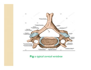
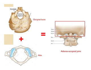
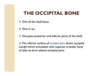
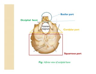
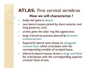
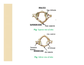
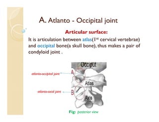
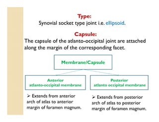
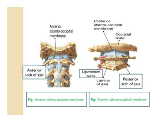
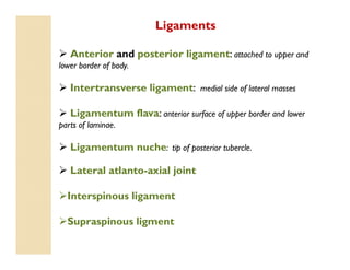
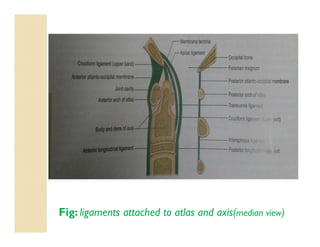
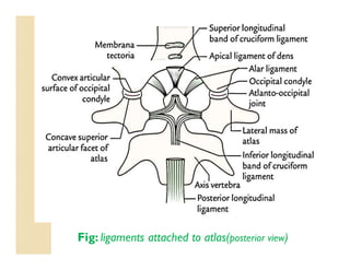
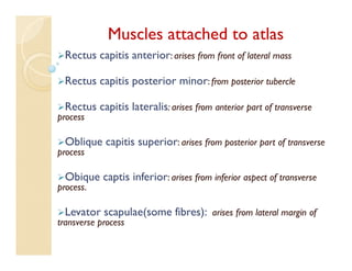
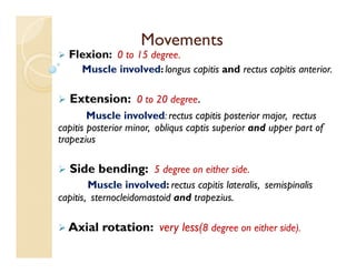
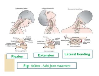
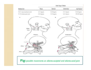
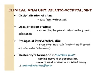
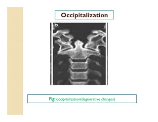
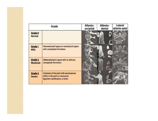
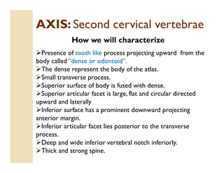
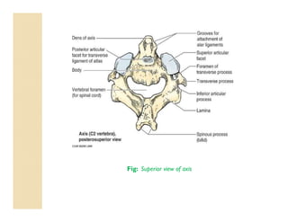
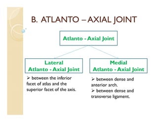
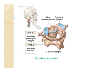
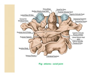
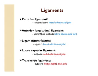
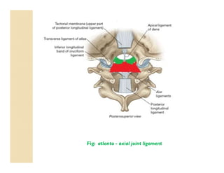
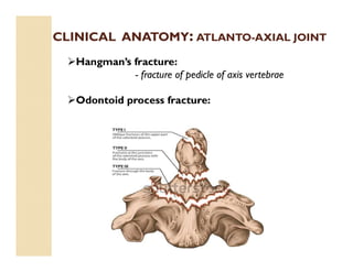
Anatomy:
>Atlas is the topmost vertebra and chief peculiarity of atlas is that it has no body, it is ring like and consist of anterior and posterior arch and two lateral masses.
>Axis, the 2nd cervical vertebra has a concave under side and convex from side to side. The most distinctive characteristic of this bone is strong odontoid process, the dens.
TheJoint:
>Atlanto-occipital joint (articulation between the atlas and the occipital bone) consists of a pair of condyloid joints.
>The atlanto-occipital joints are synovial socket-type joints
Ligaments:
> Posterior atlanto-occipital membrane: extend from anterior arch of atlas to posterior margin of foramen magnum.
>Anterior atlanto-occipital membrane: extend from anterior arch of atlas to anterior margin of foramen magnum.
>The ligamentam flavam join laminae of adjacent vertebral arches.
>The interspinous ligaments expand to form the ligamentum nuchae which inserts along the posterior foramen magnum and external occipital condyle.
> The following four ligaments stabilize these joints:
1.Apical ligament: Connects the dens to the foramen magnum of the occipital bone.
2.Alar ligaments: Connect the dens to the lateral margins of the foramen magnum.
3.Cruciate ligament: Attaches the dens to the anterior arch of the atlas and the body of the axis to the foramen magnum of the occipital bone.
4.Tectorial membrane: Starts at the skull and becomes the posterior longitudinal ligament.
>Atlanto-axial articular capsules are thick and loose, and connect the margins of the lateral masses of the atlas with those of the posterior articular surfaces of the axis.
Muscles:
>Flexion is produced mainly by the action of longis capitis, rectus capitis anterior and sternocleidomastoid (anterior fibres)
>Extension by the rectus capitis posterior major and minor, the obliquus capitis superior, the semispinalis capitis, splenius capitis, longissimus capitis, sternocleidomastoid and upper fibres of the trapezius
>The recti lateralis are concerned in the lateral movement, assisted by the trapezius, splenius capitis, semispinalis capitis, and the sternocleidomastoid of the same side, all acting together.
Movements:
>Flexion and extension in the Sagittal axis, which give rise to the ordinary forward and backward nodding of the head.
>Lateral flexion to one or other side in the Frontal axis(titling of head
>Lateral AAJ Movement: It is a synovial joint which allows only gliding
>Medial AAJ Movement: This joint allows the rotation of the atlas the axis i.e round the dens.
Clinical anatomy:
> Headaches can arise from many different sources including dysfunctional muscles, tears in the ligaments, misalignment of the vertebral bodies, injury to cervical facets and degenerative discs.
>Excessive flexion could rupture the supraspinous ligament.
>Posterior atlanto-occipital membrane ossification cause migraine headaches due to compression of artery.
2. TOPIC TO BE PRESENTED
Introduction to cervical vertebrae.
Characteristic of cervical vertebrae.
Atlanto-occipital joint.
Introduction to atlas and occipit.
Introduction to axis
Atlanto-axial joint.
Clinical anatomy
3. A. Introduction to cervical vertebrae
Identifying feature:
The cervical vertebrae are identified by the presence of
foramania transversaria in transverse process.
Fig: a typical cervical vertebrae
Foramania
transversia
4. Characteristic
These are 7 in no. of which 3rd to 6th are typical
while1st, 2nd and 7th are atypical.
Small body.
Transverse process is relatively short.
Pedicles are long and directed backward and laterally.
Lamina is long and narrow.
Short spinous process.
Vertebral foramen is larger than the body and
triangular in shape.
Flat articular facets.
8. THE OCCIPITAL BONE
One of the skull bone.
One in no.
Occupies posterior and inferior parts of the skull.
The inferior surface of condylar part shows occipital
condyl which articulates with superior articular facet
of atlas to form atlanto-occipital joint.
9. Fig: Inferior view of occipital bone
Basilar part
Occipital facet
Condylar part
Squamous part
10. ATLAS: First cervical vertebrae
How we will characterize ?
body and spine is absent.
two lateral masses joined by short anterior and
long posterior arch.
arches gives the atlas ring like appearance.
large transverse process pierced by foramen
transversarium.
Superiorly lateral mass shows an elongated
concave facet which articulates with the
corresponding condyle of occipital bone.
Inferiorly lateral masses shows a large oval facet
for articulation with the corresponding superior
articular facet of axis.
12. A. Atlanto - Occipital joint
Articular surface:
It is articulation between atlas(1st cervical vertebrae)
and occipital bone(a skull bone), thus makes a pair of
condyloid joint .
atlanto-occipital joint
atlanto-axial joint
Fig: posterior view
13. Membrane/Capsule
Posterior
atlanto occipital membrane
Anterior
atlanto-occipital membrane
Extends from anterior
arch of atlas to anterior
margin of foramen magnum.
Extends from posterior
arch of atlas to posterior
margin of foramen magnum.
Capsule:
The capsule of the atlanto-occipital joint are attached
along the margin of the corresponding facet.
Type:
Synovial socket type joint i.e. ellipsoid.
14. Fig: Anterior atlanto-occipital membrane Fig: Anterior atlanto-occipital membrane
Posterior
arch of axis
Anterior
arch of axis
Ligamentum
nuche
15. Ligaments
Anterior and posterior ligament: attached to upper and
lower border of body.
Intertransverse ligament: medial side of lateral masses
Ligamentum flava: anterior surface of upper border and lower
parts of laminae.
Ligamentum nuche: tip of posterior tubercle.
Lateral atlanto-axial joint
Interspinous ligament
Supraspinous ligment
18. Muscles attached to atlas
Rectus capitis anterior: arises from front of lateral mass
Rectus capitis posterior minor: from posterior tubercle
Rectus capitis lateralis: arises from anterior part of transverse
process
Oblique capitis superior: arises from posterior part of transverse
process
Obique captis inferior: arises from inferior aspect of transverse
process.
Levator scapulae(some fibres): arises from lateral margin of
transverse process
19. Movements
Flexion: 0 to 15 degree.
Muscle involved: longus capitis and rectus capitis anterior.
Extension: 0 to 20 degree.
Muscle involved: rectus capitis posterior major, rectus
capitis posterior minor, obliqus captis superior and upper part of
trapezius
Side bending: 5 degree on either side.
Muscle involved: rectus capitis lateralis, semispinalis
capitis, sternocleidomastoid and trapezius.
Axial rotation: very less(8 degree on either side).
22. CLINICAL ANATOMY: ATLANTO-OCCIPITAL JOINT
Occipitalization of atlas:
- atlas fuses with occipit
Decalcification of atlas:
- caused by pharyngeal and retropharyngeal
inflamation.
Prolapse of intervertebral disc:
- most often traumatic(usually 6th and 7th cervical
and upper lumbar prolase occurs).
Oesteophyte formation in ‘luschka’s joint’:
- cervical nerve root compression.
- may cause distortion of vertebral artery
i.e vertebrobasilar insufficency .
26. AXIS: Second cervical vertebrae
How we will characterize
Presence of tooth like process projecting upward from the
body called “dense or odontoid”.
The dense represent the body of the atlas.
Small transverse process.
Superior surface of body is fused with dense.
Superior articular facet is large, flat and circular directed
upward and laterally
Inferior surface has a prominent downward projecting
anterior margin.
Inferior articular facet lies posterior to the transverse
process.
Deep and wide inferior vertebral notch inferiorly.
Thick and strong spine.
28. B. ATLANTO – AXIAL JOINT
Atlanto - Axial Joint
Medial
Atlanto - Axial Joint
Lateral
Atlanto - Axial Joint
between the inferior
facet of atlas and the
superior facet of the axis.
between dense and
anterior arch.
between dense and
transverse ligament.
33. Movement
Rotation: takes around vertical plane.
Muscle involved: obliqus capitis inferior
rectus capitis posterior major
spleinus capitus
sternoclidomastoid.
Fig: atlanto - axial joint movement
34.
35. CLINICAL ANATOMY: ATLANTO-AXIAL JOINT
Hangman’s fracture:
- fracture of pedicle of axis vertebrae
Odontoid process fracture: