Downloaded 643 times

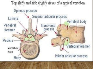
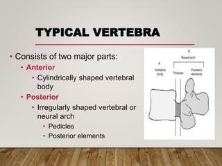
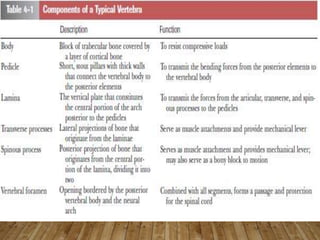
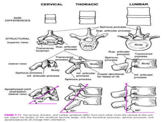
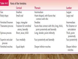
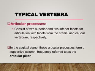
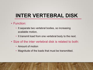
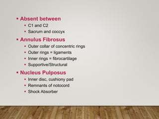
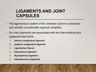
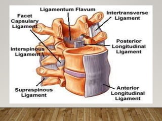
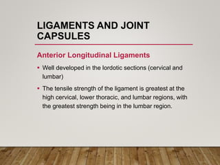


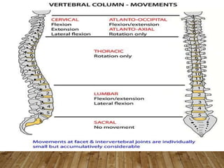
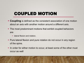
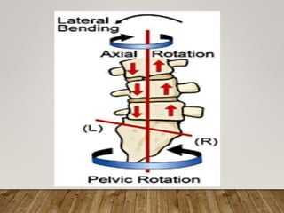
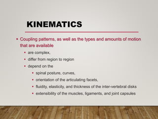


The document provides a comprehensive overview of the biomechanics of the human spine, covering its structure, spinal curves, vertebrae features, intervertebral disks, articulations, ligaments, and associated movements. It discusses primary and secondary curves, abnormal curves, and the role of various ligaments and muscle activities in spinal stability and function. Additionally, it addresses the kinetic and kinematic aspects of spinal motion, including axial compression, bending, torsion, and shear forces, along with their implications on spinal health.


![Biomechanics_of_spine[1].pptx](https://cdn.slidesharecdn.com/ss_thumbnails/biomechanicsofspine1-230804185208-4b0b1a1a-thumbnail.jpg?width=640&height=640&fit=bounds)


