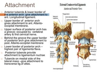
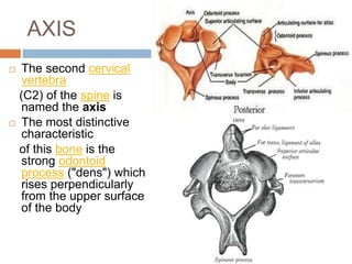
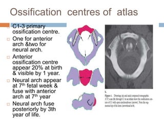
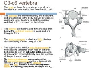
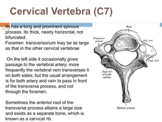
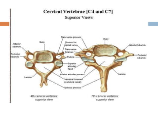
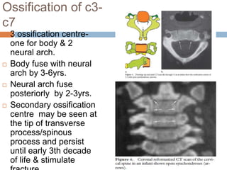
ANATOMY OF
CERVICAL SPINE
DR ASHISH KUMAR
GUPTA
Pg 2nd year
RADIODIAGNOSIS
SLIMS
Cervical vertebrae
 2 TYPES
 Atypical
 Axis
 Atlas
 C 7
 Typical
 C 3-6
Atlas
 Doesn’t Have body
&spinous process
 Its ring-like, has anterior and
a posterior arch and two
lateral masses.
 Each lateral mass has
superior articular
facet&inferior articular facet.
 Superior articular facet
articulate with occipital
condoyle- atlanto-occipital
joint.
 Inferior articular facet
articulate with axis superior
facet –atlanto-axis joint.
 Transverse process project
laterally from lateral mass
which is pierced by foramen
transversorium
Attachment
 Anterior tubercle & lower border of
the anterior arch give attachment to
ant. Longitudinal ligament.
 Upper border of anterior arch
gives attachment to ant. Atlanto-
occipital membrane.
 Upper surface of posterior arch has
a groove- occupied by vertebral
artery & first cervical nerve.
 Behind the groove the upper border
of posterior arch give attachment to
post. Atlanto-occipital membrane
 Lower border of posterior arch –
highest pair of ligamenta flava.
 Posterior tubercle provide
attachment to ligamentum nuchae
 Tubercle on medial side of the
lateral mass –give attachment to
transverse lig.of atlas.
AXIS
 The second cervical
vertebra
(C2) of the spine is
named the axis
 The most distinctive
characteristic
of this bone is the
strong odontoid
process ("dens") which
rises perpendicularly
from the upper surface
of the body
 Dens provide attachment at its apex to apical
ligament& on each side to alar ligament.
 Anterior surface of body gives attachment to
ant. Longitudinal ligament.
 Posterior surface of body gives attachment to
vertical limb of cruciate ligament , membrana
tectoria, post.longitudinal ligament.
Ligamentous Anatomy
 Anterior longitudinal ligament
 Reinforces anterior discs, limits extension
 Posterior longitudinal ligament
 Reinforces posterior discs, limits flexion
 Ligamentum nuchae =
supraspinous ligament
 Thicker than in thoracic/lumbar regions
 Limits flexion
 Interspinous/intertransverse
ligaments
 Limit flexion and rotation/limits lateral
flexion
 Ligamentum flavum
 Attach lamina of one vertebrae to
another, reinforces articular facets
 Limits flexion and rotation
Ossification centres of atlas
 C1-3 primary
ossification centre.
 One for anterior
arch &two for
neural arch.
 Anterior
ossification centre
appear 20% at birth
& visible by 1 year.
 Neural arch appear
at 7th fetal week &
fuse with anterior
arch at 7th year
 Neural arch fuse
posteriorly by 3th
year of life.
Ossification centres of axis
 C2-4 ossification centre.
 One for each neural arch,
body & odontoid process
each one.
 Odontoid process forms in
utero from 2 separate
ossification centre & fuse in
mid line by 7th fetal month.
 Secondary ossification
centre appear at apex of
odontoid process between
3 to 6 yrs & fuse by 12-13
yrs.
 Body fuse with odontoid
process by 3-6yrs.
 Neural arch fuse with body
of odontoid between 3-6 yrs
& fuse posteriorly 2-3yrs.
C3-c6 vertebra
 The body of these four vertebrae is small, and
broader from side to side than from front to back.
 The pedicles are directed laterally and backward,
and are attached to the body midway between its
upper and lower borders, so that the superior
vertebral notch is as deep as the inferior.
 The laminae are narrow, and thinner above than
below; the vertebral foramen is large, and of a
triangular form.
 The spinous process is short and bifid, the two
divisions being often of unequal size.
 The superior and inferior articular processes of
neighbouring vertebrae often fuse on either or
both sides to form an articular pillar, a column
of bone which projects laterally from the junction
of the pedicle and lamina.
 The transverse processes are each pierced by
the foramen transversorium, which, in the upper six
vertebrae, gives passage to the vertebral
artery and vein, as well as a plexus of sympathetic
nerves. Each process consists of an anterior and a
Cervical Vertebra (C7)
 .Its has a long and prominent spinous
process. Its thick, nearly horizontal, not
bifurcated.
Foramen transversorium may be as large
as that in the other cervical vertebrae
On the left side it occasionally gives
passage to the vertebral artery; more
frequently the vertebral vein transverses it
on both sides; but the usual arrangement
is for both artery and vein to pass in front
of the transverse process, and not
through the foramen.
Sometimes the anterior root of the
transverse process attains a large size
and exists as a separate bone, which is
known as a cervical rib.
Ossification of c3-
c7
 3 ossification centre-
one for body & 2
neural arch.
 Body fuse with neural
arch by 3-6yrs.
 Neural arch fuse
posteriorly by 2-3yrs.
 Secondary ossification
centre may be seen at
the tip of transverse
process/spinous
process and persist
until early 3th decade
of life & stimulate
 Spinal Nerves
 C1-c8
 Cervical Plexus
 C1-C4
 C4 -Phrenic Nerve - Breathing
 Brachial Plexus
 C5-T1
C3

Anatomy c spine

  • 1.
    ANATOMY OF CERVICAL SPINE DRASHISH KUMAR GUPTA Pg 2nd year RADIODIAGNOSIS SLIMS
  • 2.
    Cervical vertebrae  2TYPES  Atypical  Axis  Atlas  C 7  Typical  C 3-6
  • 3.
    Atlas  Doesn’t Havebody &spinous process  Its ring-like, has anterior and a posterior arch and two lateral masses.  Each lateral mass has superior articular facet&inferior articular facet.  Superior articular facet articulate with occipital condoyle- atlanto-occipital joint.  Inferior articular facet articulate with axis superior facet –atlanto-axis joint.  Transverse process project laterally from lateral mass which is pierced by foramen transversorium
  • 4.
    Attachment  Anterior tubercle& lower border of the anterior arch give attachment to ant. Longitudinal ligament.  Upper border of anterior arch gives attachment to ant. Atlanto- occipital membrane.  Upper surface of posterior arch has a groove- occupied by vertebral artery & first cervical nerve.  Behind the groove the upper border of posterior arch give attachment to post. Atlanto-occipital membrane  Lower border of posterior arch – highest pair of ligamenta flava.  Posterior tubercle provide attachment to ligamentum nuchae  Tubercle on medial side of the lateral mass –give attachment to transverse lig.of atlas.
  • 6.
    AXIS  The secondcervical vertebra (C2) of the spine is named the axis  The most distinctive characteristic of this bone is the strong odontoid process ("dens") which rises perpendicularly from the upper surface of the body
  • 7.
     Dens provideattachment at its apex to apical ligament& on each side to alar ligament.  Anterior surface of body gives attachment to ant. Longitudinal ligament.  Posterior surface of body gives attachment to vertical limb of cruciate ligament , membrana tectoria, post.longitudinal ligament.
  • 8.
    Ligamentous Anatomy  Anteriorlongitudinal ligament  Reinforces anterior discs, limits extension  Posterior longitudinal ligament  Reinforces posterior discs, limits flexion  Ligamentum nuchae = supraspinous ligament  Thicker than in thoracic/lumbar regions  Limits flexion  Interspinous/intertransverse ligaments  Limit flexion and rotation/limits lateral flexion  Ligamentum flavum  Attach lamina of one vertebrae to another, reinforces articular facets  Limits flexion and rotation
  • 10.
    Ossification centres ofatlas  C1-3 primary ossification centre.  One for anterior arch &two for neural arch.  Anterior ossification centre appear 20% at birth & visible by 1 year.  Neural arch appear at 7th fetal week & fuse with anterior arch at 7th year  Neural arch fuse posteriorly by 3th year of life.
  • 11.
    Ossification centres ofaxis  C2-4 ossification centre.  One for each neural arch, body & odontoid process each one.  Odontoid process forms in utero from 2 separate ossification centre & fuse in mid line by 7th fetal month.  Secondary ossification centre appear at apex of odontoid process between 3 to 6 yrs & fuse by 12-13 yrs.  Body fuse with odontoid process by 3-6yrs.  Neural arch fuse with body of odontoid between 3-6 yrs & fuse posteriorly 2-3yrs.
  • 12.
    C3-c6 vertebra  Thebody of these four vertebrae is small, and broader from side to side than from front to back.  The pedicles are directed laterally and backward, and are attached to the body midway between its upper and lower borders, so that the superior vertebral notch is as deep as the inferior.  The laminae are narrow, and thinner above than below; the vertebral foramen is large, and of a triangular form.  The spinous process is short and bifid, the two divisions being often of unequal size.  The superior and inferior articular processes of neighbouring vertebrae often fuse on either or both sides to form an articular pillar, a column of bone which projects laterally from the junction of the pedicle and lamina.  The transverse processes are each pierced by the foramen transversorium, which, in the upper six vertebrae, gives passage to the vertebral artery and vein, as well as a plexus of sympathetic nerves. Each process consists of an anterior and a
  • 13.
    Cervical Vertebra (C7) .Its has a long and prominent spinous process. Its thick, nearly horizontal, not bifurcated. Foramen transversorium may be as large as that in the other cervical vertebrae On the left side it occasionally gives passage to the vertebral artery; more frequently the vertebral vein transverses it on both sides; but the usual arrangement is for both artery and vein to pass in front of the transverse process, and not through the foramen. Sometimes the anterior root of the transverse process attains a large size and exists as a separate bone, which is known as a cervical rib.
  • 15.
    Ossification of c3- c7 3 ossification centre- one for body & 2 neural arch.  Body fuse with neural arch by 3-6yrs.  Neural arch fuse posteriorly by 2-3yrs.  Secondary ossification centre may be seen at the tip of transverse process/spinous process and persist until early 3th decade of life & stimulate
  • 17.
     Spinal Nerves C1-c8  Cervical Plexus  C1-C4  C4 -Phrenic Nerve - Breathing  Brachial Plexus  C5-T1 C3