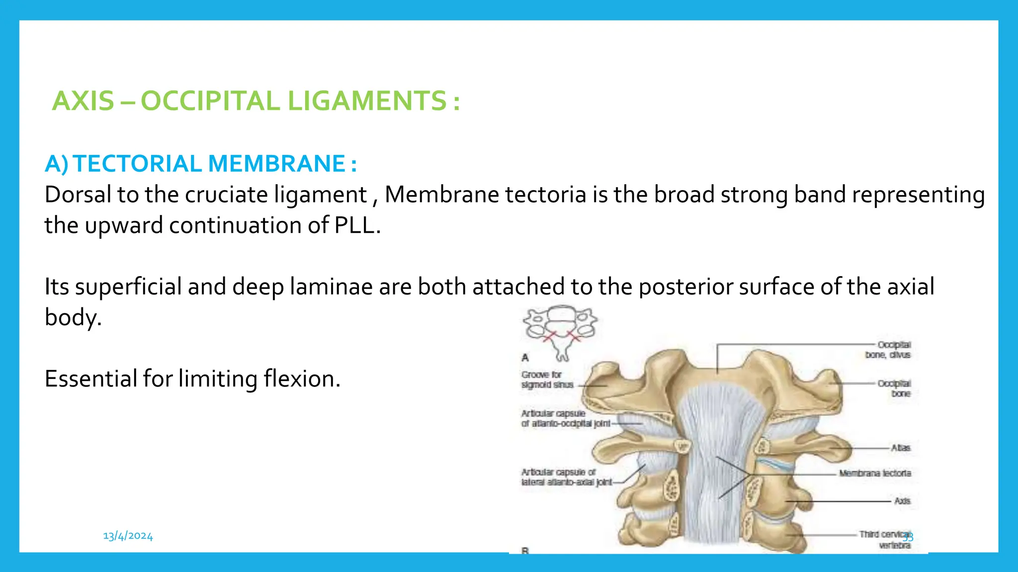
CRANIOVERTEBRAL
JUNCTION ANATOMY
Dr LAXMI KANTH
13/4/2024 1
CRANIOVERTEBRAL JUNCTION (CVJ)
The craniovertebral (craniocervical) junction is a collective term that refers to the
occiput, atlas, axis, and supporting ligaments.
It is a transition zone b/w a mobile cranium & relatively rigid spinal column.
It encloses the soft tissue structures of the cervicomedullary junction (medulla,
spinal cord, and lower cranial nerves).
13/4/2024 2
COMPONENTS OF CVJ
• Bones
• Joints
• Ligaments
13/4/2024 3
THE OCCIPITAL BONE
• Occipital bone is Composed of basi occipit, exoccipit and supraoccipital
portions.
• The Basi occiput embryologically derived from the fusion of 4 occipital
sclerotomes (also reffered as primary cranial vertebrae) forms the lower part of
clivus.
• The upper part of clivus is formed by the basi sphenoid, separated from basi
occiput by the sphenooccipital synchondrosis .
13/4/2024 4
1. Basioccipital portion
2. Exoccipital portion
3. supraoccipital portion
4 occipital sclerotomes
Sphenooccipital synchondrosis
13/4/2024 5
13/4/2024 6
OCCIPITAL BONE
Includes
• Basilar part (basiocciput): Quadrilateral part anterior to foramen magnum.
• Condylar part (exoccipital): Includes Occipital condyles; lateral to foramen magnum.
• Squamous part(supraoccipital): Large bony plate posterosuperior to foramen
magnum.
• Occipital condyles are paired, oval-shaped, obliquely oriented such that anterior part
facing medially and posterior part facing more laterally With Articular facet projects
laterally.
13/4/2024 7
BASILAR PART
CONDYLAR PART
SQUAMOUS PART
13/4/2024 8
ATLAS (C1)
• 3 primary ossification centres.
• Composed of anterior and posterior arches, with absent vertebral body.
• Contains Paired lateral masses with superior and inferior articular facets.
• Large transverse processes with transverse foramen.
• The canal diameter of the atlas is 3 cms in AP direction.
13/4/2024 9
The superior articular facet is concave anteroposteriorly
& projects medially for articulation with the occipital condyle.
The inferior articular facet is concave mediolaterally &
projects medially for articulation with the convex surface of
the superior articular facet of C2.
13/4/2024 10
13/4/2024 11
AXIS (C2)
• 5 primary & 2 secondary centers.
• Large body, superiorly projecting odontoid process and Lateral masses.
• Superior articular facet surface is convex & directed laterally and articulates with
inferior facet of C1.
• Inferior articular facet surface is typical of lower cervical vertebrae and articulates
with superior articular facet of C3.
• Superior facet is positioned relatively anterior & inferior facet is positioned
posteriorly with elongated pars interarticularis.
13/4/2024 12
The odontoid process is the embryologic centrum of C1 which is
incorporated into C2, giving C2 its unique morphology.
The odontoid process has ant & post joints for articulation
with C1.
13/4/2024 13
CVJ JOINTS
• OccipitoAtlantal joint (Co – C1 )
• Atlanto Axial joint (C1 – C2)
13/4/2024 14
• The occipito –atlantal articulation comprises two synovial joints formed b/w the
occipital condyles & lateral masses of atlas.
• Inferior articular facet of occipital condyle: Oval, Convex surface, projects laterally.
• Superior articular facet of C1 Oval, concave anteroposteriorly, projects medially.
OCCIPITO ATLANTAL JOINT
13/4/2024 15
coronal bone CT reconstructions of the craniocervical junction .
13/4/2024 16
Muscles Providing movement at Atlanto Occipital joint
• Allows mainly for Flexion – Extension, which has a total range of 15º.
• Flexion: Longus Capitis and rectus Capitis anterior
• Extension: Recti Capitis posterior major and minor, Obliqus capitis superior,
Semispinalis Capitis, Splenius capitis andTrapezius
• Rotation: Obliqus capitis superior, rectus capitis posterior minor, Splenius Capitis
and SCM
13/4/2024 17
Median atlanto-axial joints
• Pivot type joint between dens + ring formed by anterior arch + transverse ligament of C1
• Synovial cavities between transverse ligament/odontoid & atlas/odontoid articulations
Lateral atlanto-axial joints
• Inferior articular facet of C1: Concave mediolaterally, projects medially in coronal plane
• Superior articular facet of C2: Convex surface, projects laterally.
ATLANTO – AXIAL JOINT
13/4/2024 18
MOVEMENT AT ATLANTO AXIAL JOINT
• Movement is simultaneous at all 3 Joints and almost exclusively Rotation
around the Axis.
• Rotation is limited mainly by the Alar Ligaments.
• The Normal range of Atlanto-Axial Rotation is about 40º
13/4/2024 19
The atlanto-occipital jt sloping superolateral to inferomedial.
The atlanto-axial joints sloping inferolateral to superomedial.
13/4/2024 20
Occipital Condyles
Atlas
Atlanto Axial Jt
Dens
Atlanto Occipital Jt
13/4/2024 21
LIGAMENTOUS ANATOMY OF CVJ
■ Principal stabilizing ligaments of C1 -
- Transverse atlantal ligament
- Alar ligaments
■ Secondary stabilizing ligaments ofCVJ are more elastic & weaker than the primary
ligaments.
- Apical ligament
- Anterior & posteriorA-O membranes
- Tectorial membrane
- Ligamentum flavum
- ALL & PLL
- Capsular ligaments
13/4/2024 22
A-O Capsule
A-A Capsule
PLL
Cruciate Lig
Alar Lig
13/4/2024 23
LIGAMENTS AT CVJ
ATLANTO-OCCIPITAL LIGAMENTS: Anterior, Posterior and Lateral
A)AnteriorAtlanto-occipital Membrane
■ Is a broad dense fibrous structure that connects the anterior margin of FM to the
Upper border of anterior arch of Atlas.
■ Laterally blends with Joint capsule.
■ Central fibres are thicker than lateral portion.
■ Ligament is continuous caudally with the anteriorA-A ligament & to
the ALL of the spinal column.
■ It acts as 2° stabilizer at the Jt preventing hyperextension.
13/4/2024 24
13/4/2024 25
B) POSTERIORATLANTO-OCCIPITAL MEMBRANE:
• Broad but thin, connects posterior margin of FM to upper border of posterior
atlantal arch, Blending laterally with the joint capsule.
• Arches over the grooves forVertebral.Ar,Venous Plexus & first cervical nerve.
• A weak ligament containing no significant elastic tissue.
C) LATERAL ATLANTO-OCCIPITAL LIGAMENTS:
Ascending lig which reinforce the A-O joint capsules.
13/4/2024 26
13/4/2024 27
Transverse Ligament
■ Thick, strong & about 20 mm in length.
■ Atached laterally to a small, but prominent tubercle on the medial side of each
atlantal lateral mass, It broadens medially.
■ Consists of collagen fibres, central part cross one another at an angle forming an
interlacing mesh.
■ Allowing free gliding motion over the posterior facet of the dens.
13/4/2024 28
- The tranverse ligament divide the ring of atlas into unequal parts.The posterior
2/3rd surround the spinal cord and meninges & the anterior third contains the dens.
- Posterior fibres of this ligament are arcuate & the more ventral fibres are circular
in configuration.
13/4/2024 29
13/4/2024 30
ATLANTOAXIAL LIGAMENTS : Anterior and Posterior A-A Ligament
CRUCIFORM LIGAMENT
■ From upper margin of Transverse lig. a strong Median Longitudinal band arises and that
inserts in to the basilar part of occipital bone.
From its inferior surface a weaker and less consistent longitudinal band passes to posterior
surface of axis.
■ Caudal crus & Rostral crus fibres joined with transverse ligament on its dorsal aspect to
formCruciate ligament ofAtlas.
13/4/2024 31
13/4/2024 32
AXIS – OCCIPITAL LIGAMENTS :
A)TECTORIAL MEMBRANE :
Dorsal to the cruciate ligament , Membrane tectoria is the broad strong band representing
the upward continuation of PLL.
Its superficial and deep laminae are both attached to the posterior surface of the axial
body.
Essential for limiting flexion.
13/4/2024 33
B) ALAR LIGAMENTS (Check ligaments ofOdontoid) :
■ 2 strong cordsAbout 11mm long that connects the sides of dens to tubercles on
medial side of occipital condyle.
■ Fibres extend laterally & rostrally and limits the rotation of head
■ They are ventral & cranial to the transverse ligament.
C) APICAL LIGAMENT :
■ Slender band of fibres about 2-8 mm long containing small amount of collagen &
elastic fibres.
13/4/2024 34
13/4/2024 35
MUSCLESAT CVJ
■ Muscles have only a minor role related to CVJ stabilization & do not limit the
movements of the joints.
■ Their principal function is one of the initiating & maintaining movement at
theCVJ.
13/4/2024 36
13/4/2024 37
NEURAL STRUCTURES RELATEDTO CVJ
■ Caudal portion of brainstem (Medulla)
Cerebellum
Fourth ventricle
Rostral part of spinal cord
Lower cranial & upper cervical nerves
■ In cerebellum, only the tonsils, biventral lobules & the lower part of the
vermis (nodule, uvula & pyramid) are related toCVJ.
13/4/2024 38
CRANIAL NERVES
■ Lower four cranial N. are closely related to CVJ.
■ 9th & 10th cranial N arises in the groove b/w the inferior olivary nucleus & the
inferior cerebellar peduncle and are separated by dural sheath .
■ The accessory N is the only cranial N that passes through the FM.
■ HYPOGLOSSAL N : formed by the rootlets arising from the medulla along
the anterior margin of the olive & pass behind the vertebral artery.
■ Its rootlets are collected in to two bundles which perforate the dura mater
separately, pass via the hypoglossal canal & then unite.
13/4/2024 39
ACCESSORY NERVE
■ The cranial part or the accessory portion (ramus internus) is the smaller of the two &
is accessory to the vagus.
■ It arises from the medulla, is composed of multiple rootlets.
■ The major portion (ramus externus) is the spinal portion formed by a series of
rootlets arising from the lower medulla & upper spinal cord.
■ The rootlets may arise as low as the C7 root level.
■ Major trunk enters the skull through FM and leaves the skull through jugular foramen.
13/4/2024 40
SPINAL NERVE ROOTS :
■ The C1, C2, and C3 nerves, distal to the ganglion, divide into dorsal and ventral
rami.
■ The first cervical nerve located just below the foramen magnum.
■ The C1 ventral root (SUBOCCIPITAL NERVE) is composed of four to eight
rootlets
■ The dorsal rami supply skin and muscles of the posterior region of the neck.
13/4/2024 41
LYMPHATICS
■ The lymphatic drainage of the O-A-A joints is primarily into the retropharyngeal
LN & then into the deep cervical chain.
■ These LN’s also drain the nasopharynx & hence retrograde infection may
affect the synovial lining of the CVJ complex with resultant neck stiffness &
instability.
13/4/2024 42
ARTERIAL RELATION
■ The major arteries related to CVJ are vertebral, posteroinferior cerebellar
arteries (PICA), the meningeal branches of the vertebral ar., and external and
internal carotid arteries.
VERTEBRAL ARTERY
■ Vertebral artery arises from the posterior part of the first segment of the
subclavian artery in the neck.
■ PICA is the largest branch of vertebral arteries.
■ Each artery is divided into intradural and extradural parts.
13/4/2024 43
■ The extradural segment of the vertebral artery gives rise to the posterior
meningeal, posterior spinal arteries, & branches to the deep cervical
musculature, and infrequently the PICA.
■ The intradural segment begins at the dural foramina & is divided into lateral and
anterior medullary segments.
■ The first cervical nerve exits the spinal canal, and the posterior spinal artery enters the
spinal canal through this dural foramen with the vertebral artery.
■ The branches arising from the vertebral artery in the region of the FM are the posterior
spinal, anterior spinal, PICA, and anterior and posterior meningeal arteries.
13/4/2024 44
■ The tonsillomedullary PICA segment, which forms the caudal loop related to the
lower part of the tonsil, is most intimately related to the foramen magnum.
■ The paired posterior spinal arteries usually arise from the posteromedial
surface of the vertebral arteries, just outside the dura mater, but they may also
arise from the intradural part of the vertebral arteries, or from the PICA.
■ The anterior spinal artery is formed by the union of the paired anterior ventral
spinal arteries, which originate from the anterior medullary segment of the
vertebral arteries.
13/4/2024 45
MENINGEAL ARTERIES :
■ The dura mater around the Foramen Magnum is supplied by the anterior and
posterior meningeal branches of the vertebral artery, and the meningeal
branches of the ascending pharyngeal and occipital arteries.
13/4/2024 46
ROLE OF RADIODIAGNOSIS IN CVJ
• Plain radiograph, CT , MRI can be used for detecting CVJ structural relations and its
abnormalities.
• To determine presence of bony and soft tissue anomalies
• To determine associated instability
• To determine spinal canal compromise
13/4/2024 47
PLAIN RADIOGRAPH
• Lateral view
• AP view (Open Mouth view)
• Oblique
• Fuch’s method
13/4/2024 48
LATERALVIEW
• Place the patient in supine position with arms along the
sides of the body and adjust the shoulders to lie in same
horizontal plane with slight extension of neck.
• IR in vertical position and in contact with upper neck,
centering done at level ofAtlantoaxial articulation (1 inch
distal to the tip of mastoid process)
• Structures shows a lateral projection of the atlas and axis
13/4/2024 49
• Usually the occipital condyles are hidden by the mastoid processes.
• The tip of the dens usually lies on a plane formed by the upper surface of the atlas
anterior & posterior arches.
• The superior articular facet of the axis overlies the junction of the dens & the axis
body.
• Translateral views of skull including the upper cervical spine (flexion & extension)
can also be done as a reference for various radiological baselines and anomalies.
13/4/2024 50
Lateral radiograph of craniocervical junction.There is smooth alignment of the posterior
vertebral body margins, and the posterior spino-laminar line of the posterior elements.
13/4/2024 51
B) ANTERO-POSTERIOR (AP)-OPEN MOUTH VIEW
• Place the patient in the supine position with arms along the sides of body
• Adjust the patient’s head so that the midsagittal plane is perperndicular to the
plane of the table
• Have the patient open the mouth as wide as possible, then adjust the head so that
lower edge of the upper incisors to the tip of the mastoid process is perpendicular
to the IR. And advise to keep mouth wide open and to phonate “ah” softly during
the exposure.
• Additionally detects dens anomalies & its lateral displacement.
13/4/2024 52
AP open mouth view of odontoid process.With proper positioning, the odontoid process is
visualized in the midline with symmetrically placed lateral C1 masses on either side.The atlanto-
occipital and atlanto-axial joints are visible bilaterally, with smooth cortical margins.
13/4/2024 53
C) OBLIQUEVIEWS :
• Allows the visualization of homolateral superior and inferior facets as well as a small
portion of the contralateral superior facet.
• Intervertebral neural canal for the 3rd cervical nerve also evident.
D) FUCH’S METHOD
• Recommended the AP projection to show the dens when its upper half is not
clearly shown in the open-mouth position.
• With the patient in supine position with chin extended until the tip of chin and the
tip of the mastoid process are vertical.
• Shows Dens lying within the circular foramen magnum.
13/4/2024 54
13/4/2024 55
CRANIOMETRY
■ Craniometry of the CVJ uses a series of lines, planes & angles to define the normal
anatomic relationships of the CVJ.
■ These measurements can be taken on plain Xrays, 3DCT or on MRI.
■ There is a certain disadvantage in all of these measurements because the
anatomic structures and planes vary within a normal range.
■ Understanding of the important land marks and accurate assessment of the lines
and angles is crucial in the evaluation of craniovertebral junction anomalies.
13/4/2024 56
LATERAL PROJECTION OF SKULL X-RAY :
■ Palato – occipital (Chamberlain’s line)
■ Palato – suboccipital line (McGregor line)
■ Foramen magnum line (McRae line)
■ Height of the posterior cranial fossa (Klaus Index)
■ Wackenheim’s clival canal line
■ Bull’s angle (Atlanto-palatal angle)
13/4/2024 57
FRONTAL PROJECTIONOF SKULL X-RAY :
■ Bimastoid line (Fischgold & Metzer)
■ Bidigastric line (Fischgold & Metzer)
■ Condylar angle (Schmidt & Fischer)
FOR DIAGNOSIS OF PLATYBASIA :
■ Basal angle (Welcher)
■ Boogard’s angle
13/4/2024 58
NORMAL LAND MARKS OF CVJ
13/4/2024 59
WACKENHEIM CLIVUS BASELINE (BASILAR LINE)
■ The Wackenheim clivus baseline is constructed by drawing a line along the
clivus and extending it inferiorly into the upper cervical spinal canal.
■ This line should fall tangent to the posterior aspect of the tip of the odontoid
process.
■ This line intersects the dens when the skull is anterior in position.
C2
Wackenheim
Clivus Base line Chamberlain’s
■ Not more than 1/3rd of the odontoid process should extend above chamberlain’s line.
13/4/2024 60
13/4/2024 61
CHAMBERLAIN’S LINE (PALATOOCCIPITAL LINE)
■ The Chamberlain line extends between the posterior pole of the hard palate and the
opisthion (posterior margin of the foramen magnum).Tip of Dens <3mm.
13/4/2024 62
CHAMBERLAIN’S LINE
13/4/2024 63
• The McGregor line is a modification of the Chamberlain line and is used in the
evaluation of basilar invagination when the opisthion is not identified on plain
radiographs.
• It refers to a line connecting posterior edge of the hard palate to the most caudal point
of the occipital curve.Tip of Dens shld be < 4.5 mm above this line.
McGREGOR LINE
13/4/2024 64
Mc RAES LINE / FORAMEN MANGUM LINE
• Line joining anterior margin of FM with Posterior margin of FM,Tip of the
dens does not exceed this line.
• Normal diameter is around 40mm.
• The FM is enlarged to >50 mm in c/o Chiari malformation.
13/4/2024 65
McRAE'S LINE
13/4/2024 66
KLAUS HEIGHT INDEX
Distance between tip of Dens and tuberculum cruciate line.
Represents the height or depth of the posterior fossa (mean = 41 ± 4 mm)
Tuberculum Cruciate line
13/4/2024 67
SPINOLAMELLAR LINE
■ Line Drawn from the interoccipital ridge above & downward along the fused
spinous process of C2 & C3 below.
■ This curvilinear line should intersect the fused posterior arch of atlas.
■ If the atlas lies anterior to this line, posterior compression of the spinal cord may be
present.
13/4/2024 68
BULL’SANGLE (ATLANTO PALATINEANGLE)
■ Angle formed by joining hard palate posteriorly and the line joinig mid points of
Ant. & Post. arches of atlas.
■ <13o is normal.
13/4/2024 69
WELCHER BASAL ANGLE :
■ The Welcher basal angle is formed at the intersection of the nasion -tuberculum line and
the tuberculum - basion line.
■ It averages 132o & should always be less than 140o.
13/4/2024 70
BOOGARD’S ANGLE :
■ A line is drawn from basion to opisthion & another line along the plane of clivus to
the basion, intersecting the first line.
■ The normal value is 124o _ 142o .
Clivus
13/4/2024 71
FISCHGOLD BIMASTOID LINE
■ Line connecting tips of mastoid process.
■ Normally passes through the A-O joint.
■ Apex of odontoid should lie about 2mm above this line with a range b/w 3mm
below & 10mm above.
FISCHGOLD BIDIGASTRIC LINE
■ Line Joining bilateral Digastric grooves (medial to mastoid process).
■ This line is situated 10mm above the bimastoid line / A-O joint.
■ Odontoid tip normally should not project above this line.
■ Corresponds to McRae’s line on lateral radiograph.
13/4/2024 72
DIGASTRIC LINE BIMASTOID LINE
13/4/2024 73
Atlanto-occipital junction
Atlanto-occipital
assimilation
Platybasia
Basilar invagination
Occipital
• Basiocciput hypoplasia
• Occipital condyle
hypoplasia
• Condylus tertius
Atlas
• Posterior arch
anomalies
• Anterior arch anomalies
Axis
• Ossiculum terminale
• Os odontoideum
• Odontoid aplasia
Associated conditions
• Chiari malformation
• Klippel Fiel syndrome
• Osteogenesis imperfecta
• Achondroplasia
CONGENITALCVJANOMALIES-CLASSIFICATION
13/4/2024 74
ThankYou
13/4/2024 75

CRANIO VERTEBRAL JUNCTION RADIOLOGICAL ANATOMY .pptx

  • 1.
  • 2.
    CRANIOVERTEBRAL JUNCTION (CVJ) Thecraniovertebral (craniocervical) junction is a collective term that refers to the occiput, atlas, axis, and supporting ligaments. It is a transition zone b/w a mobile cranium & relatively rigid spinal column. It encloses the soft tissue structures of the cervicomedullary junction (medulla, spinal cord, and lower cranial nerves). 13/4/2024 2
  • 3.
    COMPONENTS OF CVJ •Bones • Joints • Ligaments 13/4/2024 3
  • 4.
    THE OCCIPITAL BONE •Occipital bone is Composed of basi occipit, exoccipit and supraoccipital portions. • The Basi occiput embryologically derived from the fusion of 4 occipital sclerotomes (also reffered as primary cranial vertebrae) forms the lower part of clivus. • The upper part of clivus is formed by the basi sphenoid, separated from basi occiput by the sphenooccipital synchondrosis . 13/4/2024 4
  • 5.
    1. Basioccipital portion 2.Exoccipital portion 3. supraoccipital portion 4 occipital sclerotomes Sphenooccipital synchondrosis 13/4/2024 5
  • 6.
  • 7.
    OCCIPITAL BONE Includes • Basilarpart (basiocciput): Quadrilateral part anterior to foramen magnum. • Condylar part (exoccipital): Includes Occipital condyles; lateral to foramen magnum. • Squamous part(supraoccipital): Large bony plate posterosuperior to foramen magnum. • Occipital condyles are paired, oval-shaped, obliquely oriented such that anterior part facing medially and posterior part facing more laterally With Articular facet projects laterally. 13/4/2024 7
  • 8.
  • 9.
    ATLAS (C1) • 3primary ossification centres. • Composed of anterior and posterior arches, with absent vertebral body. • Contains Paired lateral masses with superior and inferior articular facets. • Large transverse processes with transverse foramen. • The canal diameter of the atlas is 3 cms in AP direction. 13/4/2024 9
  • 10.
    The superior articularfacet is concave anteroposteriorly & projects medially for articulation with the occipital condyle. The inferior articular facet is concave mediolaterally & projects medially for articulation with the convex surface of the superior articular facet of C2. 13/4/2024 10
  • 11.
  • 12.
    AXIS (C2) • 5primary & 2 secondary centers. • Large body, superiorly projecting odontoid process and Lateral masses. • Superior articular facet surface is convex & directed laterally and articulates with inferior facet of C1. • Inferior articular facet surface is typical of lower cervical vertebrae and articulates with superior articular facet of C3. • Superior facet is positioned relatively anterior & inferior facet is positioned posteriorly with elongated pars interarticularis. 13/4/2024 12
  • 13.
    The odontoid processis the embryologic centrum of C1 which is incorporated into C2, giving C2 its unique morphology. The odontoid process has ant & post joints for articulation with C1. 13/4/2024 13
  • 14.
    CVJ JOINTS • OccipitoAtlantaljoint (Co – C1 ) • Atlanto Axial joint (C1 – C2) 13/4/2024 14
  • 15.
    • The occipito–atlantal articulation comprises two synovial joints formed b/w the occipital condyles & lateral masses of atlas. • Inferior articular facet of occipital condyle: Oval, Convex surface, projects laterally. • Superior articular facet of C1 Oval, concave anteroposteriorly, projects medially. OCCIPITO ATLANTAL JOINT 13/4/2024 15
  • 16.
    coronal bone CTreconstructions of the craniocervical junction . 13/4/2024 16
  • 17.
    Muscles Providing movementat Atlanto Occipital joint • Allows mainly for Flexion – Extension, which has a total range of 15º. • Flexion: Longus Capitis and rectus Capitis anterior • Extension: Recti Capitis posterior major and minor, Obliqus capitis superior, Semispinalis Capitis, Splenius capitis andTrapezius • Rotation: Obliqus capitis superior, rectus capitis posterior minor, Splenius Capitis and SCM 13/4/2024 17
  • 18.
    Median atlanto-axial joints •Pivot type joint between dens + ring formed by anterior arch + transverse ligament of C1 • Synovial cavities between transverse ligament/odontoid & atlas/odontoid articulations Lateral atlanto-axial joints • Inferior articular facet of C1: Concave mediolaterally, projects medially in coronal plane • Superior articular facet of C2: Convex surface, projects laterally. ATLANTO – AXIAL JOINT 13/4/2024 18
  • 19.
    MOVEMENT AT ATLANTOAXIAL JOINT • Movement is simultaneous at all 3 Joints and almost exclusively Rotation around the Axis. • Rotation is limited mainly by the Alar Ligaments. • The Normal range of Atlanto-Axial Rotation is about 40º 13/4/2024 19
  • 20.
    The atlanto-occipital jtsloping superolateral to inferomedial. The atlanto-axial joints sloping inferolateral to superomedial. 13/4/2024 20
  • 21.
    Occipital Condyles Atlas Atlanto AxialJt Dens Atlanto Occipital Jt 13/4/2024 21
  • 22.
    LIGAMENTOUS ANATOMY OFCVJ ■ Principal stabilizing ligaments of C1 - - Transverse atlantal ligament - Alar ligaments ■ Secondary stabilizing ligaments ofCVJ are more elastic & weaker than the primary ligaments. - Apical ligament - Anterior & posteriorA-O membranes - Tectorial membrane - Ligamentum flavum - ALL & PLL - Capsular ligaments 13/4/2024 22
  • 23.
    A-O Capsule A-A Capsule PLL CruciateLig Alar Lig 13/4/2024 23
  • 24.
    LIGAMENTS AT CVJ ATLANTO-OCCIPITALLIGAMENTS: Anterior, Posterior and Lateral A)AnteriorAtlanto-occipital Membrane ■ Is a broad dense fibrous structure that connects the anterior margin of FM to the Upper border of anterior arch of Atlas. ■ Laterally blends with Joint capsule. ■ Central fibres are thicker than lateral portion. ■ Ligament is continuous caudally with the anteriorA-A ligament & to the ALL of the spinal column. ■ It acts as 2° stabilizer at the Jt preventing hyperextension. 13/4/2024 24
  • 25.
  • 26.
    B) POSTERIORATLANTO-OCCIPITAL MEMBRANE: •Broad but thin, connects posterior margin of FM to upper border of posterior atlantal arch, Blending laterally with the joint capsule. • Arches over the grooves forVertebral.Ar,Venous Plexus & first cervical nerve. • A weak ligament containing no significant elastic tissue. C) LATERAL ATLANTO-OCCIPITAL LIGAMENTS: Ascending lig which reinforce the A-O joint capsules. 13/4/2024 26
  • 27.
  • 28.
    Transverse Ligament ■ Thick,strong & about 20 mm in length. ■ Atached laterally to a small, but prominent tubercle on the medial side of each atlantal lateral mass, It broadens medially. ■ Consists of collagen fibres, central part cross one another at an angle forming an interlacing mesh. ■ Allowing free gliding motion over the posterior facet of the dens. 13/4/2024 28
  • 29.
    - The tranverseligament divide the ring of atlas into unequal parts.The posterior 2/3rd surround the spinal cord and meninges & the anterior third contains the dens. - Posterior fibres of this ligament are arcuate & the more ventral fibres are circular in configuration. 13/4/2024 29
  • 30.
  • 31.
    ATLANTOAXIAL LIGAMENTS :Anterior and Posterior A-A Ligament CRUCIFORM LIGAMENT ■ From upper margin of Transverse lig. a strong Median Longitudinal band arises and that inserts in to the basilar part of occipital bone. From its inferior surface a weaker and less consistent longitudinal band passes to posterior surface of axis. ■ Caudal crus & Rostral crus fibres joined with transverse ligament on its dorsal aspect to formCruciate ligament ofAtlas. 13/4/2024 31
  • 32.
  • 33.
    AXIS – OCCIPITALLIGAMENTS : A)TECTORIAL MEMBRANE : Dorsal to the cruciate ligament , Membrane tectoria is the broad strong band representing the upward continuation of PLL. Its superficial and deep laminae are both attached to the posterior surface of the axial body. Essential for limiting flexion. 13/4/2024 33
  • 34.
    B) ALAR LIGAMENTS(Check ligaments ofOdontoid) : ■ 2 strong cordsAbout 11mm long that connects the sides of dens to tubercles on medial side of occipital condyle. ■ Fibres extend laterally & rostrally and limits the rotation of head ■ They are ventral & cranial to the transverse ligament. C) APICAL LIGAMENT : ■ Slender band of fibres about 2-8 mm long containing small amount of collagen & elastic fibres. 13/4/2024 34
  • 35.
  • 36.
    MUSCLESAT CVJ ■ Muscleshave only a minor role related to CVJ stabilization & do not limit the movements of the joints. ■ Their principal function is one of the initiating & maintaining movement at theCVJ. 13/4/2024 36
  • 37.
  • 38.
    NEURAL STRUCTURES RELATEDTOCVJ ■ Caudal portion of brainstem (Medulla) Cerebellum Fourth ventricle Rostral part of spinal cord Lower cranial & upper cervical nerves ■ In cerebellum, only the tonsils, biventral lobules & the lower part of the vermis (nodule, uvula & pyramid) are related toCVJ. 13/4/2024 38
  • 39.
    CRANIAL NERVES ■ Lowerfour cranial N. are closely related to CVJ. ■ 9th & 10th cranial N arises in the groove b/w the inferior olivary nucleus & the inferior cerebellar peduncle and are separated by dural sheath . ■ The accessory N is the only cranial N that passes through the FM. ■ HYPOGLOSSAL N : formed by the rootlets arising from the medulla along the anterior margin of the olive & pass behind the vertebral artery. ■ Its rootlets are collected in to two bundles which perforate the dura mater separately, pass via the hypoglossal canal & then unite. 13/4/2024 39
  • 40.
    ACCESSORY NERVE ■ Thecranial part or the accessory portion (ramus internus) is the smaller of the two & is accessory to the vagus. ■ It arises from the medulla, is composed of multiple rootlets. ■ The major portion (ramus externus) is the spinal portion formed by a series of rootlets arising from the lower medulla & upper spinal cord. ■ The rootlets may arise as low as the C7 root level. ■ Major trunk enters the skull through FM and leaves the skull through jugular foramen. 13/4/2024 40
  • 41.
    SPINAL NERVE ROOTS: ■ The C1, C2, and C3 nerves, distal to the ganglion, divide into dorsal and ventral rami. ■ The first cervical nerve located just below the foramen magnum. ■ The C1 ventral root (SUBOCCIPITAL NERVE) is composed of four to eight rootlets ■ The dorsal rami supply skin and muscles of the posterior region of the neck. 13/4/2024 41
  • 42.
    LYMPHATICS ■ The lymphaticdrainage of the O-A-A joints is primarily into the retropharyngeal LN & then into the deep cervical chain. ■ These LN’s also drain the nasopharynx & hence retrograde infection may affect the synovial lining of the CVJ complex with resultant neck stiffness & instability. 13/4/2024 42
  • 43.
    ARTERIAL RELATION ■ Themajor arteries related to CVJ are vertebral, posteroinferior cerebellar arteries (PICA), the meningeal branches of the vertebral ar., and external and internal carotid arteries. VERTEBRAL ARTERY ■ Vertebral artery arises from the posterior part of the first segment of the subclavian artery in the neck. ■ PICA is the largest branch of vertebral arteries. ■ Each artery is divided into intradural and extradural parts. 13/4/2024 43
  • 44.
    ■ The extraduralsegment of the vertebral artery gives rise to the posterior meningeal, posterior spinal arteries, & branches to the deep cervical musculature, and infrequently the PICA. ■ The intradural segment begins at the dural foramina & is divided into lateral and anterior medullary segments. ■ The first cervical nerve exits the spinal canal, and the posterior spinal artery enters the spinal canal through this dural foramen with the vertebral artery. ■ The branches arising from the vertebral artery in the region of the FM are the posterior spinal, anterior spinal, PICA, and anterior and posterior meningeal arteries. 13/4/2024 44
  • 45.
    ■ The tonsillomedullaryPICA segment, which forms the caudal loop related to the lower part of the tonsil, is most intimately related to the foramen magnum. ■ The paired posterior spinal arteries usually arise from the posteromedial surface of the vertebral arteries, just outside the dura mater, but they may also arise from the intradural part of the vertebral arteries, or from the PICA. ■ The anterior spinal artery is formed by the union of the paired anterior ventral spinal arteries, which originate from the anterior medullary segment of the vertebral arteries. 13/4/2024 45
  • 46.
    MENINGEAL ARTERIES : ■The dura mater around the Foramen Magnum is supplied by the anterior and posterior meningeal branches of the vertebral artery, and the meningeal branches of the ascending pharyngeal and occipital arteries. 13/4/2024 46
  • 47.
    ROLE OF RADIODIAGNOSISIN CVJ • Plain radiograph, CT , MRI can be used for detecting CVJ structural relations and its abnormalities. • To determine presence of bony and soft tissue anomalies • To determine associated instability • To determine spinal canal compromise 13/4/2024 47
  • 48.
    PLAIN RADIOGRAPH • Lateralview • AP view (Open Mouth view) • Oblique • Fuch’s method 13/4/2024 48
  • 49.
    LATERALVIEW • Place thepatient in supine position with arms along the sides of the body and adjust the shoulders to lie in same horizontal plane with slight extension of neck. • IR in vertical position and in contact with upper neck, centering done at level ofAtlantoaxial articulation (1 inch distal to the tip of mastoid process) • Structures shows a lateral projection of the atlas and axis 13/4/2024 49
  • 50.
    • Usually theoccipital condyles are hidden by the mastoid processes. • The tip of the dens usually lies on a plane formed by the upper surface of the atlas anterior & posterior arches. • The superior articular facet of the axis overlies the junction of the dens & the axis body. • Translateral views of skull including the upper cervical spine (flexion & extension) can also be done as a reference for various radiological baselines and anomalies. 13/4/2024 50
  • 51.
    Lateral radiograph ofcraniocervical junction.There is smooth alignment of the posterior vertebral body margins, and the posterior spino-laminar line of the posterior elements. 13/4/2024 51
  • 52.
    B) ANTERO-POSTERIOR (AP)-OPENMOUTH VIEW • Place the patient in the supine position with arms along the sides of body • Adjust the patient’s head so that the midsagittal plane is perperndicular to the plane of the table • Have the patient open the mouth as wide as possible, then adjust the head so that lower edge of the upper incisors to the tip of the mastoid process is perpendicular to the IR. And advise to keep mouth wide open and to phonate “ah” softly during the exposure. • Additionally detects dens anomalies & its lateral displacement. 13/4/2024 52
  • 53.
    AP open mouthview of odontoid process.With proper positioning, the odontoid process is visualized in the midline with symmetrically placed lateral C1 masses on either side.The atlanto- occipital and atlanto-axial joints are visible bilaterally, with smooth cortical margins. 13/4/2024 53
  • 54.
    C) OBLIQUEVIEWS : •Allows the visualization of homolateral superior and inferior facets as well as a small portion of the contralateral superior facet. • Intervertebral neural canal for the 3rd cervical nerve also evident. D) FUCH’S METHOD • Recommended the AP projection to show the dens when its upper half is not clearly shown in the open-mouth position. • With the patient in supine position with chin extended until the tip of chin and the tip of the mastoid process are vertical. • Shows Dens lying within the circular foramen magnum. 13/4/2024 54
  • 55.
  • 56.
    CRANIOMETRY ■ Craniometry ofthe CVJ uses a series of lines, planes & angles to define the normal anatomic relationships of the CVJ. ■ These measurements can be taken on plain Xrays, 3DCT or on MRI. ■ There is a certain disadvantage in all of these measurements because the anatomic structures and planes vary within a normal range. ■ Understanding of the important land marks and accurate assessment of the lines and angles is crucial in the evaluation of craniovertebral junction anomalies. 13/4/2024 56
  • 57.
    LATERAL PROJECTION OFSKULL X-RAY : ■ Palato – occipital (Chamberlain’s line) ■ Palato – suboccipital line (McGregor line) ■ Foramen magnum line (McRae line) ■ Height of the posterior cranial fossa (Klaus Index) ■ Wackenheim’s clival canal line ■ Bull’s angle (Atlanto-palatal angle) 13/4/2024 57
  • 58.
    FRONTAL PROJECTIONOF SKULLX-RAY : ■ Bimastoid line (Fischgold & Metzer) ■ Bidigastric line (Fischgold & Metzer) ■ Condylar angle (Schmidt & Fischer) FOR DIAGNOSIS OF PLATYBASIA : ■ Basal angle (Welcher) ■ Boogard’s angle 13/4/2024 58
  • 59.
    NORMAL LAND MARKSOF CVJ 13/4/2024 59
  • 60.
    WACKENHEIM CLIVUS BASELINE(BASILAR LINE) ■ The Wackenheim clivus baseline is constructed by drawing a line along the clivus and extending it inferiorly into the upper cervical spinal canal. ■ This line should fall tangent to the posterior aspect of the tip of the odontoid process. ■ This line intersects the dens when the skull is anterior in position. C2 Wackenheim Clivus Base line Chamberlain’s ■ Not more than 1/3rd of the odontoid process should extend above chamberlain’s line. 13/4/2024 60
  • 61.
  • 62.
    CHAMBERLAIN’S LINE (PALATOOCCIPITALLINE) ■ The Chamberlain line extends between the posterior pole of the hard palate and the opisthion (posterior margin of the foramen magnum).Tip of Dens <3mm. 13/4/2024 62
  • 63.
  • 64.
    • The McGregorline is a modification of the Chamberlain line and is used in the evaluation of basilar invagination when the opisthion is not identified on plain radiographs. • It refers to a line connecting posterior edge of the hard palate to the most caudal point of the occipital curve.Tip of Dens shld be < 4.5 mm above this line. McGREGOR LINE 13/4/2024 64
  • 65.
    Mc RAES LINE/ FORAMEN MANGUM LINE • Line joining anterior margin of FM with Posterior margin of FM,Tip of the dens does not exceed this line. • Normal diameter is around 40mm. • The FM is enlarged to >50 mm in c/o Chiari malformation. 13/4/2024 65
  • 66.
  • 67.
    KLAUS HEIGHT INDEX Distancebetween tip of Dens and tuberculum cruciate line. Represents the height or depth of the posterior fossa (mean = 41 ± 4 mm) Tuberculum Cruciate line 13/4/2024 67
  • 68.
    SPINOLAMELLAR LINE ■ LineDrawn from the interoccipital ridge above & downward along the fused spinous process of C2 & C3 below. ■ This curvilinear line should intersect the fused posterior arch of atlas. ■ If the atlas lies anterior to this line, posterior compression of the spinal cord may be present. 13/4/2024 68
  • 69.
    BULL’SANGLE (ATLANTO PALATINEANGLE) ■Angle formed by joining hard palate posteriorly and the line joinig mid points of Ant. & Post. arches of atlas. ■ <13o is normal. 13/4/2024 69
  • 70.
    WELCHER BASAL ANGLE: ■ The Welcher basal angle is formed at the intersection of the nasion -tuberculum line and the tuberculum - basion line. ■ It averages 132o & should always be less than 140o. 13/4/2024 70
  • 71.
    BOOGARD’S ANGLE : ■A line is drawn from basion to opisthion & another line along the plane of clivus to the basion, intersecting the first line. ■ The normal value is 124o _ 142o . Clivus 13/4/2024 71
  • 72.
    FISCHGOLD BIMASTOID LINE ■Line connecting tips of mastoid process. ■ Normally passes through the A-O joint. ■ Apex of odontoid should lie about 2mm above this line with a range b/w 3mm below & 10mm above. FISCHGOLD BIDIGASTRIC LINE ■ Line Joining bilateral Digastric grooves (medial to mastoid process). ■ This line is situated 10mm above the bimastoid line / A-O joint. ■ Odontoid tip normally should not project above this line. ■ Corresponds to McRae’s line on lateral radiograph. 13/4/2024 72
  • 73.
    DIGASTRIC LINE BIMASTOIDLINE 13/4/2024 73
  • 74.
    Atlanto-occipital junction Atlanto-occipital assimilation Platybasia Basilar invagination Occipital •Basiocciput hypoplasia • Occipital condyle hypoplasia • Condylus tertius Atlas • Posterior arch anomalies • Anterior arch anomalies Axis • Ossiculum terminale • Os odontoideum • Odontoid aplasia Associated conditions • Chiari malformation • Klippel Fiel syndrome • Osteogenesis imperfecta • Achondroplasia CONGENITALCVJANOMALIES-CLASSIFICATION 13/4/2024 74
  • 75.

Editor's Notes

  • #3 Medulla oblongata, Cerebellum, Fourth ventricle -Rostral part of cervical spinal cord -Lower cranial (X, XI, XII) and upper cervical nerves (C1,2) Arterial relationships -Vertebral artery - PICA - Meningeal branches of vertebral a., ECA and ICA Venous relationships -Extradural veins- Extraspinal or intraspinal epidural -Dural venous sinuses -Intraneural veins
  • #5 The 1st 2 occipital sclerotomes forms the basi occiput and the third occipital sclerotome forms the exoccipital bone that froms jugular tubercles. The 4th sclerotomes termed as proatlas forms the anterior tubercle of the clivus, the apical cap of the dens, anterior margin of the foramen magnum, occipital condyles,lateral atlantal masses, and the superior portion of the posterior arch of the atlas.
  • #8 CONDYLES ant more medial and posterior more laterally markedly Convex anteroposteriorly
  • #24 Capsule of atlanto occipital jt Capsule of atlanto axial jt Posterior longitudinal lig Alar ligament Cruciate lig: sup and inferior band and transverse lig of atlas
  • #35 737
  • #45 terminal extradural
  • #60 A nasion, b Tuberculum sellae, C basion, d opisthion, e posterior margin if hard palate, F Anterior arch of atlas, G posterior arch of Atlas, H odontoid process, I body of axis, J jugular tubercle, K Occipital condyles, L lateral atlas masses
  • #61 Odontoid transects the line in basilar imp The maximum distance that the odontoid process may be seen above this line is variable, ranging from 1 mm ± 3.5 mm
  • #63 Drawback of chamberlain’s line : Posterior rim of FM shows great anatomic variability & also it may be difficult to radiologically pinpoint opisthion.
  • #65 This McGregor line extends between the posterior pole of the hard palate and the lowest point of the occipital basi-squamous surface.
  • #70 Posterior Angle betn 1st line from Post tip of hard palate to post margin of foramen magnum 2nd line betn ant & post tubercles of atlas