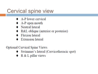
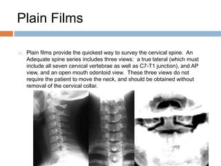
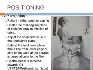
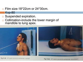
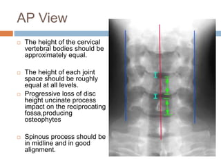
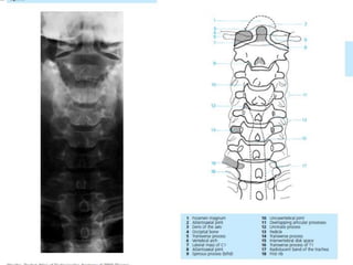
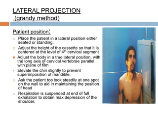
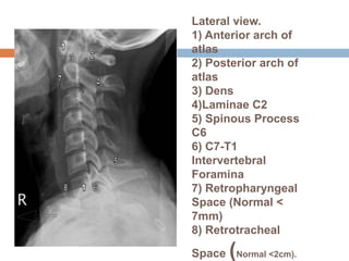
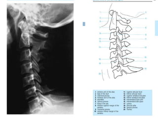
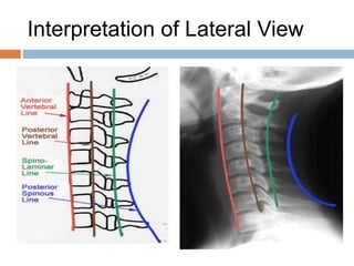
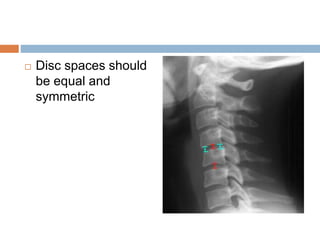
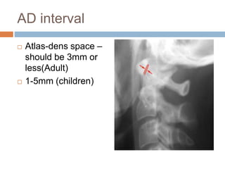
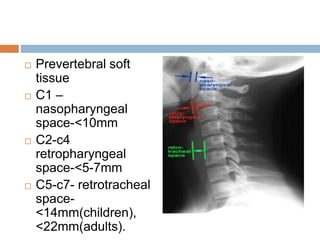
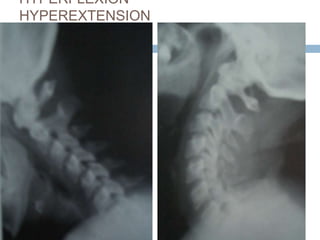
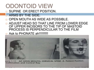
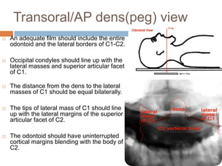
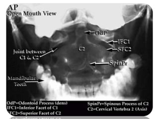
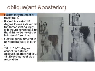
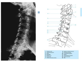
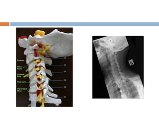
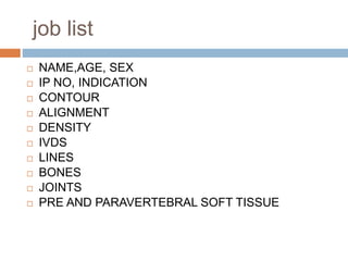
X-RAY CERVICAL
SPINE
Dr ASHISH KUMAR
GUPTA
PG 2nd YEAR
Dept. of radiodiagnosis
SLIMS
PROJECTION &
IMAGING TECHNIQUE
Cervical spine view
Plain Films
 Plain films provide the quickest way to survey the cervical spine. An
Adequate spine series includes three views: a true lateral (which must
include all seven cervical vertebrae as well as C7-T1 junction), and AP
view, and an open mouth odontoid view. These three views do not
require the patient to move the neck, and should be obtained without
removal of the cervical collar.
POSITIONING
 AP projection :
 Patient - either erect or supine
 Center the mid-sagittal plane
of patients body to mid line of
table.
 Adjust the shoulders to lie in
the transverse plane
 Extend the neck enough so
that a line from lower edge of
chin to the base of the occiput
is perpendicular to the film.
 Central beam is directed
towards C4
VERTEBRA(thyroid cartilage)
 Film size-18*22cm or 24*30cm.
 Kvp-80
 Suspended expiration.
 Collimation-include the lower margin of
mandible to lung apex.
AP View
 The height of the cervical
vertebral bodies should be
approximately equal.
 The height of each joint
space should be roughly
equal at all levels.
 Progressive loss of disc
height uncinate process
impact on the reciprocating
fossa,producing
osteophytes
 Spinous process should be
in midline and in good
alignment.
LATERAL PROJECTION
(grandy method)
Patient position:
 Place the patient in a lateral position either
seated or standing.
 Adjust the height of the cassette so that it is
centered at the level of 4th cervical segment
 Adjust the body in a true lateral position, with
the long axis of cervical vertebrae parallel
with plane of film
 Elevate the chin slightly to prevent
superimposition of mandible.
 Ask the patient too look steadily at one spot
on the wall to aid in maintaining the position
of head
 Respiration is suspended at end of full
exhalation to obtain max depression of the
shoulder.
Lateral view.
1) Anterior arch of
atlas
2) Posterior arch of
atlas
3) Dens
4)Laminae C2
5) Spinous Process
C6
6) C7-T1
Intervertebral
Foramina
7) Retropharyngeal
Space (Normal <
7mm)
8) Retrotracheal
Space (Normal <2cm).
Interpretation of Lateral View
 Disc spaces should
be equal and
symmetric
AD interval
 Atlas-dens space –
should be 3mm or
less(Adult)
 1-5mm (children)
 Prevertebral soft
tissue
 C1 –
nasopharyngeal
space-<10mm
 C2-c4
retropharyngeal
space-<5-7mm
 C5-c7- retrotracheal
space-
<14mm(children),
<22mm(adults).
Hyperflexion & hyperextension
views Used to Demonstrate normal anterioposterior movement or
fracture/subluxation or degenerative disc disease(vacuum
phenomenon).
 Spinous process are elevated and widely separated in
hyperflexion.
 Depressed and closed approximation on the hyperextension
position.
HYPERFLEXION
HYPEREXTENSION
ODONTOID VIEW
 SUPINE OR ERECT POSITION.
 ARMS BY THE SIDE.
 OPEN MOUTH AS WIDE AS POSSIBLE.
 ADJUST HEAD SO THAT LINE FROM LOWER EDGE
OF UPPER INCISORS TO THE TIP OF MASTOID
PROCESS IS PERPENDICULAR TO THE FILM
 Ask to PHONATE ah!!!!!!!!!!
Transoral/AP dens(peg) view
 An adequate film should include the entire
odontoid and the lateral borders of C1-C2.
 Occipital condyles should line up with the
lateral masses and superior articular facet
of C1.
 The distance from the dens to the lateral
masses of C1 should be equal bilaterally.
 The tips of lateral mass of C1 should line
up with the lateral margins of the superior
articular facet of C2.
 The odontoid should have uninterrupted
cortical margins blending with the body of
C2.
oblique(ant.&posterior)
 Patient may be erect or
recumbent.
 Patient is rotated 45
degree to one side –to left
for demonstrating right
side neural foramina & to
the right to demonstrate
left neural foramina.
 Central beam directed to
c6 vertebra(base of neck)
.
 Tilt of 15-20 degree
caudal for anterior
oblique& posterior oblique
15-20 degree cephalad
angulation.
job list
 NAME,AGE, SEX
 IP NO, INDICATION
 CONTOUR
 ALIGNMENT
 DENSITY
 IVDS
 LINES
 BONES
 JOINTS
 PRE AND PARAVERTEBRAL SOFT TISSUE

C spine positioning

  • 1.
    X-RAY CERVICAL SPINE Dr ASHISHKUMAR GUPTA PG 2nd YEAR Dept. of radiodiagnosis SLIMS
  • 2.
  • 3.
  • 4.
    Plain Films  Plainfilms provide the quickest way to survey the cervical spine. An Adequate spine series includes three views: a true lateral (which must include all seven cervical vertebrae as well as C7-T1 junction), and AP view, and an open mouth odontoid view. These three views do not require the patient to move the neck, and should be obtained without removal of the cervical collar.
  • 5.
    POSITIONING  AP projection:  Patient - either erect or supine  Center the mid-sagittal plane of patients body to mid line of table.  Adjust the shoulders to lie in the transverse plane  Extend the neck enough so that a line from lower edge of chin to the base of the occiput is perpendicular to the film.  Central beam is directed towards C4 VERTEBRA(thyroid cartilage)
  • 6.
     Film size-18*22cmor 24*30cm.  Kvp-80  Suspended expiration.  Collimation-include the lower margin of mandible to lung apex.
  • 7.
    AP View  Theheight of the cervical vertebral bodies should be approximately equal.  The height of each joint space should be roughly equal at all levels.  Progressive loss of disc height uncinate process impact on the reciprocating fossa,producing osteophytes  Spinous process should be in midline and in good alignment.
  • 9.
    LATERAL PROJECTION (grandy method) Patientposition:  Place the patient in a lateral position either seated or standing.  Adjust the height of the cassette so that it is centered at the level of 4th cervical segment  Adjust the body in a true lateral position, with the long axis of cervical vertebrae parallel with plane of film  Elevate the chin slightly to prevent superimposition of mandible.  Ask the patient too look steadily at one spot on the wall to aid in maintaining the position of head  Respiration is suspended at end of full exhalation to obtain max depression of the shoulder.
  • 10.
    Lateral view. 1) Anteriorarch of atlas 2) Posterior arch of atlas 3) Dens 4)Laminae C2 5) Spinous Process C6 6) C7-T1 Intervertebral Foramina 7) Retropharyngeal Space (Normal < 7mm) 8) Retrotracheal Space (Normal <2cm).
  • 12.
  • 13.
     Disc spacesshould be equal and symmetric
  • 14.
    AD interval  Atlas-densspace – should be 3mm or less(Adult)  1-5mm (children)
  • 15.
     Prevertebral soft tissue C1 – nasopharyngeal space-<10mm  C2-c4 retropharyngeal space-<5-7mm  C5-c7- retrotracheal space- <14mm(children), <22mm(adults).
  • 16.
    Hyperflexion & hyperextension viewsUsed to Demonstrate normal anterioposterior movement or fracture/subluxation or degenerative disc disease(vacuum phenomenon).  Spinous process are elevated and widely separated in hyperflexion.  Depressed and closed approximation on the hyperextension position.
  • 17.
  • 18.
    ODONTOID VIEW  SUPINEOR ERECT POSITION.  ARMS BY THE SIDE.  OPEN MOUTH AS WIDE AS POSSIBLE.  ADJUST HEAD SO THAT LINE FROM LOWER EDGE OF UPPER INCISORS TO THE TIP OF MASTOID PROCESS IS PERPENDICULAR TO THE FILM  Ask to PHONATE ah!!!!!!!!!!
  • 19.
    Transoral/AP dens(peg) view An adequate film should include the entire odontoid and the lateral borders of C1-C2.  Occipital condyles should line up with the lateral masses and superior articular facet of C1.  The distance from the dens to the lateral masses of C1 should be equal bilaterally.  The tips of lateral mass of C1 should line up with the lateral margins of the superior articular facet of C2.  The odontoid should have uninterrupted cortical margins blending with the body of C2.
  • 21.
    oblique(ant.&posterior)  Patient maybe erect or recumbent.  Patient is rotated 45 degree to one side –to left for demonstrating right side neural foramina & to the right to demonstrate left neural foramina.  Central beam directed to c6 vertebra(base of neck) .  Tilt of 15-20 degree caudal for anterior oblique& posterior oblique 15-20 degree cephalad angulation.
  • 24.
    job list  NAME,AGE,SEX  IP NO, INDICATION  CONTOUR  ALIGNMENT  DENSITY  IVDS  LINES  BONES  JOINTS  PRE AND PARAVERTEBRAL SOFT TISSUE