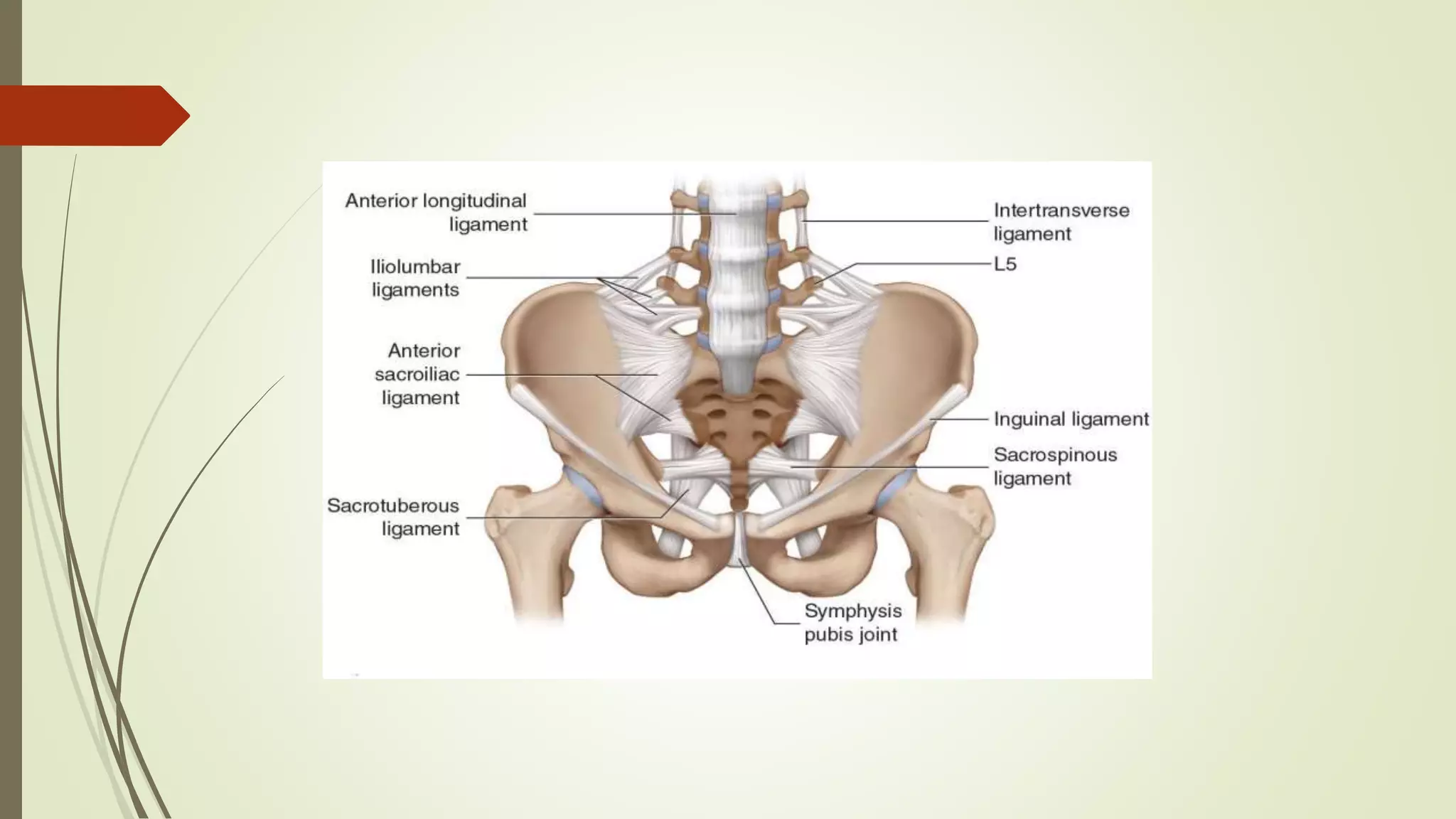
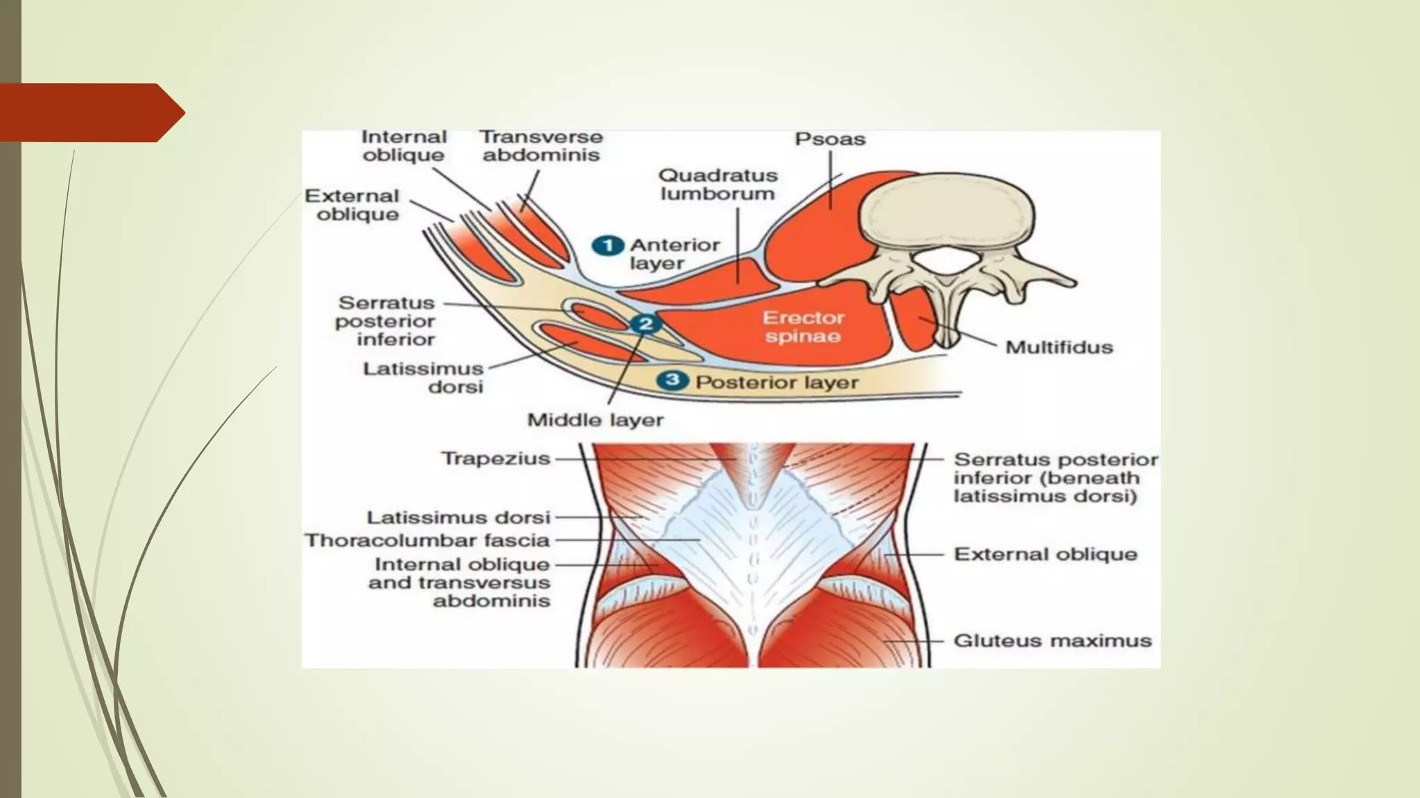
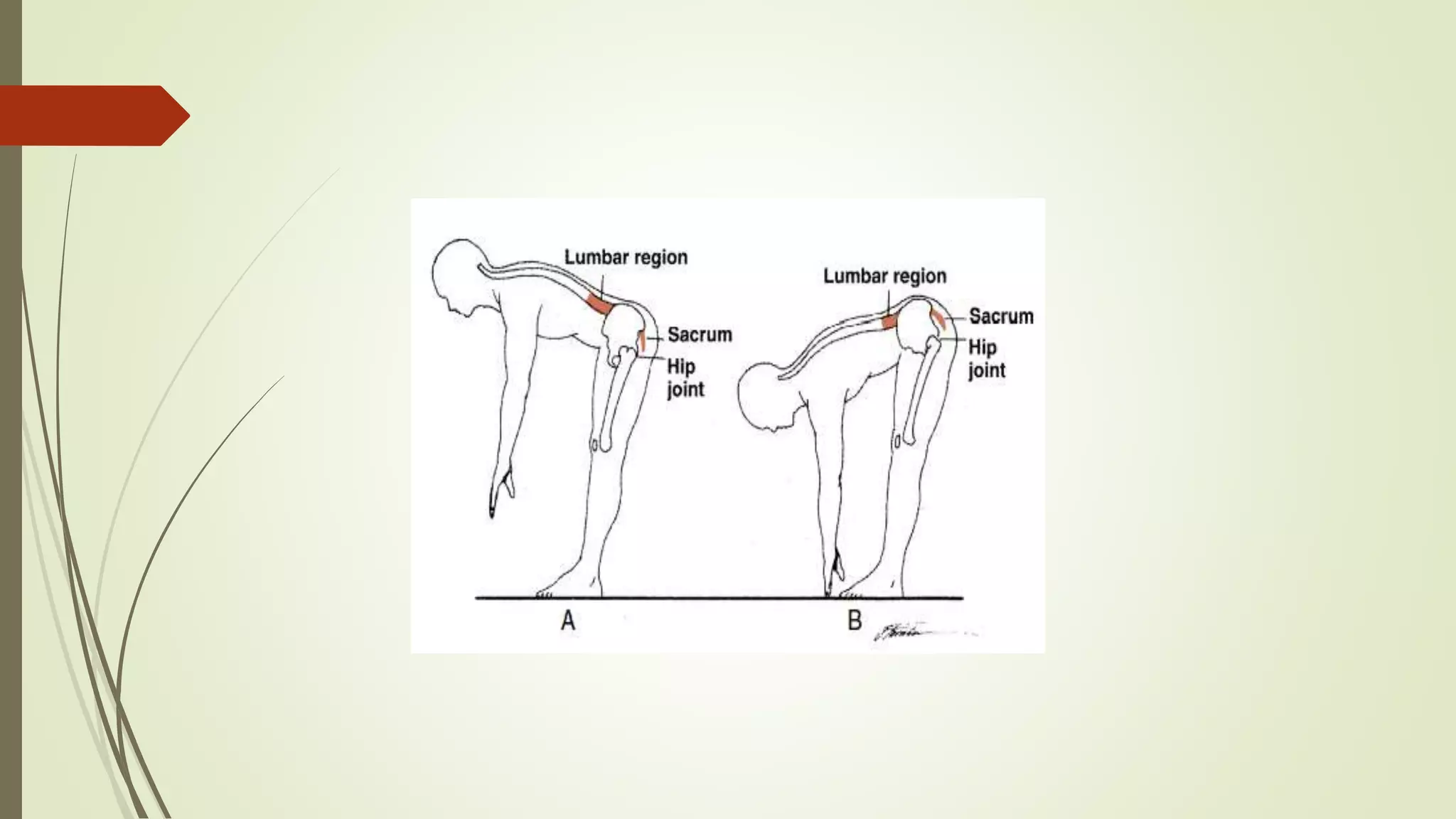
Lumbar Region
Presented By: Dr. Sagar S. Gajra
Structure:
 L1-L4 vertebrae are similar in structure.
 L5 has structural adaptations for articulation with sacrum.
Typical Lumbar Vertebra
Body:
 massive
 Transverse diameter > AP diameter & Height
 Size & shape reflect the need to support great compressive loads caused by body
weight, GRF, & muscle contraction.
Arches:
Pedicles:
 short, thick & project posterolaterally
Laminae:
 short & broad
Spinous Process :
 broad, thick & extended horizontally
Vertebral Foramen:
 triangular
 > Thoracic foramen & < cervical foramen
Zygapophyseal Articular Processes:
 both superior and inferior zygapophyseal facets vary in shape & orientation.
Mamillary Processes:
 appear as small bumps
 located on posterior edge of each superior zygapophyseal facet
 serve as attachment sites for multifidus & medial intertransverse muscles.
 inferior zygapophyseal facets - vertical, convex & face slightly anteriorly,
laterally
Transverse Process:
 long, slender & extends horizontally.
Accessory processes:
 small, irregular bony prominences
 located on the posterior surface of each transverse process near its attachment
to the pedicle.
 serve as attachment sites for multifidus & medial intertransverse muscles.
Lumbar vertebra Structure
Intervertebral Discs:
 Specific regional variations occur in the intervertebral discs of the lumbar region,
 differ from the discs of cervical region - collagen fibers of the anulus fibrosus are
arranged in sheets called lamellae.
 The lamellae are arranged in concentric rings that surround the nucleus.
 Advantage - varying fiber orientation by layer is that the anulus fibrosus is able to
resist tensile forces in nearly all directions.
 Lumbar intervertebral discs are the largest in the body.
 The shape of each disc is not purely elliptical but is concave posteriorly.
 Provides a greater cross-sectional area of anulus fibrosus posteriorly & thus
increases the ability to resist the tension that occurs here with forward bending.
5TH Lumbar Vertebra:
Body:
 it has a wedge-shaped body
 greater height of anterior portion than the posterior portion.
Disc:
 wedge-shaped(L5/S1)
 superior discal surface area of L5 is about 5% greater than L3 and L4.
 inferior discal surface area of L5 is smaller than other lumbar levels.
Spinous Process:
 smaller than other lumbar spinous processes.
Transverse Processes:
 large and directed superiorly and posteriorly.
lumbosacral articulation :
 formed by 5th lumbar vertebra & 1st sacral segment.
 1st sacral segment, inclined slightly anteriorly and inferiorly, forms an angle with
the horizontal called the lumbosacral angle.
 size of angle varies with the position of pelvis & affects the superimposed lumbar
curvature.
 increase angle - increase in lordosis of the lumbar curve - increase amount
of shearing stress LS joint.
Lumbosacral angle
Articulations
1. Interbody Joints:
 capable of translations & tilts in all directions.
2. Zygapophyseal Joints:
 true synovial joints and contain fibroadipose meniscoid structures.
 joint capsules: more lax than thoracic region & more taut than cervical region.
 anterior aspect of each joint remains in the frontal plane & posterior aspect lies
close to or in the sagittal plane
 frontal plane orientation provides resistance to anterior shear that is present in
lordotic lumbar region.
 sagittal plane orientation allows a great range of flexion and extension motion and
provides resistance to rotation pose
Ligament & Fascia
supraspinous ligament:
 well developed only in the upper lumbar region
 terminate at L3, although the most common termination site appears to be at L4.
 absent at L5/S1.
 deep layer - reinforced by tendinous fibers of the multifidus muscle.
 middle fibers - blend with the dorsal layer of the thoracolumbar fascia.
intertransverse ligaments :
 are not true ligaments in the lumbar area
 replaced by the iliolumbar ligament at L5
posterior longitudinal ligament:
 is only a thin ribbon in the lumbar region,
ligamentum flavum:
 thickened
anterior longitudinal ligament:
 strong and well developed
Iliolumbar ligament(ILL):
 consist of a series of bands that extend from tips & borders of the transverse
processes of L5 to attach bilaterally on the iliac crests of the pelvis.
 Three primary bands;
ventral(anterior) band:
 runs from the ventral caudal aspect of the transverse process of L5 to the ventral
surface of the iliac crest.
dorsal (posterior) band:
 runs from the tip of the transverse process of L5 to the more cranial part of the
iliac crest.
sacral band (lumbosacral ligament):
 runs from ventral aspect of the transverse process of L5 to ala of the sacrum and
the anterior sacroiliac ligaments.
 ILL are very strong and play a significant role in stabilizing the L5 (preventing
anterior displacement)
 & in resisting flexion, extension, axial rotation, and lateral bending of L5 on S1.
 ILL are maximally loaded at the lumbosacral junction in the absence of muscle
protection against flexion—for instance, when sitting in a relaxed, slouched
posture.
 ligaments can easily be a source of LBP when subjected to creep in this position
Thoracolumbar fascia (Lumbosacral Fascia):
 Three layers: posterior, middle, and anterior
The posterior layer:
 large, thick, and fibrous
 arises from the spinous processes and supraspinous ligaments of the thoracic,
lumbar, and sacral spines.
 Its gives rise to the latissimus dorsi cranially & continuous cranially with inferior
tendinous border of the splenius capitis and lower fibers of the splenius cervicis.
 Its travels caudally to the sacrum and ilium and blends with the fascia of the
contralateral gluteus maximus.
 Deep fibers are continuous with the sacrotuberous ligament & connected to the
posterior superior iliac spines, iliac crests, and long posterior sacroiliac ligament.
 Its travels laterally over the erector spinae muscles and forms the lateral raphe at
the lateral aspect of the erector spinae.
 The internal abdominal oblique & transversus abdominal muscles arise from the
lateral raphe.
 The posterior layer becomes the middle layer and travels medially again along the
anterior surface of the erector spinae and attaches back to the transverse processes
and intertransverse ligaments of the lumbar spine.
 These two layers completely surround the lumbar extensor muscle group.
The anterior layer:
 its derived from the fascia of the quadratus lumborum muscle, where it joins the
middle layer, inserts into the transverse processes of the lumbar spine, and blends
with the intertransverse ligaments
 anterior layer of the thoracolumbar fascia as the “passive part” and the posterior
layer as the “active part
 passive part transmit tension produced by contraction of the hip extensors to the
spinous processes.
 The active portion is activated by a contraction of the transversus abdominis
muscle, which tightens the fascia.
 The fascia transmits tension longitudinally to the tips of the spinous processes of
L1/L4 and may help the spinal extensor muscles to resist an applied load.
 the gluteus maximus and contralateral latissimus dorsi tensed the superficial layer
and provided a pathway for the mechanical transmission of forces between the
pelvis and the trunk.
Function
Kinematics:
 flexion, extension, lateral flexion, and rotation movement.
 lumbar zygapophyseal facets favor flexion and extension because of predominant
sagittal plane orientation.
 Flexion motion varies at each interspace of the lumbar vertebrae, but most of the
flexion occurs at LS joint.
 Lateral flexion and rotation,
upper lumbar region – most free
lower region – progressively diminish
 largest lateral flexion & axial rotation - between L2 and L3.
 Shape of the zygapophyseal joints is limit the ROM.
 The zygapophyseal joints is resisting axial rotation depends on the extent that the
superior facets face medially (in the sagittal plane).
 When the medial orientation of the joint surfaces is greater, the resistance to axial
rotation is greater.
 In the lumbar region, coupled motions usually occur with lateral flexion and axial
rotation.
Lumbar-Pelvic Rhythm:
 Combined lumbar and pelvic motion known as lumbar-pelvic rhythm.
 The activity of bending over to touch one’s toes with knees straight depends on
lumbar-pelvic rhythm.
 The first part of bending forward consists of lumbosacral flexion, followed by
anterior tilting of pelvis at the hip joints, return to the erect posture is initiated by
posterior tilting of pelvis at the hips, followed by extension of lumbar spine
kinetics
1. Compression:
 primary functions of the lumbar region is to provide support to the upper body
weight in static & dynamic situations.
 increased size of lumbar vertebral bodies & discs in comparison with their
counterparts in the other regions helps the lumbar structures support the additional
weight.
 Lumbar, interbody joints - 80% load
zygapophyseal facet joints - 20% load
 This percentage can change with altered mechanics
 With increased extension or lordosis, the zygapophyseal joints will assume more
of the compressive load.
 With degeneration of the intervertebral disc, the zygapophyseal joints will assume
increased compressive load.
 Lumbosacral loads
erect standing posture - 0.82 to 1.18 times body weight
walking - 1.41 to 2.07 times body weight
 Changes in body position - change the location of the body’s LOG -change the
forces acting on the lumbar spine.
2 Shear:
 In the upright standing position, the lumbar segments are subjected to anterior
shear forces caused by the lordotic position, the body weight, and GRF.
 Anterior shear or translation of the vertebra is resisted by the inferior
zygapophyseal facets of the cranial vertebra against the superior zygapophyseal
facets of the subjacent (caudal) vertebra.
 The effectiveness of the zygapophyseal joint in providing resistance to anterior
translation depends on the caudal vertebra’s superior facets lie in the frontal plane
and face posteriorly.
 More the superior zygapophyseal facets of the caudal vertebra face posteriorly, the
greater the resistance they are able to provide to forward displacement, because
the posteriorly facing facets lock against the inferior facets of the cranial vertebra.

Biomechanics of Lumbar spine

  • 1.
    Lumbar Region Presented By:Dr. Sagar S. Gajra
  • 2.
    Structure:  L1-L4 vertebraeare similar in structure.  L5 has structural adaptations for articulation with sacrum.
  • 3.
    Typical Lumbar Vertebra Body: massive  Transverse diameter > AP diameter & Height  Size & shape reflect the need to support great compressive loads caused by body weight, GRF, & muscle contraction.
  • 4.
    Arches: Pedicles:  short, thick& project posterolaterally Laminae:  short & broad Spinous Process :  broad, thick & extended horizontally Vertebral Foramen:  triangular  > Thoracic foramen & < cervical foramen
  • 5.
    Zygapophyseal Articular Processes: both superior and inferior zygapophyseal facets vary in shape & orientation. Mamillary Processes:  appear as small bumps  located on posterior edge of each superior zygapophyseal facet  serve as attachment sites for multifidus & medial intertransverse muscles.  inferior zygapophyseal facets - vertical, convex & face slightly anteriorly, laterally
  • 6.
    Transverse Process:  long,slender & extends horizontally. Accessory processes:  small, irregular bony prominences  located on the posterior surface of each transverse process near its attachment to the pedicle.  serve as attachment sites for multifidus & medial intertransverse muscles.
  • 7.
  • 8.
    Intervertebral Discs:  Specificregional variations occur in the intervertebral discs of the lumbar region,  differ from the discs of cervical region - collagen fibers of the anulus fibrosus are arranged in sheets called lamellae.  The lamellae are arranged in concentric rings that surround the nucleus.
  • 9.
     Advantage -varying fiber orientation by layer is that the anulus fibrosus is able to resist tensile forces in nearly all directions.  Lumbar intervertebral discs are the largest in the body.  The shape of each disc is not purely elliptical but is concave posteriorly.  Provides a greater cross-sectional area of anulus fibrosus posteriorly & thus increases the ability to resist the tension that occurs here with forward bending.
  • 11.
    5TH Lumbar Vertebra: Body: it has a wedge-shaped body  greater height of anterior portion than the posterior portion. Disc:  wedge-shaped(L5/S1)  superior discal surface area of L5 is about 5% greater than L3 and L4.  inferior discal surface area of L5 is smaller than other lumbar levels.
  • 12.
    Spinous Process:  smallerthan other lumbar spinous processes. Transverse Processes:  large and directed superiorly and posteriorly.
  • 14.
    lumbosacral articulation : formed by 5th lumbar vertebra & 1st sacral segment.  1st sacral segment, inclined slightly anteriorly and inferiorly, forms an angle with the horizontal called the lumbosacral angle.  size of angle varies with the position of pelvis & affects the superimposed lumbar curvature.  increase angle - increase in lordosis of the lumbar curve - increase amount of shearing stress LS joint.
  • 15.
  • 17.
    Articulations 1. Interbody Joints: capable of translations & tilts in all directions. 2. Zygapophyseal Joints:  true synovial joints and contain fibroadipose meniscoid structures.  joint capsules: more lax than thoracic region & more taut than cervical region.
  • 18.
     anterior aspectof each joint remains in the frontal plane & posterior aspect lies close to or in the sagittal plane  frontal plane orientation provides resistance to anterior shear that is present in lordotic lumbar region.  sagittal plane orientation allows a great range of flexion and extension motion and provides resistance to rotation pose
  • 20.
    Ligament & Fascia supraspinousligament:  well developed only in the upper lumbar region  terminate at L3, although the most common termination site appears to be at L4.  absent at L5/S1.  deep layer - reinforced by tendinous fibers of the multifidus muscle.  middle fibers - blend with the dorsal layer of the thoracolumbar fascia.
  • 21.
    intertransverse ligaments : are not true ligaments in the lumbar area  replaced by the iliolumbar ligament at L5 posterior longitudinal ligament:  is only a thin ribbon in the lumbar region, ligamentum flavum:  thickened anterior longitudinal ligament:  strong and well developed
  • 22.
    Iliolumbar ligament(ILL):  consistof a series of bands that extend from tips & borders of the transverse processes of L5 to attach bilaterally on the iliac crests of the pelvis.  Three primary bands; ventral(anterior) band:  runs from the ventral caudal aspect of the transverse process of L5 to the ventral surface of the iliac crest. dorsal (posterior) band:  runs from the tip of the transverse process of L5 to the more cranial part of the iliac crest.
  • 23.
    sacral band (lumbosacralligament):  runs from ventral aspect of the transverse process of L5 to ala of the sacrum and the anterior sacroiliac ligaments.  ILL are very strong and play a significant role in stabilizing the L5 (preventing anterior displacement)  & in resisting flexion, extension, axial rotation, and lateral bending of L5 on S1.
  • 24.
     ILL aremaximally loaded at the lumbosacral junction in the absence of muscle protection against flexion—for instance, when sitting in a relaxed, slouched posture.  ligaments can easily be a source of LBP when subjected to creep in this position
  • 26.
  • 27.
     Three layers:posterior, middle, and anterior The posterior layer:  large, thick, and fibrous  arises from the spinous processes and supraspinous ligaments of the thoracic, lumbar, and sacral spines.  Its gives rise to the latissimus dorsi cranially & continuous cranially with inferior tendinous border of the splenius capitis and lower fibers of the splenius cervicis.
  • 28.
     Its travelscaudally to the sacrum and ilium and blends with the fascia of the contralateral gluteus maximus.  Deep fibers are continuous with the sacrotuberous ligament & connected to the posterior superior iliac spines, iliac crests, and long posterior sacroiliac ligament.  Its travels laterally over the erector spinae muscles and forms the lateral raphe at the lateral aspect of the erector spinae.
  • 29.
     The internalabdominal oblique & transversus abdominal muscles arise from the lateral raphe.  The posterior layer becomes the middle layer and travels medially again along the anterior surface of the erector spinae and attaches back to the transverse processes and intertransverse ligaments of the lumbar spine.  These two layers completely surround the lumbar extensor muscle group.
  • 30.
    The anterior layer: its derived from the fascia of the quadratus lumborum muscle, where it joins the middle layer, inserts into the transverse processes of the lumbar spine, and blends with the intertransverse ligaments  anterior layer of the thoracolumbar fascia as the “passive part” and the posterior layer as the “active part  passive part transmit tension produced by contraction of the hip extensors to the spinous processes.
  • 31.
     The activeportion is activated by a contraction of the transversus abdominis muscle, which tightens the fascia.  The fascia transmits tension longitudinally to the tips of the spinous processes of L1/L4 and may help the spinal extensor muscles to resist an applied load.  the gluteus maximus and contralateral latissimus dorsi tensed the superficial layer and provided a pathway for the mechanical transmission of forces between the pelvis and the trunk.
  • 34.
    Function Kinematics:  flexion, extension,lateral flexion, and rotation movement.  lumbar zygapophyseal facets favor flexion and extension because of predominant sagittal plane orientation.  Flexion motion varies at each interspace of the lumbar vertebrae, but most of the flexion occurs at LS joint.  Lateral flexion and rotation, upper lumbar region – most free lower region – progressively diminish  largest lateral flexion & axial rotation - between L2 and L3.
  • 35.
     Shape ofthe zygapophyseal joints is limit the ROM.  The zygapophyseal joints is resisting axial rotation depends on the extent that the superior facets face medially (in the sagittal plane).  When the medial orientation of the joint surfaces is greater, the resistance to axial rotation is greater.  In the lumbar region, coupled motions usually occur with lateral flexion and axial rotation.
  • 36.
    Lumbar-Pelvic Rhythm:  Combinedlumbar and pelvic motion known as lumbar-pelvic rhythm.  The activity of bending over to touch one’s toes with knees straight depends on lumbar-pelvic rhythm.  The first part of bending forward consists of lumbosacral flexion, followed by anterior tilting of pelvis at the hip joints, return to the erect posture is initiated by posterior tilting of pelvis at the hips, followed by extension of lumbar spine
  • 38.
    kinetics 1. Compression:  primaryfunctions of the lumbar region is to provide support to the upper body weight in static & dynamic situations.  increased size of lumbar vertebral bodies & discs in comparison with their counterparts in the other regions helps the lumbar structures support the additional weight.  Lumbar, interbody joints - 80% load zygapophyseal facet joints - 20% load  This percentage can change with altered mechanics
  • 39.
     With increasedextension or lordosis, the zygapophyseal joints will assume more of the compressive load.  With degeneration of the intervertebral disc, the zygapophyseal joints will assume increased compressive load.  Lumbosacral loads erect standing posture - 0.82 to 1.18 times body weight walking - 1.41 to 2.07 times body weight  Changes in body position - change the location of the body’s LOG -change the forces acting on the lumbar spine.
  • 40.
    2 Shear:  Inthe upright standing position, the lumbar segments are subjected to anterior shear forces caused by the lordotic position, the body weight, and GRF.  Anterior shear or translation of the vertebra is resisted by the inferior zygapophyseal facets of the cranial vertebra against the superior zygapophyseal facets of the subjacent (caudal) vertebra.  The effectiveness of the zygapophyseal joint in providing resistance to anterior translation depends on the caudal vertebra’s superior facets lie in the frontal plane and face posteriorly.  More the superior zygapophyseal facets of the caudal vertebra face posteriorly, the greater the resistance they are able to provide to forward displacement, because the posteriorly facing facets lock against the inferior facets of the cranial vertebra.