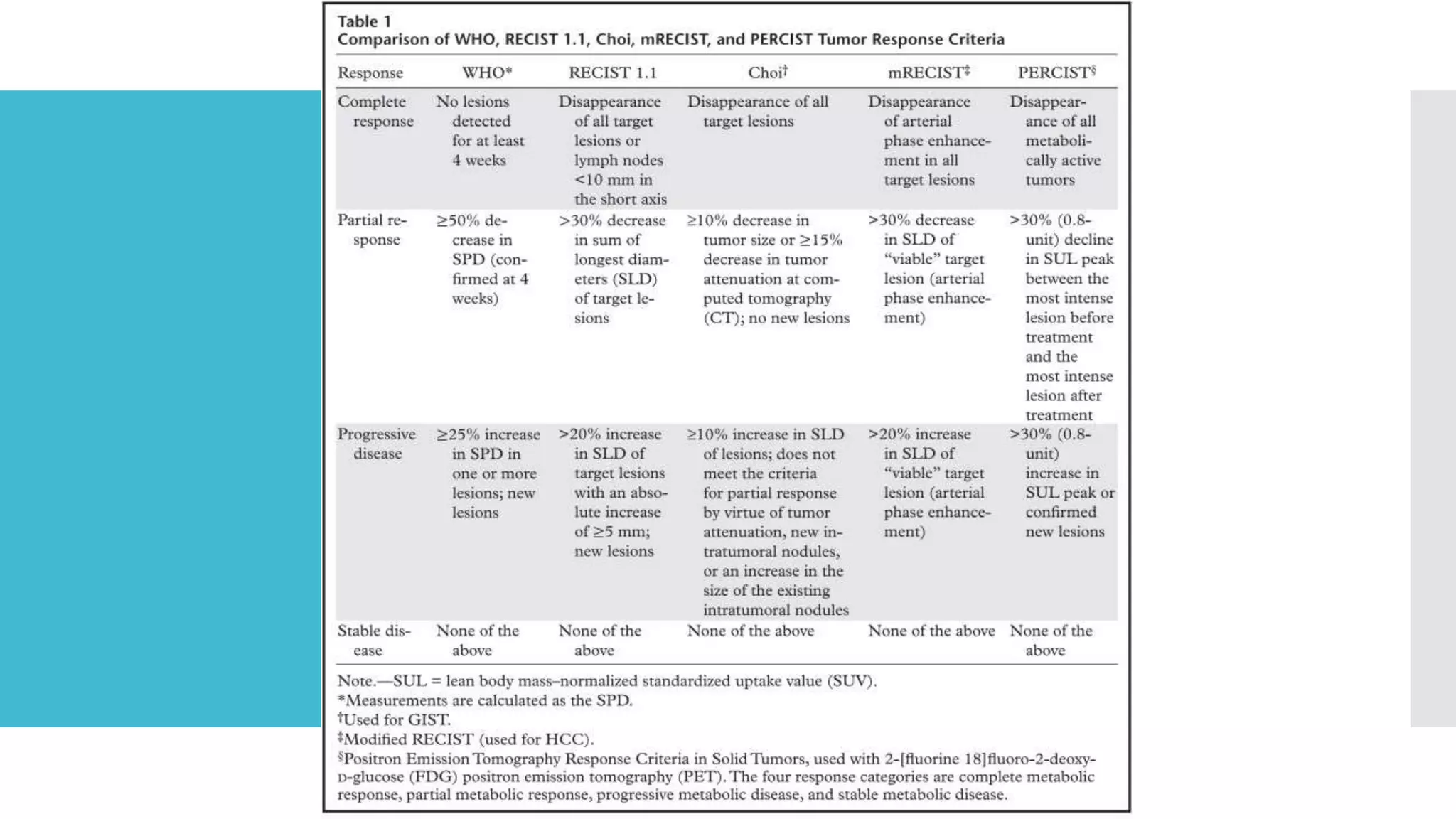
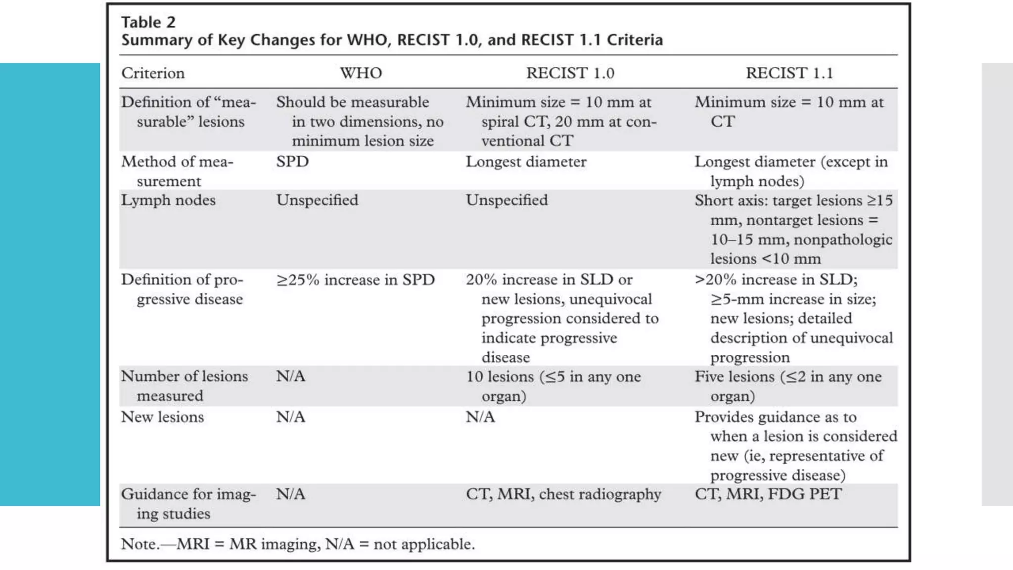
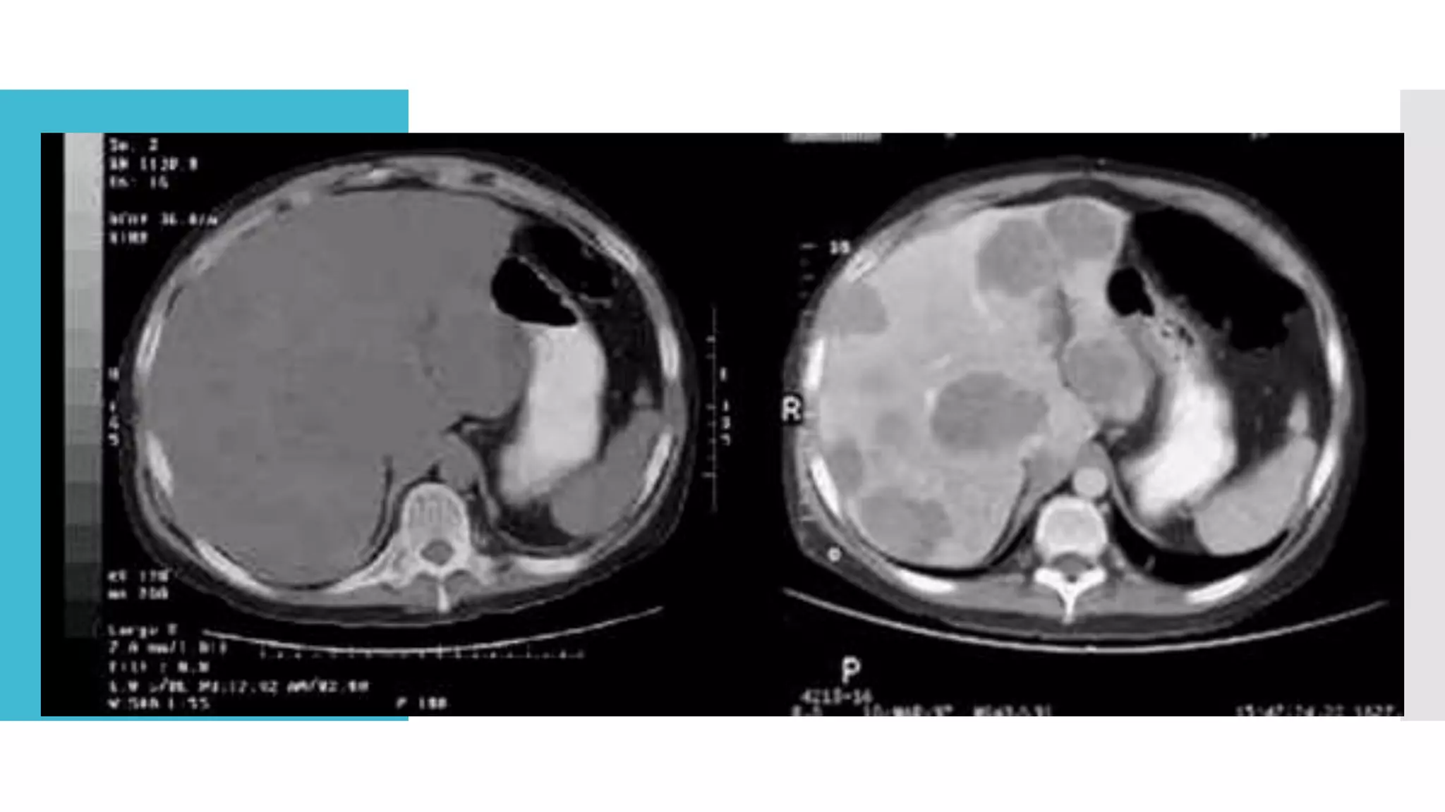
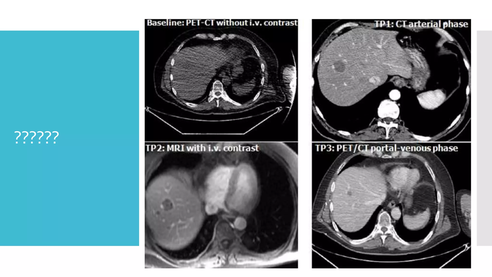
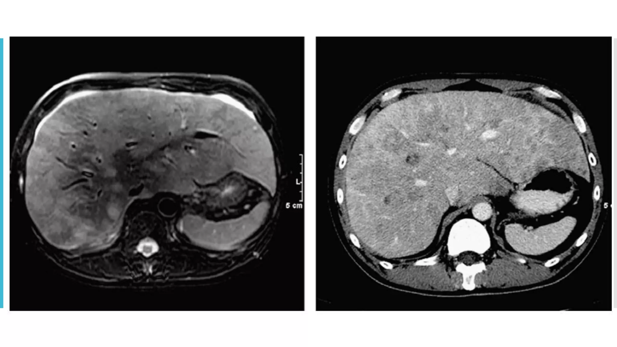
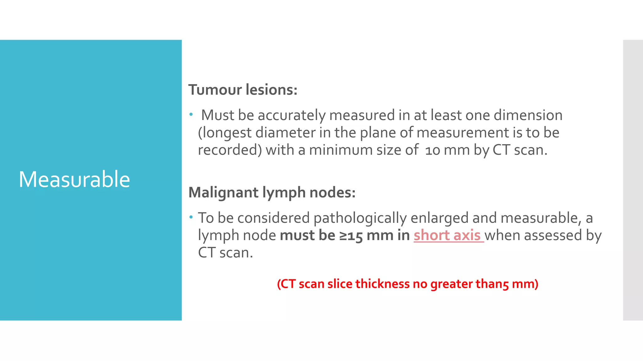
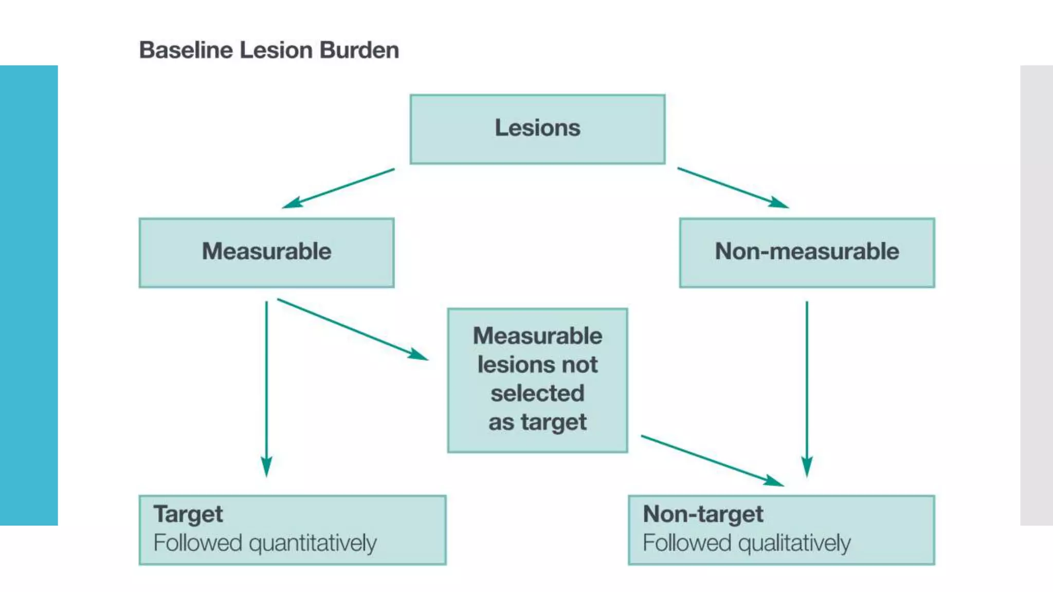
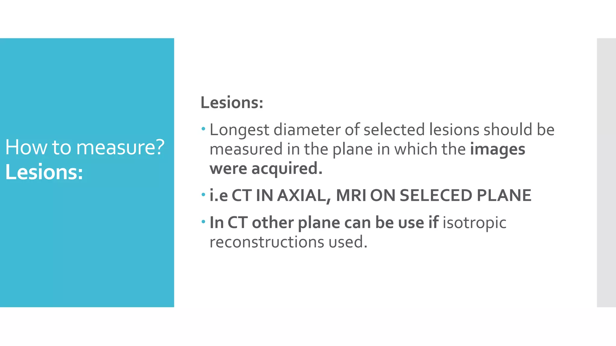
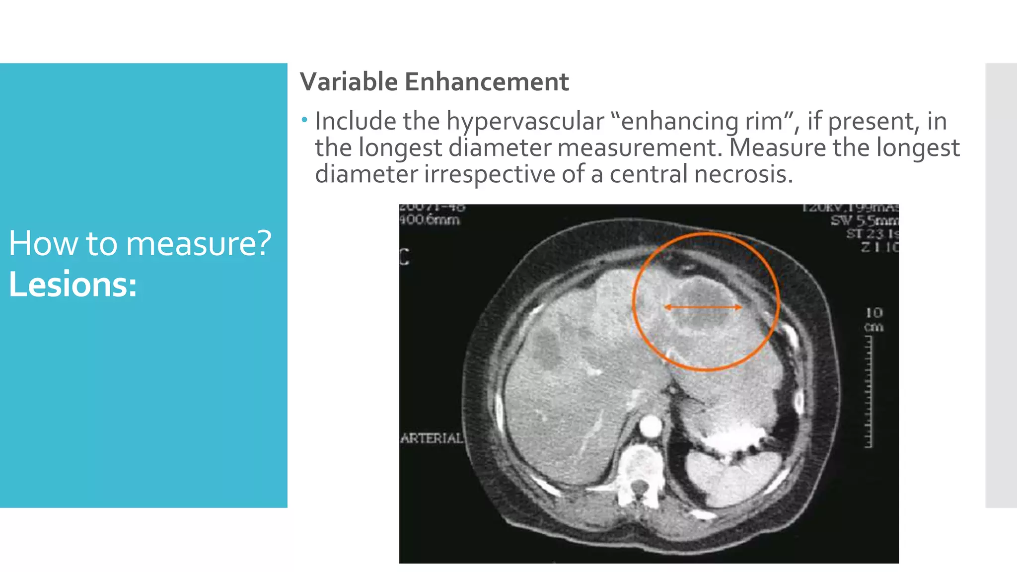
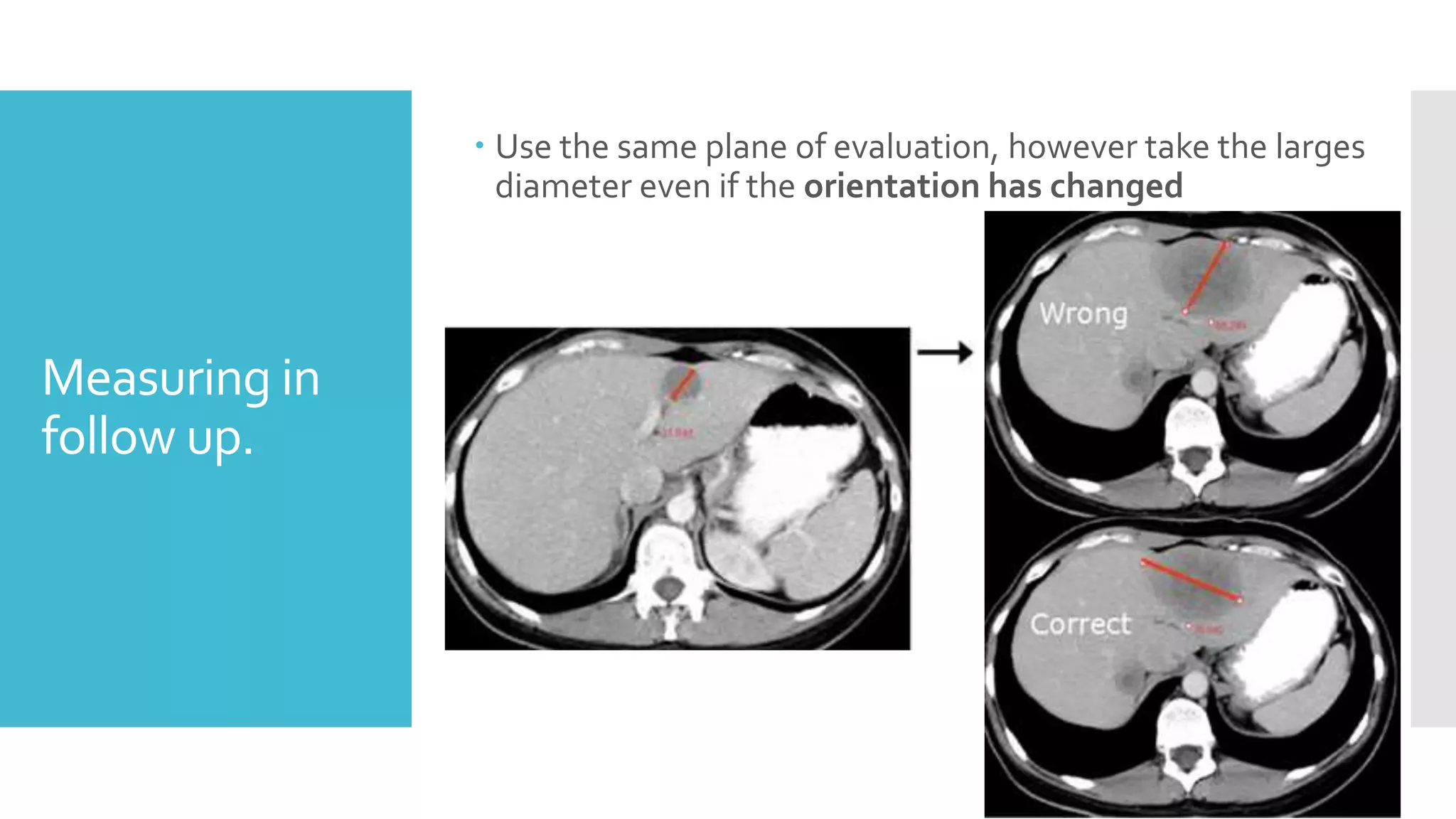
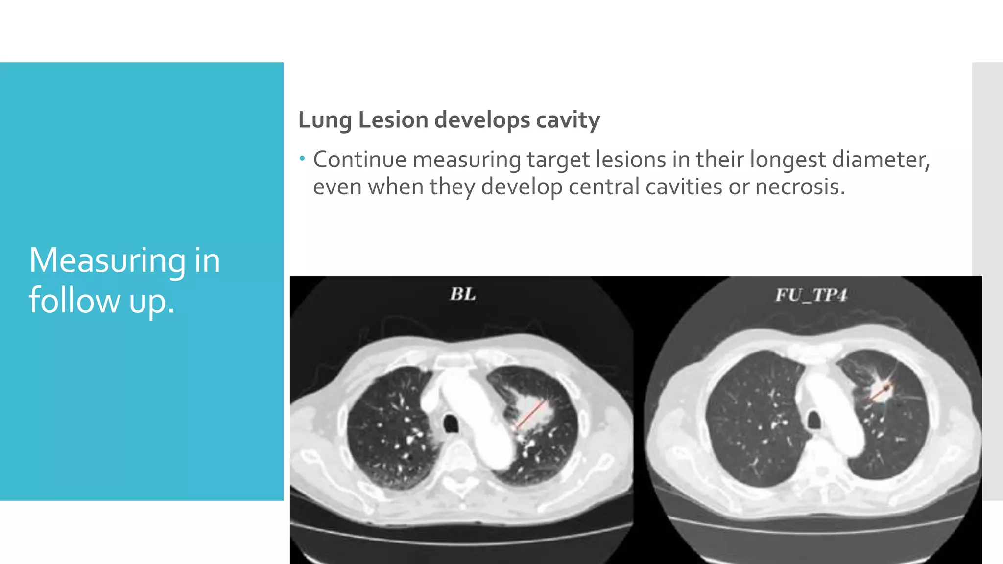
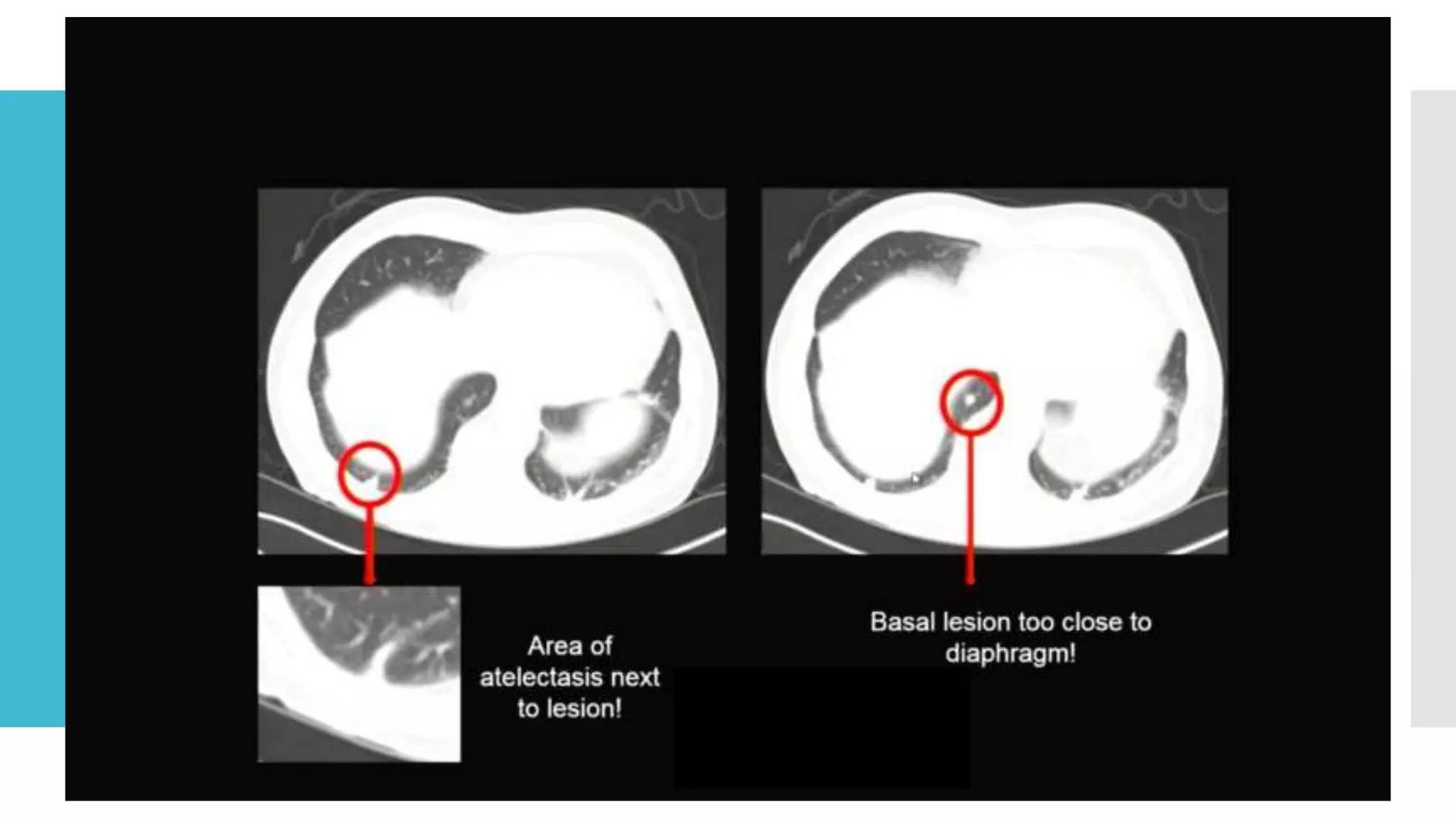
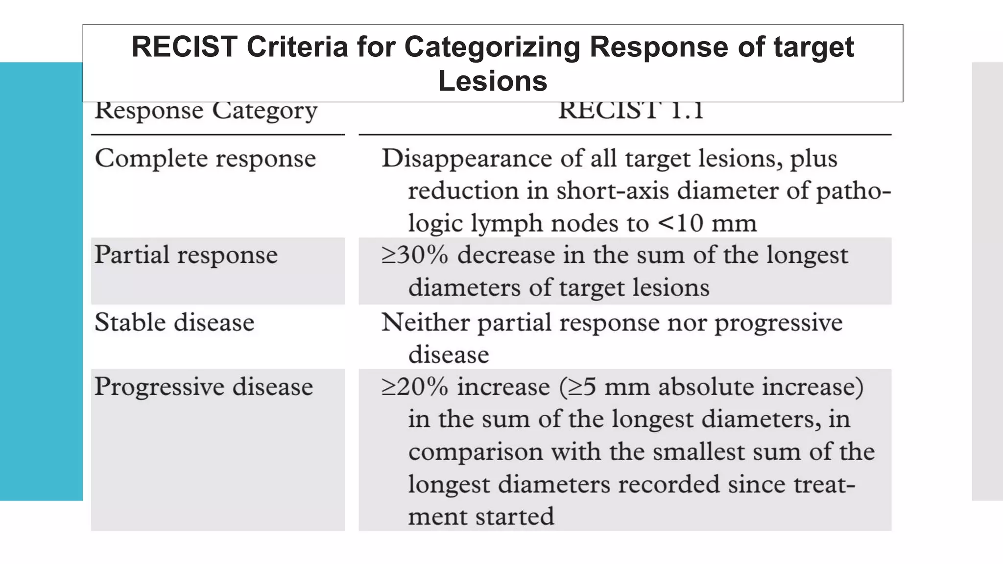
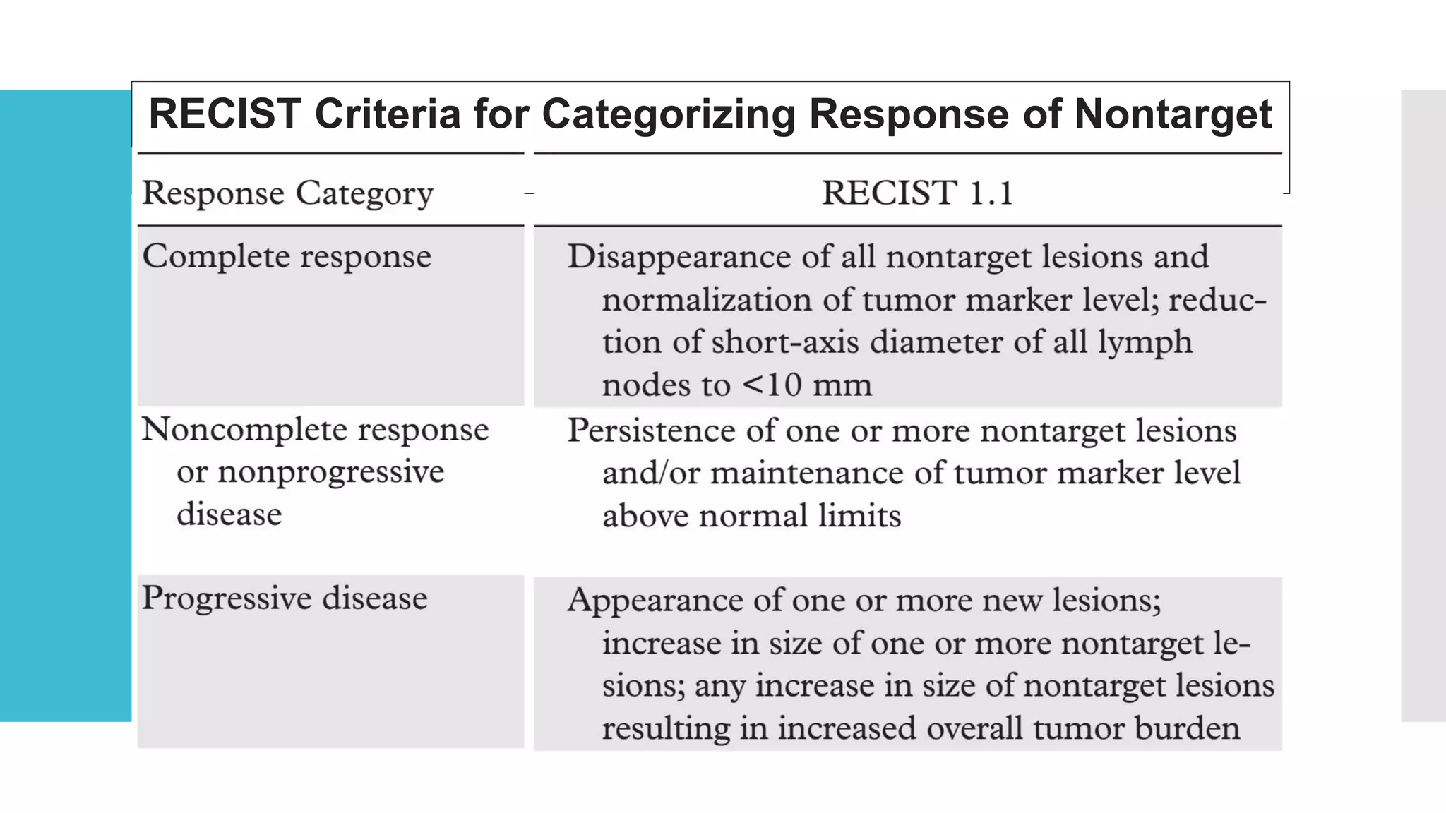
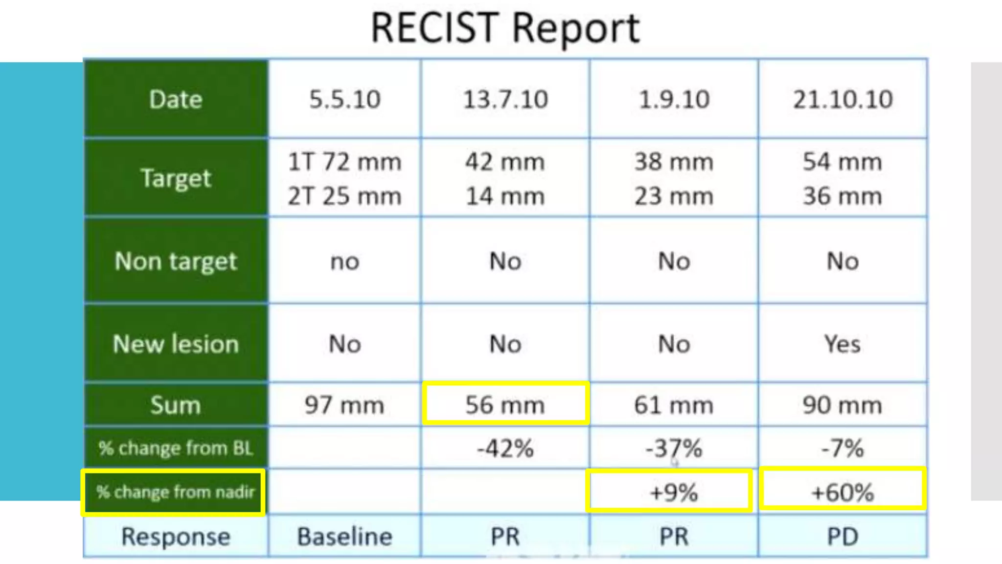
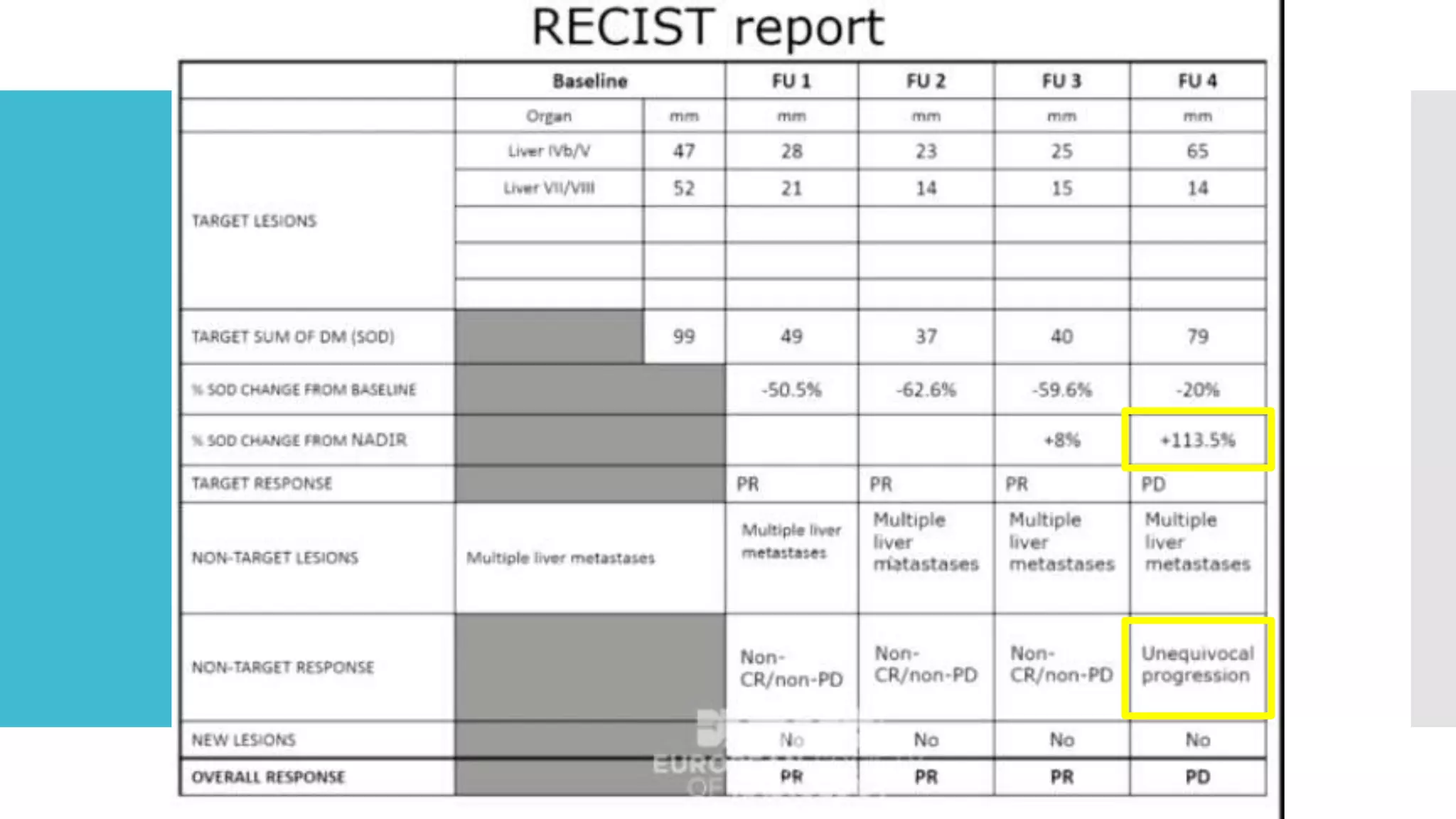
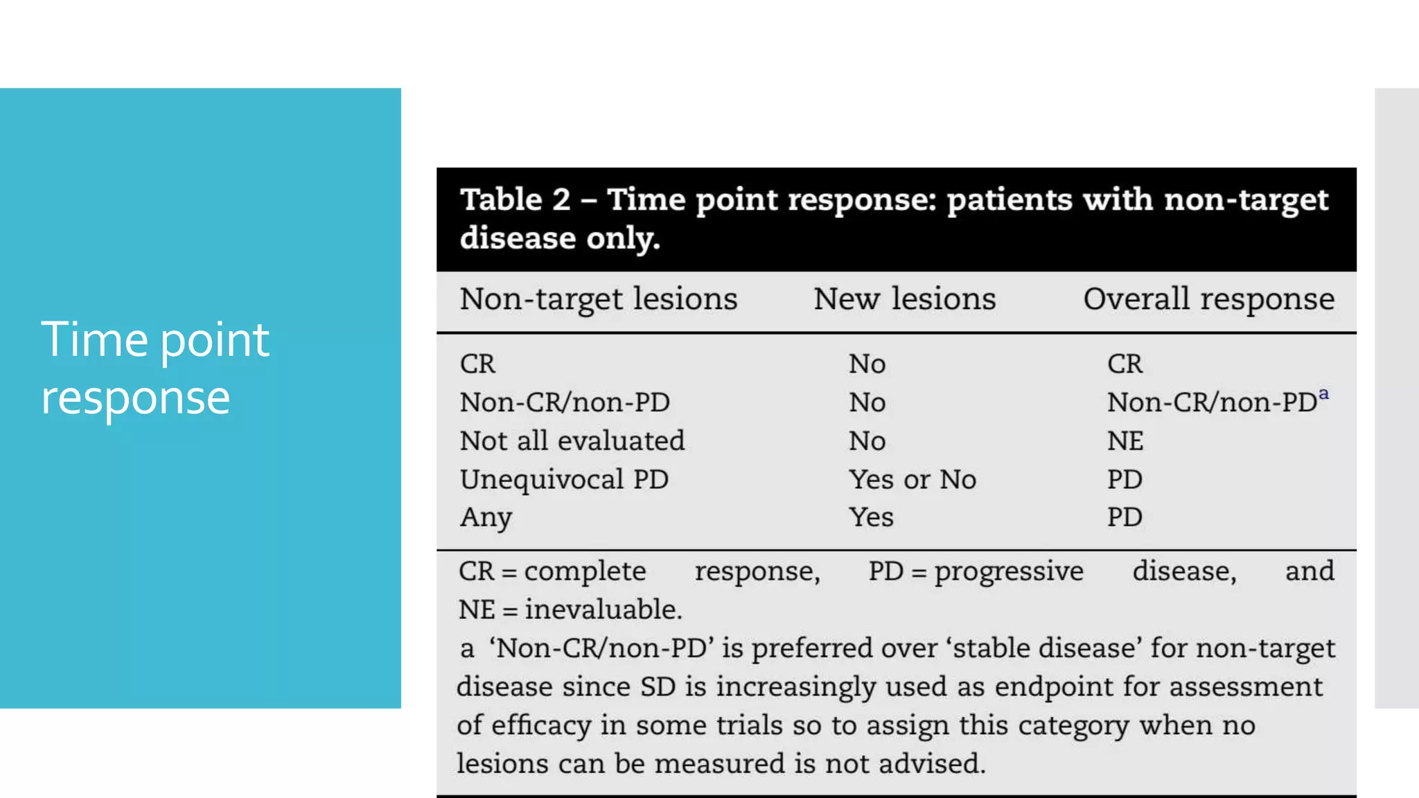
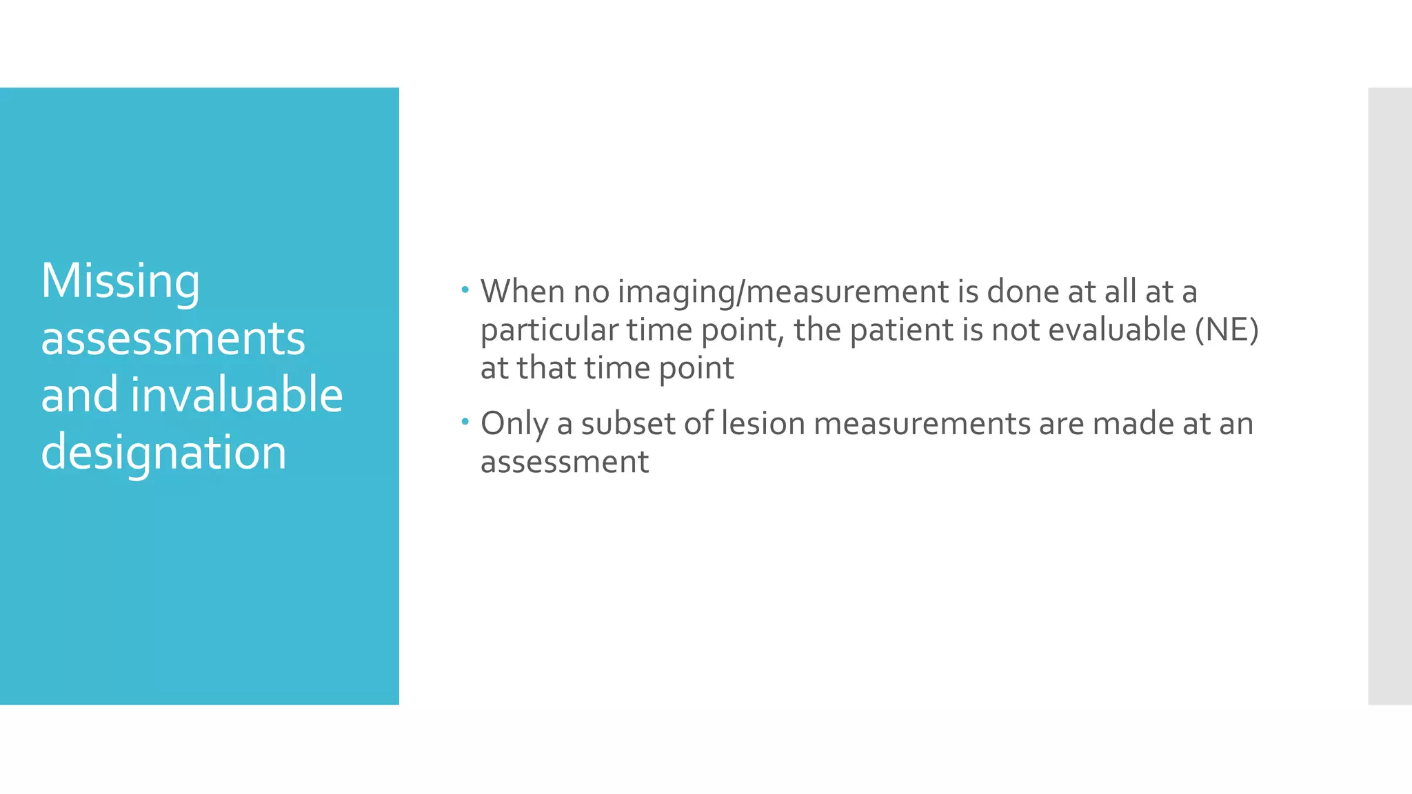
This is a made easy summary of ICRU 89 guidelines for gynecological brachytherapy. Extra practical questions for MD/DNB Radiotherapy exams are also attached.
Contouring Guidelines for Gynecological MalignancyJyotirup Goswami
A brief overview of gynecological malignancy contouring guidelines (teletherapy & brachytherapy), including a discussion of problems and inadequacies of the present guidelines
Co-Chairs Kurt A. Schalper, MD, PhD, and Vamsidhar Velcheti, MD, prepared useful Practice Aids pertaining to solid tumor for this CME/MOC activity titled “PATHway to Decoding the Impact of Cancer Immunotherapy: Latest Advances in Biomarker Testing and Pathologic Response Assessment.” For the full presentation, downloadable Practice Aids, and complete CME/MOC information, and to apply for credit, please visit us at https://bit.ly/3SsmiMV. CME/MOC credit will be available until February 14, 2025.
Co-Chairs, Nasser Altorki, MD, and Jonathan D. Spicer, MD, PhD, FRCSC, prepared useful Practice Aids pertaining to NSCLC for this CME/MOC activity titled “Realizing the Promise of Perioperative Immunotherapy in Resectable NSCLC: How to Modernize Best Practices Based on New Evidence and Better Multidisciplinary Alliances.” For the full presentation, downloadable Practice Aids, and complete CME/MOC information, and to apply for credit, please visit us at http://bit.ly/3Gqancr. CME/MOC credit will be available until February 20, 2024.
Comparision of different imaging techniques used for chronic woundseSAT Journals
Abstract Many people suffer from chronic wounds .It is a major problem in healthcare .worldwide. The treatments of chronic wounds include monitoring colour and size (area or volume) of the wound at regular intervals. This evaluation is often based on qualitative observation and manual measurements of the wound. Now a days there several researchers are developing technologies to assess the clinical improvement of chronic wounds. This paper aims to provide a study on imaging technologies applied to chronic wounds. A study on imaging technologies applied to chronic wounds is presented. Their reliability, precision, and usage are compared. The methods are divided into three categories: planimetric techniques, volumetric Technique and tissue classification. Keywords: chronic wounds, 2D, 3D, imaging, techniques, tissue classification
IJRET : International Journal of Research in Engineering and Technology is an international peer reviewed, online journal published by eSAT Publishing House for the enhancement of research in various disciplines of Engineering and Technology. The aim and scope of the journal is to provide an academic medium and an important reference for the advancement and dissemination of research results that support high-level learning, teaching and research in the fields of Engineering and Technology. We bring together Scientists, Academician, Field Engineers, Scholars and Students of related fields of Engineering and Technology.
First of its kind in South India GE IQ PET/CT at MIOT HospitalsMIOT Hospitals
The GE IQ PET/CT at MIOT International (1st of its kind in South India) offers Superior Image Quality and Intelligent Quantitation enabling the Physician to discover and diagnose cancer confidently. Now, we can detect even minute abnormalities and offer accurate diagnoses and monitoring to our patients.
8 th edition TNM classification and significance of depth of invasionishita1994
Diagnosis of oral cancer is completed for:
Initial diagnosis
Staging
Treatment planning
A complete history, and clinical examination is first completed, then a wedge of tissue is cut from the suspicious lesion for tissue diagnosis. In this procedure, the surgeon cuts all, or a piece of the tissue, to have it examined under a microscope by a pathologist.
Similar to New response evaluation criteria in solid tumours (20)
Basavarajeeyam is an important text for ayurvedic physician belonging to andhra pradehs. It is a popular compendium in various parts of our country as well as in andhra pradesh. The content of the text was presented in sanskrit and telugu language (Bilingual). One of the most famous book in ayurvedic pharmaceutics and therapeutics. This book contains 25 chapters called as prakaranas. Many rasaoushadis were explained, pioneer of dhatu druti, nadi pareeksha, mutra pareeksha etc. Belongs to the period of 15-16 century. New diseases like upadamsha, phiranga rogas are explained.
Knee anatomy and clinical tests 2024.pdfvimalpl1234
This includes all relevant anatomy and clinical tests compiled from standard textbooks, Campbell,netter etc..It is comprehensive and best suited for orthopaedicians and orthopaedic residents.
micro teaching on communication m.sc nursing.pdfAnurag Sharma
Microteaching is a unique model of practice teaching. It is a viable instrument for the. desired change in the teaching behavior or the behavior potential which, in specified types of real. classroom situations, tends to facilitate the achievement of specified types of objectives.
Title: Sense of Smell
Presenter: Dr. Faiza, Assistant Professor of Physiology
Qualifications:
MBBS (Best Graduate, AIMC Lahore)
FCPS Physiology
ICMT, CHPE, DHPE (STMU)
MPH (GC University, Faisalabad)
MBA (Virtual University of Pakistan)
Learning Objectives:
Describe the primary categories of smells and the concept of odor blindness.
Explain the structure and location of the olfactory membrane and mucosa, including the types and roles of cells involved in olfaction.
Describe the pathway and mechanisms of olfactory signal transmission from the olfactory receptors to the brain.
Illustrate the biochemical cascade triggered by odorant binding to olfactory receptors, including the role of G-proteins and second messengers in generating an action potential.
Identify different types of olfactory disorders such as anosmia, hyposmia, hyperosmia, and dysosmia, including their potential causes.
Key Topics:
Olfactory Genes:
3% of the human genome accounts for olfactory genes.
400 genes for odorant receptors.
Olfactory Membrane:
Located in the superior part of the nasal cavity.
Medially: Folds downward along the superior septum.
Laterally: Folds over the superior turbinate and upper surface of the middle turbinate.
Total surface area: 5-10 square centimeters.
Olfactory Mucosa:
Olfactory Cells: Bipolar nerve cells derived from the CNS (100 million), with 4-25 olfactory cilia per cell.
Sustentacular Cells: Produce mucus and maintain ionic and molecular environment.
Basal Cells: Replace worn-out olfactory cells with an average lifespan of 1-2 months.
Bowman’s Gland: Secretes mucus.
Stimulation of Olfactory Cells:
Odorant dissolves in mucus and attaches to receptors on olfactory cilia.
Involves a cascade effect through G-proteins and second messengers, leading to depolarization and action potential generation in the olfactory nerve.
Quality of a Good Odorant:
Small (3-20 Carbon atoms), volatile, water-soluble, and lipid-soluble.
Facilitated by odorant-binding proteins in mucus.
Membrane Potential and Action Potential:
Resting membrane potential: -55mV.
Action potential frequency in the olfactory nerve increases with odorant strength.
Adaptation Towards the Sense of Smell:
Rapid adaptation within the first second, with further slow adaptation.
Psychological adaptation greater than receptor adaptation, involving feedback inhibition from the central nervous system.
Primary Sensations of Smell:
Camphoraceous, Musky, Floral, Pepperminty, Ethereal, Pungent, Putrid.
Odor Detection Threshold:
Examples: Hydrogen sulfide (0.0005 ppm), Methyl-mercaptan (0.002 ppm).
Some toxic substances are odorless at lethal concentrations.
Characteristics of Smell:
Odor blindness for single substances due to lack of appropriate receptor protein.
Behavioral and emotional influences of smell.
Transmission of Olfactory Signals:
From olfactory cells to glomeruli in the olfactory bulb, involving lateral inhibition.
Primitive, less old, and new olfactory systems with different path
Title: Sense of Taste
Presenter: Dr. Faiza, Assistant Professor of Physiology
Qualifications:
MBBS (Best Graduate, AIMC Lahore)
FCPS Physiology
ICMT, CHPE, DHPE (STMU)
MPH (GC University, Faisalabad)
MBA (Virtual University of Pakistan)
Learning Objectives:
Describe the structure and function of taste buds.
Describe the relationship between the taste threshold and taste index of common substances.
Explain the chemical basis and signal transduction of taste perception for each type of primary taste sensation.
Recognize different abnormalities of taste perception and their causes.
Key Topics:
Significance of Taste Sensation:
Differentiation between pleasant and harmful food
Influence on behavior
Selection of food based on metabolic needs
Receptors of Taste:
Taste buds on the tongue
Influence of sense of smell, texture of food, and pain stimulation (e.g., by pepper)
Primary and Secondary Taste Sensations:
Primary taste sensations: Sweet, Sour, Salty, Bitter, Umami
Chemical basis and signal transduction mechanisms for each taste
Taste Threshold and Index:
Taste threshold values for Sweet (sucrose), Salty (NaCl), Sour (HCl), and Bitter (Quinine)
Taste index relationship: Inversely proportional to taste threshold
Taste Blindness:
Inability to taste certain substances, particularly thiourea compounds
Example: Phenylthiocarbamide
Structure and Function of Taste Buds:
Composition: Epithelial cells, Sustentacular/Supporting cells, Taste cells, Basal cells
Features: Taste pores, Taste hairs/microvilli, and Taste nerve fibers
Location of Taste Buds:
Found in papillae of the tongue (Fungiform, Circumvallate, Foliate)
Also present on the palate, tonsillar pillars, epiglottis, and proximal esophagus
Mechanism of Taste Stimulation:
Interaction of taste substances with receptors on microvilli
Signal transduction pathways for Umami, Sweet, Bitter, Sour, and Salty tastes
Taste Sensitivity and Adaptation:
Decrease in sensitivity with age
Rapid adaptation of taste sensation
Role of Saliva in Taste:
Dissolution of tastants to reach receptors
Washing away the stimulus
Taste Preferences and Aversions:
Mechanisms behind taste preference and aversion
Influence of receptors and neural pathways
Impact of Sensory Nerve Damage:
Degeneration of taste buds if the sensory nerve fiber is cut
Abnormalities of Taste Detection:
Conditions: Ageusia, Hypogeusia, Dysgeusia (parageusia)
Causes: Nerve damage, neurological disorders, infections, poor oral hygiene, adverse drug effects, deficiencies, aging, tobacco use, altered neurotransmitter levels
Neurotransmitters and Taste Threshold:
Effects of serotonin (5-HT) and norepinephrine (NE) on taste sensitivity
Supertasters:
25% of the population with heightened sensitivity to taste, especially bitterness
Increased number of fungiform papillae
Basavarajeeyam is a Sreshta Sangraha grantha (Compiled book ), written by Neelkanta kotturu Basavaraja Virachita. It contains 25 Prakaranas, First 24 Chapters related to Rogas& 25th to Rasadravyas.
263778731218 Abortion Clinic /Pills In Harare ,sisternakatoto
263778731218 Abortion Clinic /Pills In Harare ,ABORTION WOMEN’S CLINIC +27730423979 IN women clinic we believe that every woman should be able to make choices in her pregnancy. Our job is to provide compassionate care, safety,affordable and confidential services. That’s why we have won the trust from all generations of women all over the world. we use non surgical method(Abortion pills) to terminate…Dr.LISA +27730423979women Clinic is committed to providing the highest quality of obstetrical and gynecological care to women of all ages. Our dedicated staff aim to treat each patient and her health concerns with compassion and respect.Our dedicated group ABORTION WOMEN’S CLINIC +27730423979 IN women clinic we believe that every woman should be able to make choices in her pregnancy. Our job is to provide compassionate care, safety,affordable and confidential services. That’s why we have won the trust from all generations of women all over the world. we use non surgical method(Abortion pills) to terminate…Dr.LISA +27730423979women Clinic is committed to providing the highest quality of obstetrical and gynecological care to women of all ages. Our dedicated staff aim to treat each patient and her health concerns with compassion and respect.Our dedicated group of receptionists, nurses, and physicians have worked together as a teamof receptionists, nurses, and physicians have worked together as a team wwww.lisywomensclinic.co.za/
Lung Cancer: Artificial Intelligence, Synergetics, Complex System Analysis, S...Oleg Kshivets
RESULTS: Overall life span (LS) was 2252.1±1742.5 days and cumulative 5-year survival (5YS) reached 73.2%, 10 years – 64.8%, 20 years – 42.5%. 513 LCP lived more than 5 years (LS=3124.6±1525.6 days), 148 LCP – more than 10 years (LS=5054.4±1504.1 days).199 LCP died because of LC (LS=562.7±374.5 days). 5YS of LCP after bi/lobectomies was significantly superior in comparison with LCP after pneumonectomies (78.1% vs.63.7%, P=0.00001 by log-rank test). AT significantly improved 5YS (66.3% vs. 34.8%) (P=0.00000 by log-rank test) only for LCP with N1-2. Cox modeling displayed that 5YS of LCP significantly depended on: phase transition (PT) early-invasive LC in terms of synergetics, PT N0—N12, cell ratio factors (ratio between cancer cells- CC and blood cells subpopulations), G1-3, histology, glucose, AT, blood cell circuit, prothrombin index, heparin tolerance, recalcification time (P=0.000-0.038). Neural networks, genetic algorithm selection and bootstrap simulation revealed relationships between 5YS and PT early-invasive LC (rank=1), PT N0—N12 (rank=2), thrombocytes/CC (3), erythrocytes/CC (4), eosinophils/CC (5), healthy cells/CC (6), lymphocytes/CC (7), segmented neutrophils/CC (8), stick neutrophils/CC (9), monocytes/CC (10); leucocytes/CC (11). Correct prediction of 5YS was 100% by neural networks computing (area under ROC curve=1.0; error=0.0).
CONCLUSIONS: 5YS of LCP after radical procedures significantly depended on: 1) PT early-invasive cancer; 2) PT N0--N12; 3) cell ratio factors; 4) blood cell circuit; 5) biochemical factors; 6) hemostasis system; 7) AT; 8) LC characteristics; 9) LC cell dynamics; 10) surgery type: lobectomy/pneumonectomy; 11) anthropometric data. Optimal diagnosis and treatment strategies for LC are: 1) screening and early detection of LC; 2) availability of experienced thoracic surgeons because of complexity of radical procedures; 3) aggressive en block surgery and adequate lymph node dissection for completeness; 4) precise prediction; 5) adjuvant chemoimmunoradiotherapy for LCP with unfavorable prognosis.
Adv. biopharm. APPLICATION OF PHARMACOKINETICS : TARGETED DRUG DELIVERY SYSTEMSAkankshaAshtankar
MIP 201T & MPH 202T
ADVANCED BIOPHARMACEUTICS & PHARMACOKINETICS : UNIT 5
APPLICATION OF PHARMACOKINETICS : TARGETED DRUG DELIVERY SYSTEMS By - AKANKSHA ASHTANKAR
ARTIFICIAL INTELLIGENCE IN HEALTHCARE.pdfAnujkumaranit
Artificial intelligence (AI) refers to the simulation of human intelligence processes by machines, especially computer systems. It encompasses tasks such as learning, reasoning, problem-solving, perception, and language understanding. AI technologies are revolutionizing various fields, from healthcare to finance, by enabling machines to perform tasks that typically require human intelligence.
These simplified slides by Dr. Sidra Arshad present an overview of the non-respiratory functions of the respiratory tract.
Learning objectives:
1. Enlist the non-respiratory functions of the respiratory tract
2. Briefly explain how these functions are carried out
3. Discuss the significance of dead space
4. Differentiate between minute ventilation and alveolar ventilation
5. Describe the cough and sneeze reflexes
Study Resources:
1. Chapter 39, Guyton and Hall Textbook of Medical Physiology, 14th edition
2. Chapter 34, Ganong’s Review of Medical Physiology, 26th edition
3. Chapter 17, Human Physiology by Lauralee Sherwood, 9th edition
4. Non-respiratory functions of the lungs https://academic.oup.com/bjaed/article/13/3/98/278874
Flu Vaccine Alert in Bangalore Karnatakaaddon Scans
As flu season approaches, health officials in Bangalore, Karnataka, are urging residents to get their flu vaccinations. The seasonal flu, while common, can lead to severe health complications, particularly for vulnerable populations such as young children, the elderly, and those with underlying health conditions.
Dr. Vidisha Kumari, a leading epidemiologist in Bangalore, emphasizes the importance of getting vaccinated. "The flu vaccine is our best defense against the influenza virus. It not only protects individuals but also helps prevent the spread of the virus in our communities," he says.
This year, the flu season is expected to coincide with a potential increase in other respiratory illnesses. The Karnataka Health Department has launched an awareness campaign highlighting the significance of flu vaccinations. They have set up multiple vaccination centers across Bangalore, making it convenient for residents to receive their shots.
To encourage widespread vaccination, the government is also collaborating with local schools, workplaces, and community centers to facilitate vaccination drives. Special attention is being given to ensuring that the vaccine is accessible to all, including marginalized communities who may have limited access to healthcare.
Residents are reminded that the flu vaccine is safe and effective. Common side effects are mild and may include soreness at the injection site, mild fever, or muscle aches. These side effects are generally short-lived and far less severe than the flu itself.
Healthcare providers are also stressing the importance of continuing COVID-19 precautions. Wearing masks, practicing good hand hygiene, and maintaining social distancing are still crucial, especially in crowded places.
Protect yourself and your loved ones by getting vaccinated. Together, we can help keep Bangalore healthy and safe this flu season. For more information on vaccination centers and schedules, residents can visit the Karnataka Health Department’s official website or follow their social media pages.
Stay informed, stay safe, and get your flu shot today!
2. What is RECIST?
RECIST (Response Evaluation Criteria In Solid
Tumors) is a set of published rules that define
when cancer patients improve ("respond"), stay
the same ("stable") or worsen ("progression")
during treatments.
RECIST 1.1, published in January 2009, is an
update to the original criteria.Today, the
majority of clinical trials evaluating cancer
treatments for objective response in solid
tumors are using RECIST.
9. Modalities
Standardization of imaging requirements and image
acquisition parameters.
X-ray chest- not recommended
Ultrasound - should not be used
CT with contrast is ideal
MRI remains a complex issue: (IF IT USED AS A
BASE LINE IMAGINGTECHNIQUETHE IT MUST
TO BE USED IN FOLLOW UPWITHTHE SAME
TYPE OF SCANNER SHOULD BE USED ANDTHE
IMAGE ACQUISITION PROTOCOL SHOULD BE
FOLLOWEDAS CLOSELYAS POSSIBLETO PRIOR
SCANS)
11. Anatomic
Coverage.
Optimal anatomic coverage for most solid tumours is the
chest, abdomen and pelvis.
Additionally investigate areas that may be involved based
on signs and symptoms of individual patients.
12. IV contrast
administration
Dose and rate as well as timing of scanning should be standardized
Recommend triphasic CT for hepatocellular and neuroendocrine
tumors
If contraindication present (think of using non contrst or MRI), should
be guided by the tumour type under investigation and the anatomic
location of the disease.
13.
14. Slice thickness
and
reconstruction
interval
recommended that CT scans be
performed at 5 mm contiguous slice
thickness.
Variations in slice thickness can have an
impact on lesion measurement and on
detection of new lesions.
If reconstruction is used it should be
Isotropic
15.
16. INSUMMARY
Modality Should allow an accurate comparison of size and number
of lesions
Standardization of protocol.
CT is the preferred, MRI an alternative.
≤ 5 mm slice thickness.
Use contrast (if there is no contraindication).
If contraindication present (think of using non contrst or
MRI).
Follow up with the same modality.
Use the same imaging protocol.
Image the same part of body.
19. Measurability
of tumour at
baseline
At baseline, tumour lesions/lymph nodes will be
categorized measurable or non-measurable
Definitions:
Measurable lesions:
Target and non target lesions
Non-measurable lesions.
Special considerations regarding lesion measurability.
How to measure?
How/which, what to document?
20. Measurable
Tumour lesions:
Must be accurately measured in at least one dimension
(longest diameter in the plane of measurement is to be
recorded) with a minimum size of 10 mm by CT scan.
Malignant lymph nodes:
To be considered pathologically enlarged and measurable, a
lymph node must be ≥15 mm in short axis when assessed by
CT scan.
(CT scan slice thickness no greater than5 mm)
22. Non target
lesions
Other measurable lesion that are not
selected are consider non target lesions
Other non-measurable lesions
Lymph node ≥10 -≤14mm (not normal
node that <10 mm)
23. Non-
measurable
Small lesions (longest diameter <10 mm)
Pathological lymph nodes with ≥10 to ≤15 mm (short axis)
Non-measurable lesions :
leptomeningeal disease, ascites, pleural or pericardial
effusion, inflammatory breast disease, lymphangitic
involvement of skin or lung, abdominal masses/abdominal
organomegaly identified by physical exam that is not
measurable by reproducible imaging techniques. .
26. Special
considerations
Bone lesions:
Bone scan, PET scan or plain films are not considered adequate
imaging techniques to measure bone lesions. But can be used to
confirm the presence or disappearance of bone lesions.
Lytic bone lesions or mixed lytic-blastic lesions:
measurable lesions if the soft tissue component PRESENT AND
meets the definition of measurability described above.
Blastic bone lesions are non-measurable
27. Special
considerations
Cystic lesions:
Simple cysts should not be considered as malignant
lesions
(neither measurable nor non-measurable)
If noncystic lesions (i.e solid )are present in the same
patient, these are preferred for selection as target
lesions.
Cystic lesions’ thought to represent cystic metastases
can
be considered as measurable lesions, if they meet the
definition of measurability described above.
28. Lesions with
prior local
treatment.
Tumour lesions situated in a previously irradiated area.
In an area subjected to other loco-regional therapy.
Are usually not considered measurable unless there
has been demonstrated progression in the lesion.
29. How to measure?
Lesions:
Lesions:
Longest diameter of selected lesions should be
measured in the plane in which the images
were acquired.
i.e CT IN AXIAL, MRI ON SELECED PLANE
In CT other plane can be use if isotropic
reconstructions used.
30. Target Measurement Rules
Do not measure lesions across normal, non-tumor tissue
How to measure?
Lesions:
31. Variable Enhancement
Include the hypervascular “enhancing rim”, if present, in
the longest diameter measurement. Measure the longest
diameter irrespective of a central necrosis.
How to measure?
Lesions:
32. Use the proper window/window sitting
How to measure?
Lesions:
33.
34. Lymph node
First find and measure the long diameter. Draw a line
perpendicular to it (this is the short axis)
How to measure?
Lymph nodes
35. How to measure?
Too Small to Measure:
All target lesions (nodal and non-nodal) recorded at baseline
should have their actual measurements recorded at each
subsequent evaluation, even when very small (e.g. 2 mm)
However, if target lesions or lymph nodes become so faint on
CT scan that the radiologist may not feel comfortable
assigning an exact measure and may report them as being
‘too small to measure’ and a default value of 5 mm should
be assigned.
36. Measuring in
follow up.
Use the same plane of evaluation, however take the larges
diameter even if the orientation has changed
37. Measuring in
follow up.
Splitting Lesions:
If a target lesion separates, measure the longest diameter of each lesion
separately.
The individual longest diameters of all the resulting lesions shall contribute
to the sum of diameters (SOD)
38. Measuring in
follow up.
Merging Lesions:
If target lesions become confluent, calculate the longest diameter
of the resulting lesion
The resulting longest diameter accounts for the contribution of ALL
involved target lesions to the sum of diameters (SOD)
39.
40. Measuring in
follow up.
Lung Lesion develops cavity
Continue measuring target lesions in their longest diameter,
even when they develop central cavities or necrosis.
41. How/which/
and what to
document?
The date, the protocol/plane of measurement must be
documented.
Do not necessarily select the largest lesions as targets.
Lesions should be those that lend themselves to
reproducible repeated measurements
Sum of the diameters (longest for non-nodal lesions, short
axis for nodal lesions) for all target lesions will be calculated
and reported as the baseline sum diameters.
All other lesions (or sites of disease) including pathological
lymph nodes should be identified as non-target lesions and
should also be recorded at baseline. Measurements are not
required and these lesions should be followed as ‘present’,
‘absent’, or in rare cases ‘unequivocal progression.
It is possible to record multiple nontarget lesions involving the
same organ as a single item on the case record form (e.g.
‘multiple enlarged pelvic lymph nodes’ or ‘multiple liver
metastases’).
52. NEW LESIONS
The appearance of new malignant lesions
denotes disease progression; therefore, some
comments on detection of new lesions are
important.
Finding of a new lesion should be unequivocal:
i.e. not attributable to differences in scanning
technique, change in imaging modality or
findings thought to represent something other
than tumor.This is particularly important when
patient’s baseline lesions show PR or CR
When in doubt, subsequent timepoint should be
evaluated
Lesion seen in anatomical region which was not
imaged at baseline = new lesion
53. IMPORTAT
POINTS
Repapering of Disappearing lesions:
It should continue to be measured
Response depend on other lesions
If patient reached a CR status and the lesion reappeared,
then the patient would be considered PD at the time of
reappearance.
If the patient status was a PR or SD and one lesion which
had disappeared then reappears, its maximal diameter
should be added to the sum of the remaining lesions for a
calculated response (i.e. IS NOT CONSEDER PD BY IT SELF)
it requires the sum of all lesions to meet the PD criteria).
54. IMPORTAT
POINTS
Unequivocal progression of existing non-target lesions
defined as:
Overall level of substantial worsening in non-target disease
Such that, even in presence of SD or PR in target disease, the
overall tumor burden has increased sufficiently to merit
discontinuation of therapy.
In the absence of measurable disease, change in non-
measurable disease comparable in magnitude to the increase
that would be required to declare PD for measurable disease.
Examples include an increase in a pleural effusion from ‘trace’
to ‘large’, an increase in lymphangitic disease from localized to
widespread.
61. Missing
assessments
and invaluable
designation
When no imaging/measurement is done at all at a
particular time point, the patient is not evaluable (NE)
at that time point
Only a subset of lesion measurements are made at an
assessment
An example of thisis the patient who has visceral disease at baseline and while onstudy has a CTor MRI brain ordered which reveals metastases.The patient’s brain metastases are considered to be evidence ofPD even if he/she did not have brain imaging at baseline.