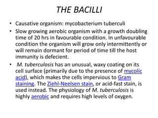
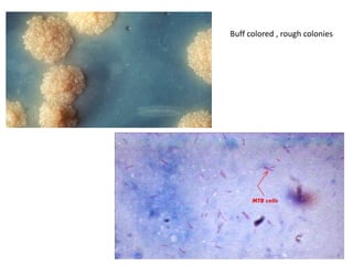
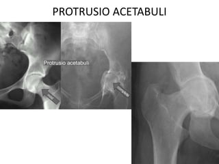
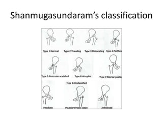
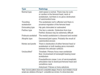
TUBERCULOSIS OF THE HIP
DR.ABHINAV KESARKAR
THE BACILLI
• Causative organism: mycobacterium tuberculi
• Slow growing aerobic organism with a growth doubling
time of 20 hrs in favourable condition. In unfavourable
condition the organism will grow only intermittently or
will remain dormant for period of time till the host
immunity is defecient.
• M. tuberculosis has an unusual, waxy coating on its
cell surface (primarily due to the presence of mycolic
acid), which makes the cells impervious to Gram
staining. The Ziehl-Neelsen stain, or acid-fast stain, is
used instead. The physiology of M. tuberculosis is
highly aerobic and requires high levels of oxygen.
Buff colored , rough colonies
• Ideally , diagnosis of tuberculosis is confirmed
on demonstration of the bacilli in skeletal
tuberculosis lesion. However , this is not
possible in numerous case series because ,
skeletal tuberculosis is supposed to be a
paucibacillary condition.
• Bacilli load in osteoarticular TB is less than 105
THE TUBERCLE
• The initial response is in the
reticuloendothelial depots of the skeletal
tissue.
• PMNs Macrophages & Monocytes
• The tubercle bacilli are phagocytosed and
broken down and their lipid is dispersed
throughout the cytoplasm of mononuclear
cells transforming them into epitheloid cells.
• The epitheloid cells fuse together to form the
langhans giant cells when caesation occurs.
• Lympphocytes form a ring around the
langhans cells with caesation necrosis.
• This mass formed by the reticuloendothelial
cells together with caseous material is known
as the tubercle.
• Tubercle with caesation called ‘soft tubercle’
• Tubercle without caesation called ‘hard
tubercle’.
TUBERCULAR SEQUESTRA
• Osseous destruction occurs by lysis of
bone,which softens and yields under gravity &
muscle action leading to compression ,
collapse or deformation of bone.
• Necrosis occurs due to thromboembolic
phenomenon , endarteritis and periarteritis.
• As a result of ischaemic changes , sequestra
occurs and appears as ‘coarse sand’ pattern
which is radiologically difficult to see.
• Sometimes , the adjacent articular cartilage or
disc gets involved and becomes part of the
sequestra.
• ‘ Feathery Sequestra’ occurs when caseous
material becomes calcified.
‘TB HIP’
EPIDEMOLOGY
• Bones and joints and affected in ~5% of pts
with TB
• Commonest is spinal TB in ~50% of cases
• Hip – 15% of all osteoarticular TB
• Can occur in any age group but is more
common in children.
• Next common after spinal TB
PATHOLOGY
• M.TB entry – inhalation, ingestion, skin innoculation
• Primary complex, secondary spread and tertiary lesion.
osseoarticular TB is haematogenus
• Always starts in bone, rarely synovium –granulomatous
reaction
• The anatomical sites of the lesions:
1.The superior rim of the acetabulam
2. Epiphysis
3. Babcock's triangle
4. Greater trochanter.
5. Rarely, purely synovial in location.
• In hip joint head and neck are intracapsular so a bony
lesion invades the joint early
BABCOCKS
TRIANGLE
CLINICAL PICTURE
• h/o previous TB infection or contact
• Insidious onset, chronic course
• Most pts are children
• Prior constitutional symptoms
• First symptom stiffness of hip with a limp
• Pain may be absent in early stages
• Pain worse at night – “night cries”
EXAMINATION
• INSPECTION
- Antalgic / stiff / trendelenberg
- Muscle wasting
- Discharging sinus / cold abscess
- Limb length discrepecency, FFD
• PALPATION
- Local tempreature
- Pelvic tilt
- Tenderness
• MOVEMENTS
- Restricted movements
STAGES OF TB HIP
• STAGE -1 ( STAGE OF SYNOVITIS)
- effusion in the joint that demands the hip in
position of maximun capacity i.e
FLEXION , ABDUCTION & EXTERNAL ROTATION
causing APPARENT LENGTHENING
• STAGE -2 (STAGE OF ARTHRITIS)
- Articular cartilage is involved leading to spasm
of the strong muscles of the hip i.e. the flexors
and the adductors.
- FLEXION ADDUCTION & INTERNAL ROTATION.
Causing APPARENT SHORTENING.
• STAGE – 3 ( ADVANCED ARTHRITIS)
- Further destruction of hip elements leading to
exaggeration of FADIR , restriction of
movements, muscle wasting and limb
shortenings.
• STAGE – 4 (ADVANCED ARTHRITIS WITH
SUBLUXATION & DISLOCATION)
- More destruction of hip leading to displacement
of the femoral head in acetabulum leaving its
lower part empty and broken shentons arc.
- Generally , movements are restricted but gross
destruction may cause collapse and certain
radiological appearance may retain fairly good
ROM.
The subluxated or dislocated hip
Occurs due to
capsular laxity and
synovial hypertrophy
‘Wandering’ Acetabuli
‘Mortar & Pestle’
PROTRUSIO ACETABULI
Shanmugasundaram’s classification
IMPORTANT OBSERVATIONS
A] Childhood TB hip (growing period) chronic
hyperemia would lead to enlargement of
femoral head epiphysis and metaphysis
leading to COXA MAGNA.
B] Thromboembolic phenomena of selective
terminal vasculature create Perthe’s like
changes and reduced blood supply due to
effusion (tamponad effect) causing decrease
size of femoral head and neck – COXA BREVA.
C] Restricted growth of femoral capital epiphysis
with normal growth of trochanteric growth
plate lead to – COXA VARA.
D] Restricted growth of trochanteric physis with
normal growth of femoral epiphysis lead to -
COXA VALGA.
E] A triad of radiologic abnormalities (Phemister
triad);
– periarticular osteoporosis
– peripherally located osseous erosion
– gradual diminution of joint space suggests the dx
of TB
F] Occasionally, wedge-shaped areas of necrosis
(kissing sequestra) in joint margin. These
marginal erosions may simulate RA
TREATMENT
• Rest
• Chemotherapy
• Arthroplasty
• Arthrodesis
• Osteotomy
• Thomas urged that TB should be treated by
rest – which had to be
‘prolonged,
uninterrupted,
rigid and enforced’.
HUGH OWEN THOMAS
TRACTION
–Provides rest to the joint
–Relieves muscle spasm
–Prevents and corrects deformity
–Maintains joint space
–Minimises chance of developing wandering
acetabulum
CHEMOTHERAPY
TB disease category Intensive phase
(2 months)
Continuation phase
(4 months)
All forms of PTB and
EPTB except TB meningitis
and osteoarticular TB
2RHZE 4RH
TB meningitis,
osteoarticular TB
2RHZE 10RH
Courtesy: 2009TBguidelinesbyministryof health
ARTHROPLASTY
THR
• RESERVED FOR ADULT TB
ISSUES
• Reactivation of disease
• Duration of dz free interval before arthroplasty
• Anti-TB use peri-arthroplasty
EXCISION ARTHROPLASTY
ARTHRODESIS
optimal positioning for function and
limited effect on adjacent joints
200 – 350 FLEXION
0 – 50 ADDUCTION
50 – 100 EXTERNAL ROTATION
* avoid abduction as it creates pelvic
obliquity and increased back pain
BRITTAINS ARTHRODESIS
In, TB hip , ischium is not reached in disease
progress, thus , extra articular arthrodesis
can be done. Used in children with fibrous
ankylosis to achieve painless hip. Studies
showed upto 88 % favourable results.
Tuberculosis of the hip

Tuberculosis of the hip

  • 1.
    TUBERCULOSIS OF THEHIP DR.ABHINAV KESARKAR
  • 2.
    THE BACILLI • Causativeorganism: mycobacterium tuberculi • Slow growing aerobic organism with a growth doubling time of 20 hrs in favourable condition. In unfavourable condition the organism will grow only intermittently or will remain dormant for period of time till the host immunity is defecient. • M. tuberculosis has an unusual, waxy coating on its cell surface (primarily due to the presence of mycolic acid), which makes the cells impervious to Gram staining. The Ziehl-Neelsen stain, or acid-fast stain, is used instead. The physiology of M. tuberculosis is highly aerobic and requires high levels of oxygen.
  • 3.
    Buff colored ,rough colonies
  • 4.
    • Ideally ,diagnosis of tuberculosis is confirmed on demonstration of the bacilli in skeletal tuberculosis lesion. However , this is not possible in numerous case series because , skeletal tuberculosis is supposed to be a paucibacillary condition. • Bacilli load in osteoarticular TB is less than 105
  • 5.
    THE TUBERCLE • Theinitial response is in the reticuloendothelial depots of the skeletal tissue. • PMNs Macrophages & Monocytes • The tubercle bacilli are phagocytosed and broken down and their lipid is dispersed throughout the cytoplasm of mononuclear cells transforming them into epitheloid cells. • The epitheloid cells fuse together to form the langhans giant cells when caesation occurs.
  • 6.
    • Lympphocytes forma ring around the langhans cells with caesation necrosis. • This mass formed by the reticuloendothelial cells together with caseous material is known as the tubercle. • Tubercle with caesation called ‘soft tubercle’ • Tubercle without caesation called ‘hard tubercle’.
  • 7.
    TUBERCULAR SEQUESTRA • Osseousdestruction occurs by lysis of bone,which softens and yields under gravity & muscle action leading to compression , collapse or deformation of bone. • Necrosis occurs due to thromboembolic phenomenon , endarteritis and periarteritis. • As a result of ischaemic changes , sequestra occurs and appears as ‘coarse sand’ pattern which is radiologically difficult to see.
  • 8.
    • Sometimes ,the adjacent articular cartilage or disc gets involved and becomes part of the sequestra. • ‘ Feathery Sequestra’ occurs when caseous material becomes calcified.
  • 9.
  • 10.
    EPIDEMOLOGY • Bones andjoints and affected in ~5% of pts with TB • Commonest is spinal TB in ~50% of cases • Hip – 15% of all osteoarticular TB • Can occur in any age group but is more common in children. • Next common after spinal TB
  • 11.
    PATHOLOGY • M.TB entry– inhalation, ingestion, skin innoculation • Primary complex, secondary spread and tertiary lesion. osseoarticular TB is haematogenus • Always starts in bone, rarely synovium –granulomatous reaction • The anatomical sites of the lesions: 1.The superior rim of the acetabulam 2. Epiphysis 3. Babcock's triangle 4. Greater trochanter. 5. Rarely, purely synovial in location. • In hip joint head and neck are intracapsular so a bony lesion invades the joint early
  • 12.
  • 13.
    CLINICAL PICTURE • h/oprevious TB infection or contact • Insidious onset, chronic course • Most pts are children • Prior constitutional symptoms • First symptom stiffness of hip with a limp • Pain may be absent in early stages • Pain worse at night – “night cries”
  • 14.
    EXAMINATION • INSPECTION - Antalgic/ stiff / trendelenberg - Muscle wasting - Discharging sinus / cold abscess - Limb length discrepecency, FFD
  • 15.
    • PALPATION - Localtempreature - Pelvic tilt - Tenderness • MOVEMENTS - Restricted movements
  • 16.
    STAGES OF TBHIP • STAGE -1 ( STAGE OF SYNOVITIS) - effusion in the joint that demands the hip in position of maximun capacity i.e FLEXION , ABDUCTION & EXTERNAL ROTATION causing APPARENT LENGTHENING
  • 18.
    • STAGE -2(STAGE OF ARTHRITIS) - Articular cartilage is involved leading to spasm of the strong muscles of the hip i.e. the flexors and the adductors. - FLEXION ADDUCTION & INTERNAL ROTATION. Causing APPARENT SHORTENING.
  • 20.
    • STAGE –3 ( ADVANCED ARTHRITIS) - Further destruction of hip elements leading to exaggeration of FADIR , restriction of movements, muscle wasting and limb shortenings.
  • 21.
    • STAGE –4 (ADVANCED ARTHRITIS WITH SUBLUXATION & DISLOCATION) - More destruction of hip leading to displacement of the femoral head in acetabulum leaving its lower part empty and broken shentons arc. - Generally , movements are restricted but gross destruction may cause collapse and certain radiological appearance may retain fairly good ROM.
  • 22.
    The subluxated ordislocated hip Occurs due to capsular laxity and synovial hypertrophy
  • 23.
  • 24.
  • 25.
  • 26.
  • 28.
    IMPORTANT OBSERVATIONS A] ChildhoodTB hip (growing period) chronic hyperemia would lead to enlargement of femoral head epiphysis and metaphysis leading to COXA MAGNA. B] Thromboembolic phenomena of selective terminal vasculature create Perthe’s like changes and reduced blood supply due to effusion (tamponad effect) causing decrease size of femoral head and neck – COXA BREVA.
  • 29.
    C] Restricted growthof femoral capital epiphysis with normal growth of trochanteric growth plate lead to – COXA VARA. D] Restricted growth of trochanteric physis with normal growth of femoral epiphysis lead to - COXA VALGA.
  • 30.
    E] A triadof radiologic abnormalities (Phemister triad); – periarticular osteoporosis – peripherally located osseous erosion – gradual diminution of joint space suggests the dx of TB F] Occasionally, wedge-shaped areas of necrosis (kissing sequestra) in joint margin. These marginal erosions may simulate RA
  • 31.
    TREATMENT • Rest • Chemotherapy •Arthroplasty • Arthrodesis • Osteotomy
  • 32.
    • Thomas urgedthat TB should be treated by rest – which had to be ‘prolonged, uninterrupted, rigid and enforced’. HUGH OWEN THOMAS
  • 33.
    TRACTION –Provides rest tothe joint –Relieves muscle spasm –Prevents and corrects deformity –Maintains joint space –Minimises chance of developing wandering acetabulum
  • 34.
    CHEMOTHERAPY TB disease categoryIntensive phase (2 months) Continuation phase (4 months) All forms of PTB and EPTB except TB meningitis and osteoarticular TB 2RHZE 4RH TB meningitis, osteoarticular TB 2RHZE 10RH Courtesy: 2009TBguidelinesbyministryof health
  • 35.
    ARTHROPLASTY THR • RESERVED FORADULT TB ISSUES • Reactivation of disease • Duration of dz free interval before arthroplasty • Anti-TB use peri-arthroplasty
  • 37.
  • 38.
    ARTHRODESIS optimal positioning forfunction and limited effect on adjacent joints 200 – 350 FLEXION 0 – 50 ADDUCTION 50 – 100 EXTERNAL ROTATION * avoid abduction as it creates pelvic obliquity and increased back pain
  • 39.
    BRITTAINS ARTHRODESIS In, TBhip , ischium is not reached in disease progress, thus , extra articular arthrodesis can be done. Used in children with fibrous ankylosis to achieve painless hip. Studies showed upto 88 % favourable results.