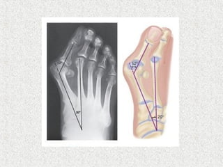
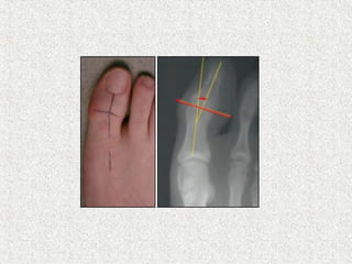
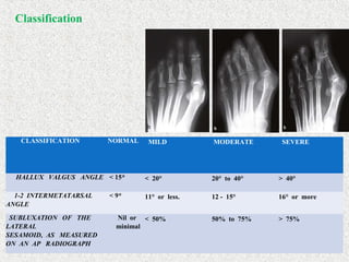
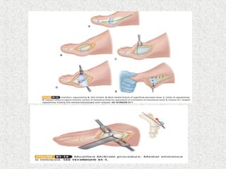
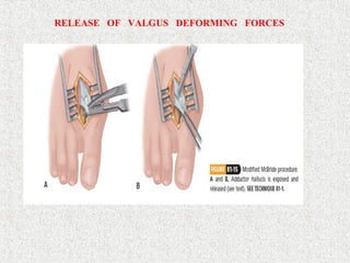
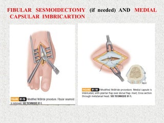
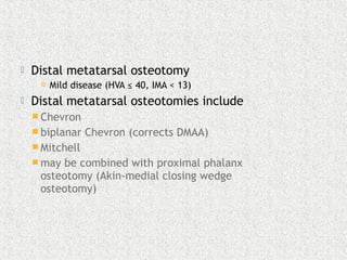
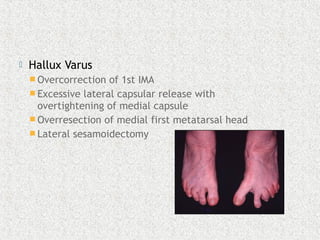
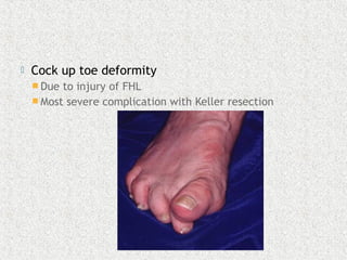
Dr (Major) Parthasarathy S
Pg Resident,MS Orthopaedics
Stanley Medical College,Chennai
Ref:Campbell’s operative orhopaedics 13th
edition
 Valgus of great toe & varus of 1rst MT
 Carl Hueterto defined the deformity
 Controversy
 Not a single disorder,complex deformity of
the first ray
 Deformity & symptoms in lesser toes
 Hammer toe 2nd
 Forefoot Splaying
 Corns
 Bursal hypertrophy 1rst MT head(BUNION)
 Osteoarthritis
 metatarsalgia
 Two forms exist
 adult hallux valgus 
 adolescent & juvenile hallux valgus
 Common in female
 Familial 70%
 Role of footwear
 Hypermobility
 Pes planus
 Abnormal posterior tibial tendon insertion
 Increased obliquity of medial cuneiform MTjt
 Abnormal long 1rst ray
 Incongrous articular surface 1rst MTP jt
 RA
 Cerebral palsy
 2nd
toe amputation
 Cystic degneration of medial capsule
 Achilles Contracture
 Hyperpronated 1st
ray
 Valgus deviation of phalanx promotes varus
position of metatarsal
 MT head displaces medially, leaving the
sesamoid complex laterally
 Sesamoids remain within the respective head
of the flexor hallucis brevis tendon and are
attached to the base of the proximal phalanx
via the sesamoido-phalangeal ligament
 This lateral displacement can lead to
transfer metatarsalgia due to shift in weight
bearing
LIGAMENTOUS ATTATCHMENTS AROUND HALLUX
 Medial MTP joint capsule becomes stretched
and attenuated while the lateral capsule
becomes contracted
 Adductor tendon becomes deforming force
 inserts on fibular sesamoid and lateral aspect of
proximal phalanx
 Lateral deviation of EHL further contributes
to deformity
 Plantar and lateral migration of the abductor
hallucis causes muscle to plantar flex and
pronate phalanx
o Asymptomatic
o Pain- the primary symptom of hallux
valgus is PAIN over the medial
eminence.
o Pressure from footwear is the most
frequent cause of this discomfort.
o Valgus deformity
o Aesthetic or cosmetic concerns
 Standard radiograph
 Weight bearing
AP
Lateral
 Non standing
Oblique
Axial sesamoid view
HALLUX VALGUS ANGLE
Normal <15
Mild <20
Moderate 20-40
Severe >40
1st
/2nd
Inter Metatarsal Angle (IMA)
Normal <9
Mild <11
Moderate 11-16
Severe >16
CLASSIFICATION NORMAL MILD MODERATE SEVERE
HALLUX VALGUS ANGLE < 15* < 20° 20° to 40° > 40°
1-2 INTERMETATARSAL
ANGLE
< 9* 11° or less. 12 - 15° 16° or more
SUBLUXATION OF THE
LATERAL
SESAMOID, AS MEASURED
ON AN AP RADIOGRAPH
Nil or
minimal
< 50% 50% to 75% > 75%
Classification
Conservative Management :
 
o FOOT WEAR MODIFICATION
•   First  line  of  treatment
•   Widening  of   toe  box 
•   Decreasing  the  heel height 
•   Enhanced  arch  support  may  negate  effects  of  pes      
planus.
 
o TA STRETCHING EXERCISES AND TA
LENGTHENING. 
o THERMOPLASTIC NIGHT SPLINTS 
o BUNION AIDS AND STRAPPING –
Bunion  pads  (like  a  Polo/doughnut  shape)  can  help  to  offload  
the  tender  bunion,  
 
 To correct all pathologic elements and
yet maintain a biomechanically functional
fore foot
 To obtain a pliable plantigrade and
cosmetically acceptable foot
 Modified McBride
 Goal is to correct an incongruent MTP joint
(phalanx not lined up with articular cartilage of
MT head)
 HVA <25 degrees
 IMA deformity <15 degrees
 Patient 30-50 years of age
 Rarely appropriate in isolation
 Usually performed in conjunction with
 medial eminence resection
 MT osteotomy
 1st TMT arthrodesis (Lapidus procedure)
 Release of adductor from lateral
sesamoid/proximal phalanx
 Lateral capsulotomy
 Medial capsular imbrication
 original McBride included lateral
sesamoidectomy
MEDIAL CAPSULAR INCISION , JOINT REDUCTION
AND MEDIAL EMINENCE REMOVAL.
RELEASE OF VALGUS DEFORMING FORCES
FIBULAR SESMOIDECTOMY (if needed) AND MEDIAL
CAPSULAR IMBRICARTION
 Distal metatarsal osteotomy
 Mild disease (HVA ≤ 40, IMA < 13)
 Distal metatarsal osteotomies include
 Chevron
 biplanar Chevron (corrects DMAA)
 Mitchell
 may be combined with proximal phalanx
osteotomy (Akin-medial closing wedge
osteotomy)
 Proximal metatarsal osteotomy
 Moderate disease (HVA >40°, IMA >13°)
 proximal metatarsal osteotomies include
 crescentic osteotomy
 Broomstick osteotomy
 Ludloff
 Scarf
 double (proximal and distal) osteotomy
 Severe disease (HVA 41-50°, IMA 16-20°)
 First cuneiform osteotomy
 severe deformity in young patient with open
physis
 Proximal phalanx osteotomies
 Akin osteotomy
 Hallux valgus interphalangeus
 congruent joint with DMAA <10°
 As a secondary procedure if a primary procedure (e.g.,
chevron or distal soft-tissue procedure) did not
provide sufficient correction due to a large DMAA or
HVI
 Lapidus procedure (1st metatarsocuneiform
arthrodesis with modified McBride)
 Severe deformity (very large IMA)
 Arthritis at 1st TMT
 metatarsus primus varus
 hypermobile 1st TMT joint
 concomitant pes planus
 MTP Arthrodesis
 Cerebral palsy
 Down's syndrome
 Rheumatoid arthritis
 Gout
 Severe DJD
 Ehler-Danlos
 proximal phalanx (Keller) resection
arthroplasty
 Largely abandoned
 Rarely indicated in some elderly patient with
reduced function demands
 Recurrence
Most common cause of failure is insufficient
preoperative assessment and failure to follow
indications
 e.g., failure to recognize DMAA > 10°
Inadequate correction of IMA
Failure to do adequate distal soft tissue realignment
More common in juvenile/adolescent population
Noncompliant patient that bears weight
 Avascular necrosis
 Medial capsulotomy is primary insult to blood
flow to metatarsal head
 Distal metatarsal oseotomy and lateral soft
tissue release inconjunction do not increase risk
for AVN
 Dorsal malunion with transfer metatarsalgia
 Due to overload of lesser metatarsal heads
 Risk associated with shortening of hallux MT
 Lapidus
 Proximal crescentric osteotomies
 Hallux Varus
 Overcorrection of 1st IMA
 Excessive lateral capsular release with
overtightening of medial capsule
 Overresection of medial first metatarsal head
 Lateral sesamoidectomy
 Cock up toe deformity
 Due to injury of FHL
 Most severe complication with Keller resection
 2nd MT transfer metatarsalgia
 Often seen concomitant with hallux valgus
 Shortening metatarsal osteotomy Neuropraxia
 Painful incisional neuromas
 Frequently involve the medial branch of the
dorsal cutaneous nerve-a terminal branch of the
superficial peroneal nerve
 It is most commonly injured during the medial
approach for capsular imbrication or metatarsal
osteotomy.
Hallux valgus

Hallux valgus

  • 1.
    Dr (Major) ParthasarathyS Pg Resident,MS Orthopaedics Stanley Medical College,Chennai Ref:Campbell’s operative orhopaedics 13th edition
  • 2.
     Valgus ofgreat toe & varus of 1rst MT  Carl Hueterto defined the deformity  Controversy  Not a single disorder,complex deformity of the first ray  Deformity & symptoms in lesser toes  Hammer toe 2nd  Forefoot Splaying  Corns  Bursal hypertrophy 1rst MT head(BUNION)  Osteoarthritis  metatarsalgia
  • 4.
     Two formsexist  adult hallux valgus   adolescent & juvenile hallux valgus  Common in female
  • 5.
     Familial 70% Role of footwear  Hypermobility  Pes planus  Abnormal posterior tibial tendon insertion  Increased obliquity of medial cuneiform MTjt  Abnormal long 1rst ray  Incongrous articular surface 1rst MTP jt  RA  Cerebral palsy
  • 6.
     2nd toe amputation Cystic degneration of medial capsule  Achilles Contracture  Hyperpronated 1st ray
  • 8.
     Valgus deviationof phalanx promotes varus position of metatarsal  MT head displaces medially, leaving the sesamoid complex laterally  Sesamoids remain within the respective head of the flexor hallucis brevis tendon and are attached to the base of the proximal phalanx via the sesamoido-phalangeal ligament  This lateral displacement can lead to transfer metatarsalgia due to shift in weight bearing
  • 9.
  • 11.
     Medial MTPjoint capsule becomes stretched and attenuated while the lateral capsule becomes contracted  Adductor tendon becomes deforming force  inserts on fibular sesamoid and lateral aspect of proximal phalanx  Lateral deviation of EHL further contributes to deformity  Plantar and lateral migration of the abductor hallucis causes muscle to plantar flex and pronate phalanx
  • 13.
    o Asymptomatic o Pain-the primary symptom of hallux valgus is PAIN over the medial eminence. o Pressure from footwear is the most frequent cause of this discomfort. o Valgus deformity o Aesthetic or cosmetic concerns
  • 14.
     Standard radiograph Weight bearing AP Lateral  Non standing Oblique Axial sesamoid view
  • 17.
    HALLUX VALGUS ANGLE Normal<15 Mild <20 Moderate 20-40 Severe >40
  • 19.
    1st /2nd Inter Metatarsal Angle(IMA) Normal <9 Mild <11 Moderate 11-16 Severe >16
  • 28.
    CLASSIFICATION NORMAL MILDMODERATE SEVERE HALLUX VALGUS ANGLE < 15* < 20° 20° to 40° > 40° 1-2 INTERMETATARSAL ANGLE < 9* 11° or less. 12 - 15° 16° or more SUBLUXATION OF THE LATERAL SESAMOID, AS MEASURED ON AN AP RADIOGRAPH Nil or minimal < 50% 50% to 75% > 75% Classification
  • 29.
    Conservative Management :   oFOOT WEAR MODIFICATION •   First  line  of  treatment •   Widening  of   toe  box  •   Decreasing  the  heel height  •   Enhanced  arch  support  may  negate  effects  of  pes       planus.   o TA STRETCHING EXERCISES AND TA LENGTHENING.  o THERMOPLASTIC NIGHT SPLINTS  o BUNION AIDS AND STRAPPING – Bunion  pads  (like  a  Polo/doughnut  shape)  can  help  to  offload   the  tender  bunion,    
  • 30.
     To correctall pathologic elements and yet maintain a biomechanically functional fore foot  To obtain a pliable plantigrade and cosmetically acceptable foot
  • 33.
     Modified McBride Goal is to correct an incongruent MTP joint (phalanx not lined up with articular cartilage of MT head)  HVA <25 degrees  IMA deformity <15 degrees  Patient 30-50 years of age  Rarely appropriate in isolation  Usually performed in conjunction with  medial eminence resection  MT osteotomy  1st TMT arthrodesis (Lapidus procedure)
  • 34.
     Release ofadductor from lateral sesamoid/proximal phalanx  Lateral capsulotomy  Medial capsular imbrication  original McBride included lateral sesamoidectomy
  • 35.
    MEDIAL CAPSULAR INCISION, JOINT REDUCTION AND MEDIAL EMINENCE REMOVAL.
  • 37.
    RELEASE OF VALGUSDEFORMING FORCES
  • 38.
    FIBULAR SESMOIDECTOMY (ifneeded) AND MEDIAL CAPSULAR IMBRICARTION
  • 39.
     Distal metatarsalosteotomy  Mild disease (HVA ≤ 40, IMA < 13)  Distal metatarsal osteotomies include  Chevron  biplanar Chevron (corrects DMAA)  Mitchell  may be combined with proximal phalanx osteotomy (Akin-medial closing wedge osteotomy)
  • 40.
     Proximal metatarsalosteotomy  Moderate disease (HVA >40°, IMA >13°)  proximal metatarsal osteotomies include  crescentic osteotomy  Broomstick osteotomy  Ludloff  Scarf
  • 41.
     double (proximaland distal) osteotomy  Severe disease (HVA 41-50°, IMA 16-20°)  First cuneiform osteotomy  severe deformity in young patient with open physis
  • 42.
     Proximal phalanxosteotomies  Akin osteotomy  Hallux valgus interphalangeus  congruent joint with DMAA <10°  As a secondary procedure if a primary procedure (e.g., chevron or distal soft-tissue procedure) did not provide sufficient correction due to a large DMAA or HVI
  • 43.
     Lapidus procedure(1st metatarsocuneiform arthrodesis with modified McBride)  Severe deformity (very large IMA)  Arthritis at 1st TMT  metatarsus primus varus  hypermobile 1st TMT joint  concomitant pes planus
  • 44.
     MTP Arthrodesis Cerebral palsy  Down's syndrome  Rheumatoid arthritis  Gout  Severe DJD  Ehler-Danlos
  • 45.
     proximal phalanx(Keller) resection arthroplasty  Largely abandoned  Rarely indicated in some elderly patient with reduced function demands
  • 46.
     Recurrence Most commoncause of failure is insufficient preoperative assessment and failure to follow indications  e.g., failure to recognize DMAA > 10° Inadequate correction of IMA Failure to do adequate distal soft tissue realignment More common in juvenile/adolescent population Noncompliant patient that bears weight
  • 47.
     Avascular necrosis Medial capsulotomy is primary insult to blood flow to metatarsal head  Distal metatarsal oseotomy and lateral soft tissue release inconjunction do not increase risk for AVN  Dorsal malunion with transfer metatarsalgia  Due to overload of lesser metatarsal heads  Risk associated with shortening of hallux MT  Lapidus  Proximal crescentric osteotomies
  • 48.
     Hallux Varus Overcorrection of 1st IMA  Excessive lateral capsular release with overtightening of medial capsule  Overresection of medial first metatarsal head  Lateral sesamoidectomy
  • 49.
     Cock uptoe deformity  Due to injury of FHL  Most severe complication with Keller resection
  • 50.
     2nd MTtransfer metatarsalgia  Often seen concomitant with hallux valgus  Shortening metatarsal osteotomy Neuropraxia  Painful incisional neuromas  Frequently involve the medial branch of the dorsal cutaneous nerve-a terminal branch of the superficial peroneal nerve  It is most commonly injured during the medial approach for capsular imbrication or metatarsal osteotomy.