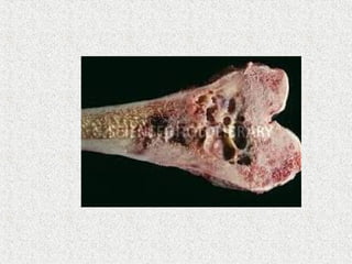
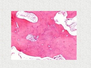
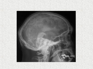
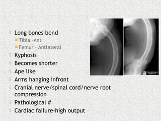
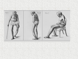
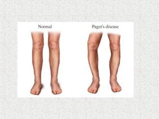
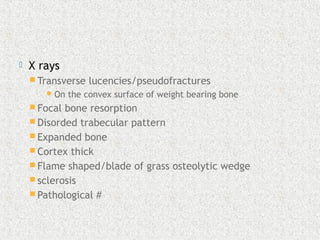
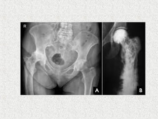
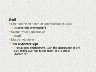
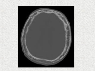
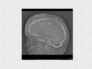
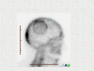
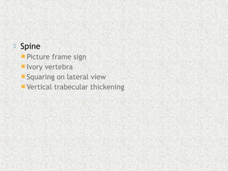
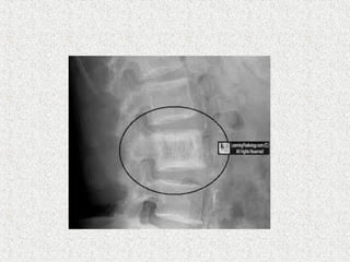
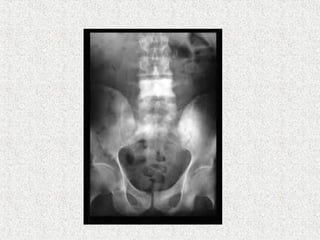
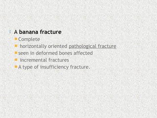
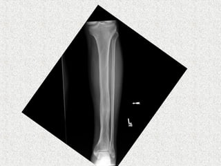
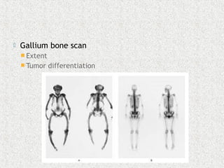
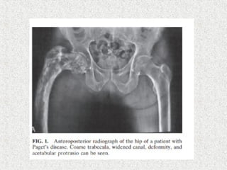
Dr (Major) Parthasarathy S
Junior Resident,MS Orthopaedics
Stanley Medical College,Chennai
Ref: Chapman’s orthopaedic surgery 3rd
edn
Apley’s system of orthopaedics and
fractures 9th
edn
Davidson’s textbook of medicine
Harshmohan textbook of pathology
 Osteitis deformans
 Disturbance in rate of bone turn over
 Initiated by early increase in bone
resorption(osteoclasts)
 Excessive & pathologic bone
formation(osteoblast)
 Result
 Distorted bone
 Thick cortex
 Coarse rabeculations
 Immature woven appearance
Can affect any bone
Commonly affects
Pelvis & tibia (commonest)
Femur
Spine 75%
 Lumbar common
 Cervical rare
Cranium
Monostotic or polyostotic
Symtomatic or asymptomatic
 Unknown
 Familial clustering
 14%-30% + family history
 Autosomal dominant
 Some HLA correlation
 ? Viral etiology –Rebel et al 1980
 Inclusion bodies resembling nuclear
capsule(osteoclast)
 Resembles paramyxo/measles virus
 D/D
 Familial expansile osteolysis
 Expansile skeletal hyperphosphatasia
 Idiopathic hyperphosphatasia
 Early onset PDB
 3:2 sex ratio M:F
 >50 yrs
 3-4% >45yrs / 8% >80yrs prevalence in USA
 Common in Europe(north> south)/North
America/Australia/Newzealand
 Uncommon in Asians
 Starts at one end & progresses leaving a trail
of altered architecture
 Tends to start at muscle insertion points on
bone
 Marked increase in osteoclast & osteoblast
activity
Burned out pattern
 Steal syndrome –blood diverted from vital
organs to skeletal circulation
 Cerebral impairement
 Spinal cord ischaemia
 Asymptomatic 10-20%
 Bone pain
 Rest
 Motion
 Warmth of bone/skin over bone
 Back pain
 Spinal stenosis
 Compression #
 Paraplegia -secondary GCT
 Skull size increased
 Hearing loss(otosclerosis)
 Long bones bend
 Tibia –Ant
 Femur – Antlateral
 Kyphosis
 Becomes shorter
 Ape like
 Arms hanging infront
 Cranial nerve/spinal cord/nerve root
compression
 Pathological #
 Cardiac failure-high output
 Pathological #
 10% patients
 most common complication
 Femur tibia forearm
 Nonunion common
 Femoral neck/subtrochanteric #
 75-90% non-union rate
 With these exceptions # heals by abundant callus
 Secondary sarcoma # 5-20%
 Short/oblique/transverse
 Delayed/non-union-common in burned out phase
 2-4% in symptomatic
 0.1% overall
 Humerus –common site for malignant change
 Benign tumor
 GCT
 Sensitive to steroid therapy
 Malignant
 Osteogenic sarcoma(poor prognosis)
 Fibrosarcoma
 chondrosarcoma
 Signs
 More painful/tender/swollen
 New pain
 New site
 New Lytic area in sclerotic bone
 OA-atrophic
 Nerve compression
 High output cardiac failure
 Hypercalcemia
 Lab
 Serum Ca normal
 Serum phosphorous normal
 Hypercalcemia(immobilised patients)
 Serum ALP increased(osteoblast)
 90% cases
 May be normal-single bone
 24 hour urinary hydroxyproline & N
Telopeptide(osteoclast)
 Serum Pyridinoline increased
 X rays
 Transverse lucencies/pseudofractures
On the convex surface of weight bearing bone
 Focal bone resorption
 Disorded trabecular pattern
 Expanded bone
 Cortex thick
 Flame shaped/blade of grass osteolytic wedge
 sclerosis
 Pathological #
 Skull
 Circumscribed patch of osteoporosis in skull
 Osteoporosis circumscripta
 Cotton wool appearance
 Mixed
 Diploic widening
 Tam o'Shanter sign
 frontal bone enlargement, with the appearance of the
skull falling over the facial bones, like a Tam o'
Shanter hat
 Spine
 Picture frame sign
 Ivory vertebra
 Squaring on lateral view
 Vertical trabecular thickening
 A banana fracture 
 Complete
 horizontally oriented pathological fracture 
 seen in deformed bones affected
  incremental fractures 
 A type of insufficiency fracture.
 Gallium bone scan
 Extent
 Tumor differentiation
 Biopsy
 Definitive
 Rarely indicated
 only when malignant transformation is suspected
 Asymptomatic
 Who do not have disease in weight bearing bone
 Periodic monitoring
 Symptomatic
 Pain
 Neurologic deficit
 Surgery
 Cardiac failure
 Severe polyostotic
 ALP/bone marker elevated twice normal with
disease in weight bearing bone
 Biphosphonates
 Preferred
 Inhibit bone resorption
 Bind to hydroxyapatite crystals
 prolonged remission
 Ca & Vit D given along(Osteomalacia)
 Calcitonin
 Decrease number & activity of osteoclast
 Daily dose till pain controlled & ALP reduced &
stabilised
 Maintenance once/twice weekly
 Injection/nasal spray
 Dry nares 2%
 Mithramycin
 Extremely cytotoxic
 Strong inhibitor of
 Osteoblast
 Osteoclast
 Recommended only in Paget’s paraplegia
 15mg/kg/day for 10days
 Osteotomy to treat deformity
 Fracture
 NOF
 Prosthetic replacement
 Subtrochateric femur
 Intramedullary system
 Corrective osteotomy
 Most pathological long bone #
 Intramedullary system
 Even after union protective bracing for 3-6
months
 Because remodelling is slow
 Severe disabling arthritis
 THR
 Significant varus deformity
 Bowing
 Acetabular protrusion
 Increased blood loss
 Distorted medullary cavity
 Sclerosis
 Heterotrophic ossification
paget's disease
paget's disease

paget's disease

  • 1.
    Dr (Major) ParthasarathyS Junior Resident,MS Orthopaedics Stanley Medical College,Chennai Ref: Chapman’s orthopaedic surgery 3rd edn Apley’s system of orthopaedics and fractures 9th edn Davidson’s textbook of medicine Harshmohan textbook of pathology
  • 3.
     Osteitis deformans Disturbance in rate of bone turn over  Initiated by early increase in bone resorption(osteoclasts)  Excessive & pathologic bone formation(osteoblast)  Result  Distorted bone  Thick cortex  Coarse rabeculations  Immature woven appearance
  • 4.
    Can affect anybone Commonly affects Pelvis & tibia (commonest) Femur Spine 75%  Lumbar common  Cervical rare Cranium Monostotic or polyostotic Symtomatic or asymptomatic
  • 5.
     Unknown  Familialclustering  14%-30% + family history  Autosomal dominant  Some HLA correlation  ? Viral etiology –Rebel et al 1980  Inclusion bodies resembling nuclear capsule(osteoclast)  Resembles paramyxo/measles virus
  • 6.
     D/D  Familialexpansile osteolysis  Expansile skeletal hyperphosphatasia  Idiopathic hyperphosphatasia  Early onset PDB
  • 7.
     3:2 sexratio M:F  >50 yrs  3-4% >45yrs / 8% >80yrs prevalence in USA  Common in Europe(north> south)/North America/Australia/Newzealand  Uncommon in Asians
  • 8.
     Starts atone end & progresses leaving a trail of altered architecture  Tends to start at muscle insertion points on bone  Marked increase in osteoclast & osteoblast activity
  • 9.
  • 13.
     Steal syndrome–blood diverted from vital organs to skeletal circulation  Cerebral impairement  Spinal cord ischaemia
  • 14.
     Asymptomatic 10-20% Bone pain  Rest  Motion  Warmth of bone/skin over bone  Back pain  Spinal stenosis  Compression #  Paraplegia -secondary GCT  Skull size increased  Hearing loss(otosclerosis)
  • 16.
     Long bonesbend  Tibia –Ant  Femur – Antlateral  Kyphosis  Becomes shorter  Ape like  Arms hanging infront  Cranial nerve/spinal cord/nerve root compression  Pathological #  Cardiac failure-high output
  • 19.
     Pathological # 10% patients  most common complication  Femur tibia forearm  Nonunion common  Femoral neck/subtrochanteric #  75-90% non-union rate  With these exceptions # heals by abundant callus  Secondary sarcoma # 5-20%  Short/oblique/transverse  Delayed/non-union-common in burned out phase
  • 20.
     2-4% insymptomatic  0.1% overall  Humerus –common site for malignant change  Benign tumor  GCT  Sensitive to steroid therapy  Malignant  Osteogenic sarcoma(poor prognosis)  Fibrosarcoma  chondrosarcoma
  • 23.
     Signs  Morepainful/tender/swollen  New pain  New site  New Lytic area in sclerotic bone
  • 25.
     OA-atrophic  Nervecompression  High output cardiac failure  Hypercalcemia
  • 26.
     Lab  SerumCa normal  Serum phosphorous normal  Hypercalcemia(immobilised patients)  Serum ALP increased(osteoblast)  90% cases  May be normal-single bone  24 hour urinary hydroxyproline & N Telopeptide(osteoclast)  Serum Pyridinoline increased
  • 27.
     X rays Transverse lucencies/pseudofractures On the convex surface of weight bearing bone  Focal bone resorption  Disorded trabecular pattern  Expanded bone  Cortex thick  Flame shaped/blade of grass osteolytic wedge  sclerosis  Pathological #
  • 29.
     Skull  Circumscribedpatch of osteoporosis in skull  Osteoporosis circumscripta  Cotton wool appearance  Mixed  Diploic widening  Tam o'Shanter sign  frontal bone enlargement, with the appearance of the skull falling over the facial bones, like a Tam o' Shanter hat
  • 33.
     Spine  Pictureframe sign  Ivory vertebra  Squaring on lateral view  Vertical trabecular thickening
  • 36.
     A banana fracture  Complete  horizontally oriented pathological fracture   seen in deformed bones affected   incremental fractures   A type of insufficiency fracture.
  • 38.
     Gallium bonescan  Extent  Tumor differentiation
  • 39.
     Biopsy  Definitive Rarely indicated  only when malignant transformation is suspected
  • 40.
     Asymptomatic  Whodo not have disease in weight bearing bone  Periodic monitoring  Symptomatic  Pain  Neurologic deficit  Surgery  Cardiac failure  Severe polyostotic  ALP/bone marker elevated twice normal with disease in weight bearing bone
  • 41.
     Biphosphonates  Preferred Inhibit bone resorption  Bind to hydroxyapatite crystals  prolonged remission  Ca & Vit D given along(Osteomalacia)
  • 42.
     Calcitonin  Decreasenumber & activity of osteoclast  Daily dose till pain controlled & ALP reduced & stabilised  Maintenance once/twice weekly  Injection/nasal spray  Dry nares 2%
  • 45.
     Mithramycin  Extremelycytotoxic  Strong inhibitor of  Osteoblast  Osteoclast  Recommended only in Paget’s paraplegia  15mg/kg/day for 10days  Osteotomy to treat deformity
  • 46.
     Fracture  NOF Prosthetic replacement  Subtrochateric femur  Intramedullary system  Corrective osteotomy  Most pathological long bone #  Intramedullary system  Even after union protective bracing for 3-6 months  Because remodelling is slow
  • 48.
     Severe disablingarthritis  THR  Significant varus deformity  Bowing  Acetabular protrusion  Increased blood loss  Distorted medullary cavity  Sclerosis  Heterotrophic ossification