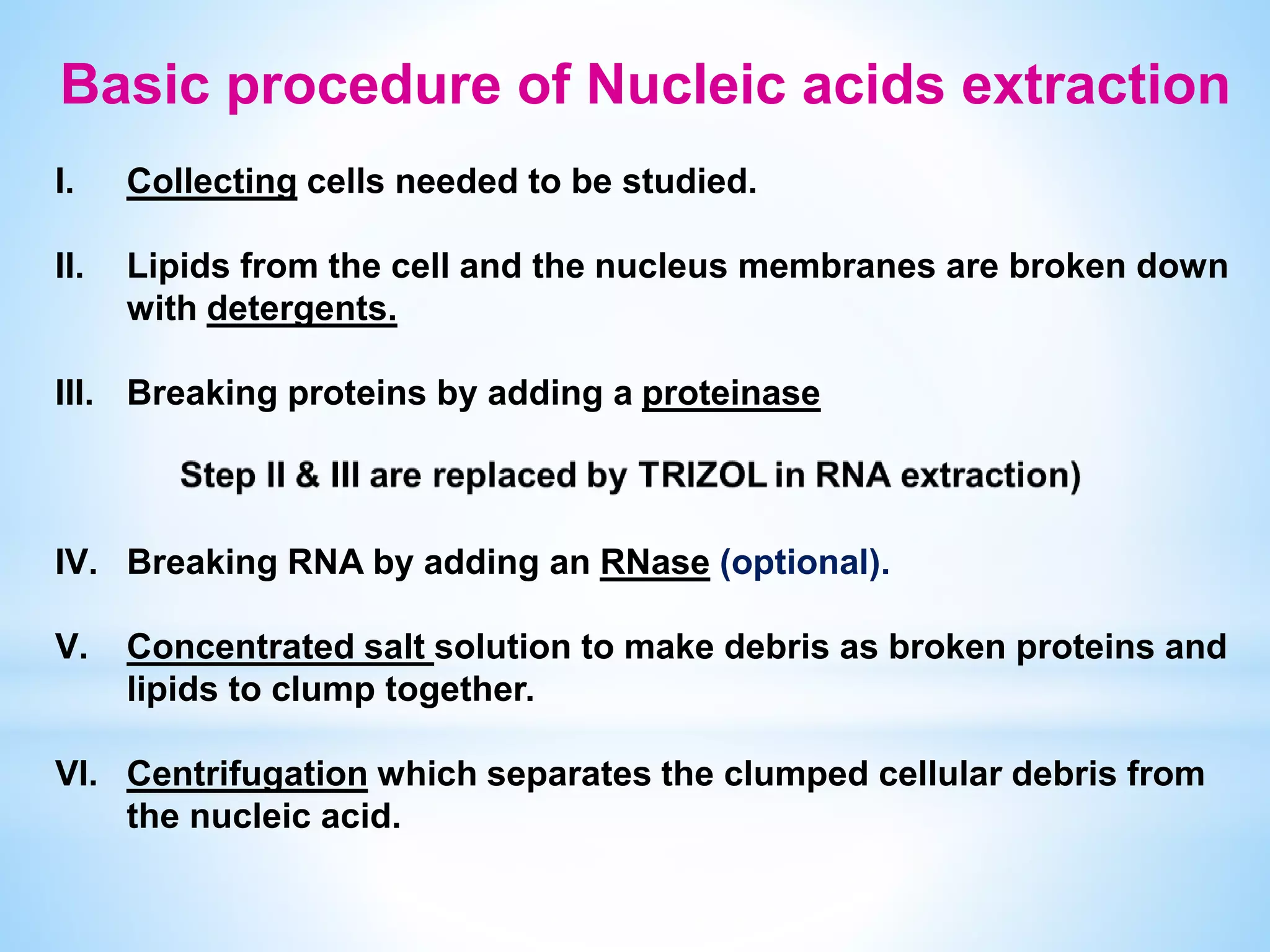
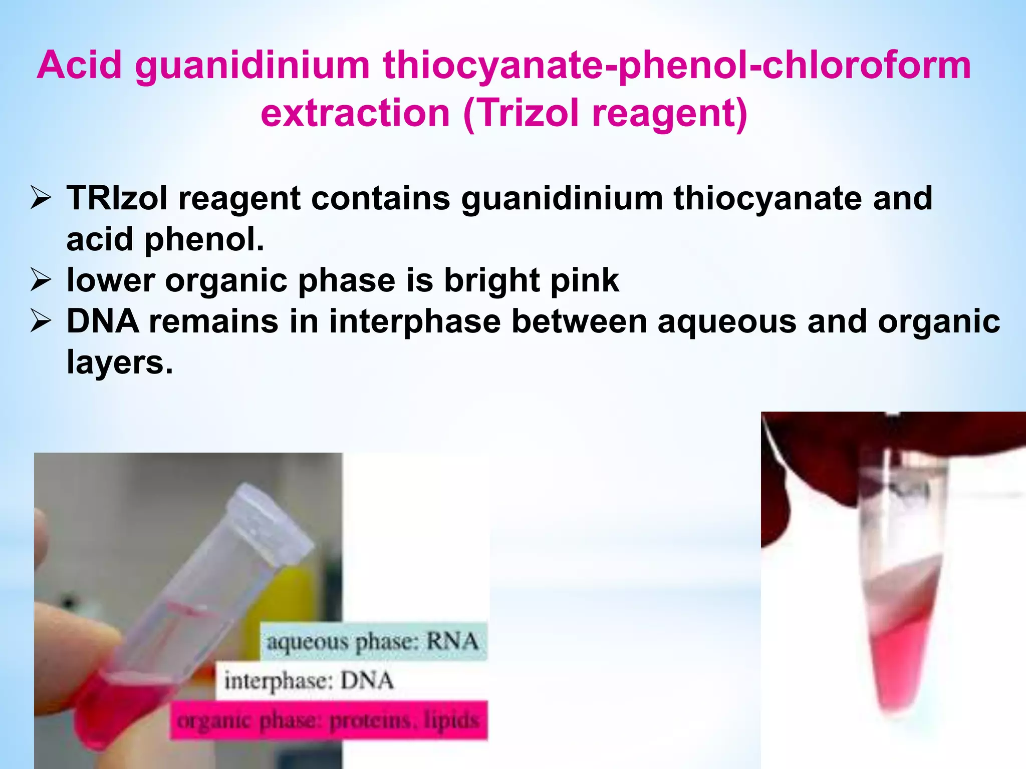
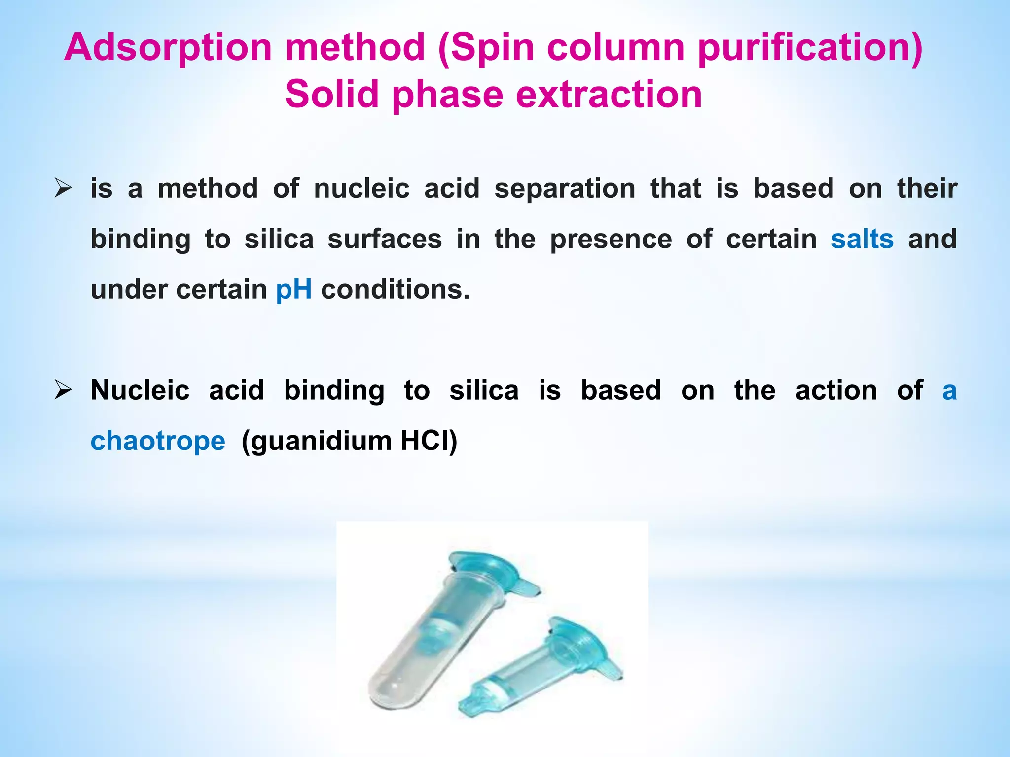
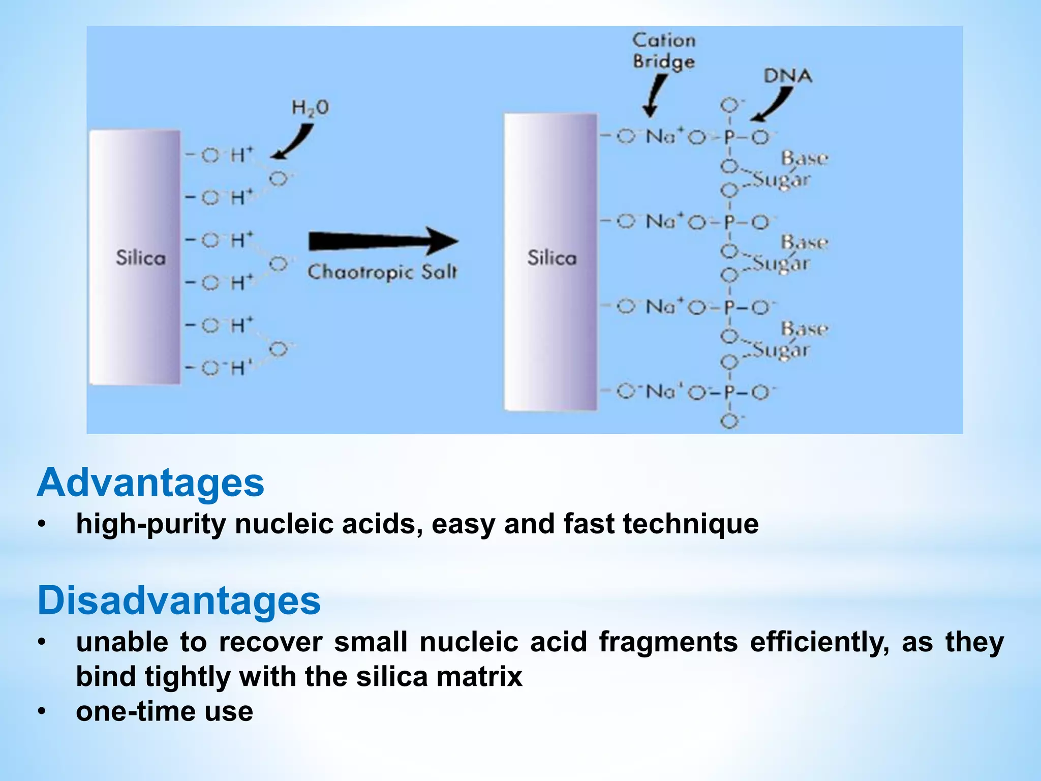
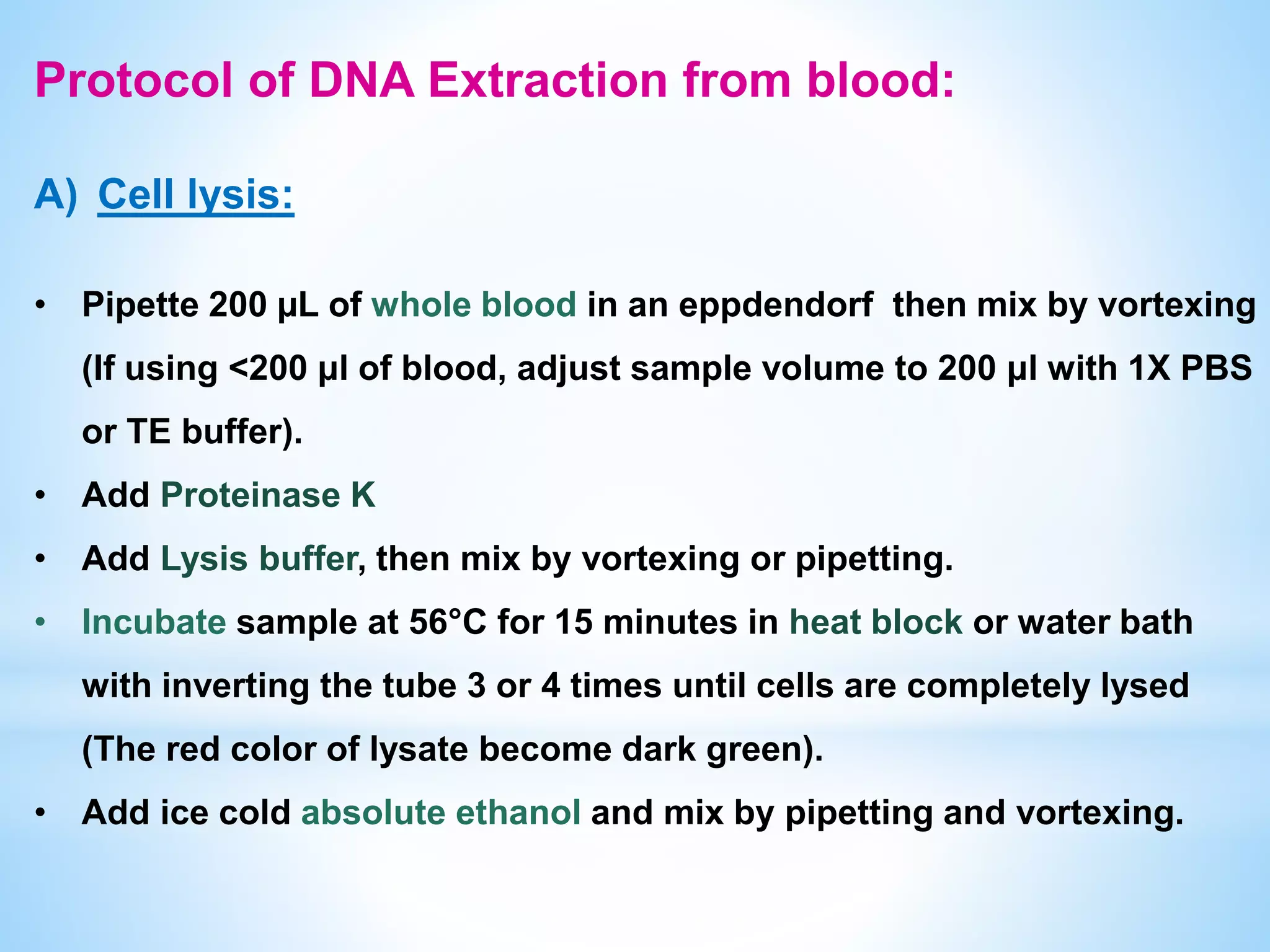
Nucleic acids Extraction
DNA Extraction
 is a process of purification of DNA from sample using
a combination of physical and chemical methods.
The first isolation of DNA was done in 1869 by Friedrich
Miescher.
Basic procedure of Nucleic acids extraction
I. Collecting cells needed to be studied.
II. Lipids from the cell and the nucleus membranes are broken down
with detergents.
III. Breaking proteins by adding a proteinase
IV. Breaking RNA by adding an RNase (optional).
V. Concentrated salt solution to make debris as broken proteins and
lipids to clump together.
VI. Centrifugation which separates the clumped cellular debris from
the nucleic acid.
RNases are EVERYWHERE!!
 RNases are naturally occurring enzymes that degrade RNA
& are common laboratory contaminants
 Released from cellular compartments during isolation of
RNA from biological samples
 Naturally occuring in the environment and human sources
(ex., RNase 7 is secreted by human skin)
 Are difficult to inactivate
 Wear powder-free latex gloves at all times
 Use RNase-free tubes and pipette tips
 Supplement reactions with RNase inhibitors
 Include a chaotropic agent (guanidine) in the procedure
(inactivate and precipitate RNases)
Protecting against RNases
Methods of Nucleic Acids
extraction
Non-Organic (Proteinase K and Salting out)
Organic (Phenol–chloroform extraction)
Adsorption method (Spin column purification)
Magnetic Beads based Isolation
 Digested proteins are precipitated by salting out with
high concentrations of salt (NaCl).
 Precipitated proteins are removed by centrifugation
 Nucleic acids remains in the supernatant.
Advantages: Does not use organic reagents such as
phenol or chloroform.
Disadvantages: if salts are not adequately removed,
problems could occur with the RFLP procedure due to
alteration of DNA mobility (band shifting).
Non-Organic Nucleic Acids Extraction
Phenol-Chloroform Extraction
 Liquid-liquid extraction technique
 Widely used for isolating DNA, RNA and protein.
 Equal volumes of a mixture of phenol-chloroform and an aqueous
sample are mixed forming a biphasic mixture.
 Chloroform facilitates separation of these solvents into 2 phases
recognized by color: a clear upper aqueous hydrophilic phase
(containing nucleic acids) and a yellow lower organic hydrophobic
phase (containing proteins dissolved in phenol and lipids
dissolved in chloroform)
 Chloroform increase density of organic phase & prevent phase
inversion
 pH is an important factor in phenol-chloroform extraction technique.
 In the case of DNA purification, a pH of 7–8 is used.
 In the case of RNA purification, a pH of around 4.5 is used.
 A pH of 4.5 has a higher conc. of H+ ions that would neutralize the -ve
phosphate charges and cause DNA (uncharged) to dissolve in the
organic phase, while RNA has additional hydroxyl group in pentose
sugar and exposed nitrogenous bases which allows the RNA to form
hydrogen bonds with water.
Advantages:
• can process small and large samples
• Inexpensive
Disadvantages:
• Time consuming
• Requires sample to be transferred to multiple tubes –
increases risk of contamination
• Involves use of hazardous (and smelly!) chemicals
Organic Extraction
 TRIzol reagent contains guanidinium thiocyanate and
acid phenol.
 lower organic phase is bright pink
 DNA remains in interphase between aqueous and organic
layers.
Acid guanidinium thiocyanate-phenol-chloroform
extraction (Trizol reagent)
Adsorption method (Spin column purification)
Solid phase extraction
 is a method of nucleic acid separation that is based on their
binding to silica surfaces in the presence of certain salts and
under certain pH conditions.
 Nucleic acid binding to silica is based on the action of a
chaotrope (guanidium HCl)
Advantages
• high-purity nucleic acids, easy and fast technique
Disadvantages
• unable to recover small nucleic acid fragments efficiently, as they
bind tightly with the silica matrix
• one-time use
Components of DNA extraction kit
 Proteinase K (lyophilised or solution)
 Lysis Solution
 RNase (optional)
 Wash Buffer I (concentrated)
 Wash Buffer II (concentrated)
 Elution Buffer
 Spin Columns
 Collection Tubes
 Perform extractions from fresh or immediately stored samples at
-20°C or -80°C.
 Read the manual carefully and re-write the protocol steps.
 Add volume of ethanol (96-100%) to Wash Buffers as indicated on
the bottle, and mark the check box on the bottle’s cap.
 Check the Lysis buffer for salt precipitation before each use. Re-
dissolve any precipitate by warming the solution to 37°C, then cool
back down to 25°C before use.
 Open heat block and adjust temp. to 56°C.
Before starting
Storage
Proteinase K solution is stable at room temperature as
long as the vial remains sealed. After the vial is opened,
proteinase K should be stored at -20°C.
Dissolved Proteinase K should be immediately stored at
-20℃.
Other components of the kit should be stored at room
temperature.
Protocol of DNA Extraction from blood:
A) Cell lysis:
• Pipette 200 µL of whole blood in an eppdendorf then mix by vortexing
(If using <200 μl of blood, adjust sample volume to 200 μl with 1X PBS
or TE buffer).
• Add Proteinase K
• Add Lysis buffer, then mix by vortexing or pipetting.
• Incubate sample at 56°C for 15 minutes in heat block or water bath
with inverting the tube 3 or 4 times until cells are completely lysed
(The red color of lysate become dark green).
• Add ice cold absolute ethanol and mix by pipetting and vortexing.
B) DNA binding to silica membrane:
 Transfer the prepared mixture to a spin column with collection
tube.
 Centrifuge, replace the collection tube by a new one
C) Membrane washing:
 Add wash buffer I (after ethanol addition) to the spin column and
centrifuge
 Discard the flow-through and return the spin column back into the
same collection tube.
 Add wash buffer II (after ethanol addition) to the column and
centrifuge.
 Discard the flow-through & centrifuge the empty spin to dry it from
residual ethanol.
D) Elution of DNA:
 Replace collection tube by an eppendorf for DNA elution.
 Add 50 μl of elution buffer to the center of the spin column
membrane, incubate for 2 min at room temperature, and then
centrifuge.
 Re-elute the same amount of elution buffer OR
divide the volume of elution buffer and repeat elution step to
increase DNA yield
 Discard the spin and store DNA at -20°C.
Proteinase K
 is able to digest hair (keratin), hence, the name "Proteinase K“
 broad-spectrum serine protease
 it very effectively inactivates DNases and RNases.
 is approximately 10 fold more active on denatured protein (i.e.
after denaturing lysis buffer).
(denaturing agents unfold the protein substrates and make them more
accessible to the protease)
 is a buffer solution used for breaking cells for use in molecular
biology experiments
 components:
 Chaotropic salts: have 2 important roles:
 They destabilize hydrogen bonds, van der Waals forces and
hydrophobic interactions, leading to destabilization of proteins,
including nucleases.
 they disrupt the association of nucleic acids with water, thereby
aiding their transfer to silica.
 Chaotropic salts include guanidium HCL, guanidium thiocyanate,
urea, and lithium perchlorate.
Lysis buffer
 Trimethamine (Tris-HCl) :
to regulate the PH (acidity) and osmolarity of the lysate.
 EDTA :
is a chelating agent of divalent cations as Mg2+ (Mg2+ is a cofactor for
nucleases). If the Mg2+ is bound up by EDTA, nucleases are
inactivated.
 Detergents :
(as Triton X- 100 or SDS) are added to break up membrane structure by
attaching to lipids & proteins.
Water has a high dielectric constant, (electrostatic forces between
charged particles are lower in aqueous solution as water molecules
form a hydration shell around them) which makes it difficult for the
Na+ and nucleic acid PO3- to come together.
i.e Water prevents +ve ions from forming stable ionic bonds with PO3-
and precipitating out of solution.
Ethanol has a much lower dielectric constant, so adding it to solution
disrupts the screening of charges by water making it much easier for
Na+ to interact with the PO3- forming stable ionic bonds causing it to
drop out of solution.
Ethanol precipitation of nucleic acids
Note that the percentage and volume of ethanol used are
important.
 Too much ethanol will bring down degraded material
that will influence UV260 readings.
 Too little ethanol may impede washing of the salt from
the membrane.
 Ethanol contamination can’t be detected by
spectrophotometer, indicators of its presence are:
 Samples don’t sink into the wells of the gel, even in
presence of loading dye.
 Samples don’t freeze at -20°C.
Wash Buffers
Wash 1 (pre-wash)
contain a low amount of chaotropic salt to remove any remaining
proteins and coloured contaminants
Wash 2
 Composed of NaCl, EDTA (pH8.0), Tris.Cl (pH 8.0), 50-70% ethanol
to remove the remaining salts.
 Salts MUST be removed for good DNA yields and purity, so Wash 2
can be repeated to ensure this.
 Correct order of using wash buffers is important.
 Composed of Tris (pH=8) ± EDTA. DNA is more stable at a slightly
basic pH and dissolve faster in a buffer than water.
(RNA can tolerate a slightly acidic pH and dissolves more in water)
 Elution (DNA yield) can be maximised by:
I. incubation for a few minutes before centrifugation.
II. divide elution volume and repeat elution or re-elute the same
volume of elution buffer
III. pre-heated EB :
• better hydration for both DNA and silica and hence better desorption of
DNA.
• the solute (the DNA) dissolve better
Elution buffer
DNA Purification from Large Volumes of Whole
Blood
For purification of DNA from samples > 200µl, it is necessary to burst
red blood cells prior to performing the cell lysis step.
Up to 500 µl of mammalian blood can be processed using following
protocol:
 Add 1 ml of ice cold nuclease free water to 500 μl of whole blood,
mix by vortexing or pipetting.
 Incubate the sample for 5 min at RT then Centrifuge
 Discard the supernatant
 Resuspend the pellet in 200 µl of 1 x PBS.
DNA Purification from Buffy Coat
Buffy coat is a leukocyte-enriched fraction of whole blood and contains about
5-10 times more DNA than an equivalent volume of whole blood.
 Prepare the buffy coat by centrifuging whole blood at 2,500 xg for 10 min
at RT.
 After centrifugation, 3 different fractions are differentiated:
• the upper clear layer containing plasma
• the intermediate buffy coat layer containing concentrated leukocytes
• the bottom layer containing concentrated erythrocytes
 Remove upper clear layer by aspiration.
 Collect 200 µl of intermediate layer in
an eppendorf tube.
 Relative centrifugal force (RCF) expressed in units of
gravity (×g).
 Many microcentrifuges only have settings for speed
(RPM), not relative centrifugal force.
 The relationship between RPM and RCF is as
follows:
g = (1.118 × 10⁻⁵) R S2
Where g is the relative centrifugal force,
R is the radius of the rotor in cm
S is the speed of the centrifuge in RPM
How to convert xg to RPM?
Smile Please, it′s a joke
Magnetic beads are Fe3O4 beads coated with a silicon
dioxide (SiO2) layer that adsorbs nucleic acids.
 Advantage:
Very fast, High purity, No need for centrifugation, can be
done manually or automated
 Disadvantage:
Very expensive
Magnetic Beads Nucleic acid Isolation
 Mince fresh, solid tissue up to 3 mm3 into small pieces (1–2 mm)
with a sterile scalpel blade.
 Process the tissue within 2 h of collection, or freeze at –80°C or in
liquid nitrogen (below -196℃) until the time of extraction to
minimize the activity of endogenous nucleases.
 Place the tissue in a clean mortar
 Using a clean pestle, grind the frozen tissue to a powder while it is
submerged in liquid nitrogen until no tissue clumps are visible.
 Allow the liquid nitrogen to evaporate, leaving a dry
frozen tissue powder in the mortar.
Nucleic Acid extraction from tissue differ only in the
first step:
 Homogenize tissue samples in 1 ml of TRIZOL Reagent per 50-100
mg of tissue.
Evaluation of Nucleic Acids
Spectrophotometrically ( by Nanodrop)
•Quantity
•purity
Agarose Gel Electrophoresis
• Quality
 The bases in nucleic acids have max. absorption at 260 nm
 Proteins have a max. absorption at 280 nm
 Polyphenols/Polysaccharides have a max. absorption at 230 nm
 The A260/A280 ratio is ~1.8 for dsDNA, ~2.0 for RNA.
(Ratios < 1.7 usually indicate protein contamination)
 The A260/A230 ratio of DNA and RNA should be roughly equal to
its A260/A280 ratio (≥ 1.8).
(Lower ratios may indicate contamination by organic compounds
e.g. phenol or carbohydrates).
How pure is your sample?
 Good quality DNA should migrate as a sharp band, with
little or no evidence of smearing.
Checking for degradation (Quality)
Low yield of DNA
Excess sample
sample not well mixed
with lysis buffer and
Proteinase K.
Sample not completely
digested
Ethanol (old stock,not
added to wash buffers)
Trouble shooting
Low Purity
Poor 260/280 ratio
Excess sample &
incomplete
solubilization
Poor 260/230 ratio
residual salt or
inadequate washing.
Purified DNA is
degraded
Sample was frozen
and thawed repeatedly
Inappropriate sample
storage conditions
RNA contamination
add RNase to the
sample prior to the
addition of lysis buffer
Inhibition of
downstream enzymatic
reaction
Residual ethanol ( re-
spin the empty column
Residual salt (correct
order for the Wash
Buffers)
Nucleic acid extraction.pptx

Nucleic acid extraction.pptx

  • 1.
  • 3.
    DNA Extraction  isa process of purification of DNA from sample using a combination of physical and chemical methods. The first isolation of DNA was done in 1869 by Friedrich Miescher.
  • 4.
    Basic procedure ofNucleic acids extraction I. Collecting cells needed to be studied. II. Lipids from the cell and the nucleus membranes are broken down with detergents. III. Breaking proteins by adding a proteinase IV. Breaking RNA by adding an RNase (optional). V. Concentrated salt solution to make debris as broken proteins and lipids to clump together. VI. Centrifugation which separates the clumped cellular debris from the nucleic acid.
  • 5.
    RNases are EVERYWHERE!! RNases are naturally occurring enzymes that degrade RNA & are common laboratory contaminants  Released from cellular compartments during isolation of RNA from biological samples  Naturally occuring in the environment and human sources (ex., RNase 7 is secreted by human skin)  Are difficult to inactivate
  • 6.
     Wear powder-freelatex gloves at all times  Use RNase-free tubes and pipette tips  Supplement reactions with RNase inhibitors  Include a chaotropic agent (guanidine) in the procedure (inactivate and precipitate RNases) Protecting against RNases
  • 7.
    Methods of NucleicAcids extraction Non-Organic (Proteinase K and Salting out) Organic (Phenol–chloroform extraction) Adsorption method (Spin column purification) Magnetic Beads based Isolation
  • 8.
     Digested proteinsare precipitated by salting out with high concentrations of salt (NaCl).  Precipitated proteins are removed by centrifugation  Nucleic acids remains in the supernatant. Advantages: Does not use organic reagents such as phenol or chloroform. Disadvantages: if salts are not adequately removed, problems could occur with the RFLP procedure due to alteration of DNA mobility (band shifting). Non-Organic Nucleic Acids Extraction
  • 9.
    Phenol-Chloroform Extraction  Liquid-liquidextraction technique  Widely used for isolating DNA, RNA and protein.  Equal volumes of a mixture of phenol-chloroform and an aqueous sample are mixed forming a biphasic mixture.
  • 10.
     Chloroform facilitatesseparation of these solvents into 2 phases recognized by color: a clear upper aqueous hydrophilic phase (containing nucleic acids) and a yellow lower organic hydrophobic phase (containing proteins dissolved in phenol and lipids dissolved in chloroform)  Chloroform increase density of organic phase & prevent phase inversion
  • 11.
     pH isan important factor in phenol-chloroform extraction technique.  In the case of DNA purification, a pH of 7–8 is used.  In the case of RNA purification, a pH of around 4.5 is used.  A pH of 4.5 has a higher conc. of H+ ions that would neutralize the -ve phosphate charges and cause DNA (uncharged) to dissolve in the organic phase, while RNA has additional hydroxyl group in pentose sugar and exposed nitrogenous bases which allows the RNA to form hydrogen bonds with water.
  • 12.
    Advantages: • can processsmall and large samples • Inexpensive Disadvantages: • Time consuming • Requires sample to be transferred to multiple tubes – increases risk of contamination • Involves use of hazardous (and smelly!) chemicals Organic Extraction
  • 13.
     TRIzol reagentcontains guanidinium thiocyanate and acid phenol.  lower organic phase is bright pink  DNA remains in interphase between aqueous and organic layers. Acid guanidinium thiocyanate-phenol-chloroform extraction (Trizol reagent)
  • 14.
    Adsorption method (Spincolumn purification) Solid phase extraction  is a method of nucleic acid separation that is based on their binding to silica surfaces in the presence of certain salts and under certain pH conditions.  Nucleic acid binding to silica is based on the action of a chaotrope (guanidium HCl)
  • 15.
    Advantages • high-purity nucleicacids, easy and fast technique Disadvantages • unable to recover small nucleic acid fragments efficiently, as they bind tightly with the silica matrix • one-time use
  • 16.
    Components of DNAextraction kit  Proteinase K (lyophilised or solution)  Lysis Solution  RNase (optional)  Wash Buffer I (concentrated)  Wash Buffer II (concentrated)  Elution Buffer  Spin Columns  Collection Tubes
  • 17.
     Perform extractionsfrom fresh or immediately stored samples at -20°C or -80°C.  Read the manual carefully and re-write the protocol steps.  Add volume of ethanol (96-100%) to Wash Buffers as indicated on the bottle, and mark the check box on the bottle’s cap.  Check the Lysis buffer for salt precipitation before each use. Re- dissolve any precipitate by warming the solution to 37°C, then cool back down to 25°C before use.  Open heat block and adjust temp. to 56°C. Before starting
  • 18.
    Storage Proteinase K solutionis stable at room temperature as long as the vial remains sealed. After the vial is opened, proteinase K should be stored at -20°C. Dissolved Proteinase K should be immediately stored at -20℃. Other components of the kit should be stored at room temperature.
  • 19.
    Protocol of DNAExtraction from blood: A) Cell lysis: • Pipette 200 µL of whole blood in an eppdendorf then mix by vortexing (If using <200 μl of blood, adjust sample volume to 200 μl with 1X PBS or TE buffer). • Add Proteinase K • Add Lysis buffer, then mix by vortexing or pipetting. • Incubate sample at 56°C for 15 minutes in heat block or water bath with inverting the tube 3 or 4 times until cells are completely lysed (The red color of lysate become dark green). • Add ice cold absolute ethanol and mix by pipetting and vortexing.
  • 20.
    B) DNA bindingto silica membrane:  Transfer the prepared mixture to a spin column with collection tube.  Centrifuge, replace the collection tube by a new one C) Membrane washing:  Add wash buffer I (after ethanol addition) to the spin column and centrifuge  Discard the flow-through and return the spin column back into the same collection tube.  Add wash buffer II (after ethanol addition) to the column and centrifuge.  Discard the flow-through & centrifuge the empty spin to dry it from residual ethanol.
  • 21.
    D) Elution ofDNA:  Replace collection tube by an eppendorf for DNA elution.  Add 50 μl of elution buffer to the center of the spin column membrane, incubate for 2 min at room temperature, and then centrifuge.  Re-elute the same amount of elution buffer OR divide the volume of elution buffer and repeat elution step to increase DNA yield  Discard the spin and store DNA at -20°C.
  • 23.
    Proteinase K  isable to digest hair (keratin), hence, the name "Proteinase K“  broad-spectrum serine protease  it very effectively inactivates DNases and RNases.  is approximately 10 fold more active on denatured protein (i.e. after denaturing lysis buffer). (denaturing agents unfold the protein substrates and make them more accessible to the protease)
  • 24.
     is abuffer solution used for breaking cells for use in molecular biology experiments  components:  Chaotropic salts: have 2 important roles:  They destabilize hydrogen bonds, van der Waals forces and hydrophobic interactions, leading to destabilization of proteins, including nucleases.  they disrupt the association of nucleic acids with water, thereby aiding their transfer to silica.  Chaotropic salts include guanidium HCL, guanidium thiocyanate, urea, and lithium perchlorate. Lysis buffer
  • 25.
     Trimethamine (Tris-HCl): to regulate the PH (acidity) and osmolarity of the lysate.  EDTA : is a chelating agent of divalent cations as Mg2+ (Mg2+ is a cofactor for nucleases). If the Mg2+ is bound up by EDTA, nucleases are inactivated.  Detergents : (as Triton X- 100 or SDS) are added to break up membrane structure by attaching to lipids & proteins.
  • 26.
    Water has ahigh dielectric constant, (electrostatic forces between charged particles are lower in aqueous solution as water molecules form a hydration shell around them) which makes it difficult for the Na+ and nucleic acid PO3- to come together. i.e Water prevents +ve ions from forming stable ionic bonds with PO3- and precipitating out of solution. Ethanol has a much lower dielectric constant, so adding it to solution disrupts the screening of charges by water making it much easier for Na+ to interact with the PO3- forming stable ionic bonds causing it to drop out of solution. Ethanol precipitation of nucleic acids
  • 27.
    Note that thepercentage and volume of ethanol used are important.  Too much ethanol will bring down degraded material that will influence UV260 readings.  Too little ethanol may impede washing of the salt from the membrane.  Ethanol contamination can’t be detected by spectrophotometer, indicators of its presence are:  Samples don’t sink into the wells of the gel, even in presence of loading dye.  Samples don’t freeze at -20°C.
  • 28.
    Wash Buffers Wash 1(pre-wash) contain a low amount of chaotropic salt to remove any remaining proteins and coloured contaminants Wash 2  Composed of NaCl, EDTA (pH8.0), Tris.Cl (pH 8.0), 50-70% ethanol to remove the remaining salts.  Salts MUST be removed for good DNA yields and purity, so Wash 2 can be repeated to ensure this.  Correct order of using wash buffers is important.
  • 29.
     Composed ofTris (pH=8) ± EDTA. DNA is more stable at a slightly basic pH and dissolve faster in a buffer than water. (RNA can tolerate a slightly acidic pH and dissolves more in water)  Elution (DNA yield) can be maximised by: I. incubation for a few minutes before centrifugation. II. divide elution volume and repeat elution or re-elute the same volume of elution buffer III. pre-heated EB : • better hydration for both DNA and silica and hence better desorption of DNA. • the solute (the DNA) dissolve better Elution buffer
  • 30.
    DNA Purification fromLarge Volumes of Whole Blood For purification of DNA from samples > 200µl, it is necessary to burst red blood cells prior to performing the cell lysis step. Up to 500 µl of mammalian blood can be processed using following protocol:  Add 1 ml of ice cold nuclease free water to 500 μl of whole blood, mix by vortexing or pipetting.  Incubate the sample for 5 min at RT then Centrifuge  Discard the supernatant  Resuspend the pellet in 200 µl of 1 x PBS.
  • 31.
    DNA Purification fromBuffy Coat Buffy coat is a leukocyte-enriched fraction of whole blood and contains about 5-10 times more DNA than an equivalent volume of whole blood.  Prepare the buffy coat by centrifuging whole blood at 2,500 xg for 10 min at RT.  After centrifugation, 3 different fractions are differentiated: • the upper clear layer containing plasma • the intermediate buffy coat layer containing concentrated leukocytes • the bottom layer containing concentrated erythrocytes  Remove upper clear layer by aspiration.  Collect 200 µl of intermediate layer in an eppendorf tube.
  • 32.
     Relative centrifugalforce (RCF) expressed in units of gravity (×g).  Many microcentrifuges only have settings for speed (RPM), not relative centrifugal force.  The relationship between RPM and RCF is as follows: g = (1.118 × 10⁻⁵) R S2 Where g is the relative centrifugal force, R is the radius of the rotor in cm S is the speed of the centrifuge in RPM How to convert xg to RPM?
  • 33.
  • 34.
    Magnetic beads areFe3O4 beads coated with a silicon dioxide (SiO2) layer that adsorbs nucleic acids.  Advantage: Very fast, High purity, No need for centrifugation, can be done manually or automated  Disadvantage: Very expensive Magnetic Beads Nucleic acid Isolation
  • 37.
     Mince fresh,solid tissue up to 3 mm3 into small pieces (1–2 mm) with a sterile scalpel blade.  Process the tissue within 2 h of collection, or freeze at –80°C or in liquid nitrogen (below -196℃) until the time of extraction to minimize the activity of endogenous nucleases.  Place the tissue in a clean mortar  Using a clean pestle, grind the frozen tissue to a powder while it is submerged in liquid nitrogen until no tissue clumps are visible.  Allow the liquid nitrogen to evaporate, leaving a dry frozen tissue powder in the mortar. Nucleic Acid extraction from tissue differ only in the first step:
  • 38.
     Homogenize tissuesamples in 1 ml of TRIZOL Reagent per 50-100 mg of tissue.
  • 39.
    Evaluation of NucleicAcids Spectrophotometrically ( by Nanodrop) •Quantity •purity Agarose Gel Electrophoresis • Quality
  • 40.
     The basesin nucleic acids have max. absorption at 260 nm  Proteins have a max. absorption at 280 nm  Polyphenols/Polysaccharides have a max. absorption at 230 nm  The A260/A280 ratio is ~1.8 for dsDNA, ~2.0 for RNA. (Ratios < 1.7 usually indicate protein contamination)  The A260/A230 ratio of DNA and RNA should be roughly equal to its A260/A280 ratio (≥ 1.8). (Lower ratios may indicate contamination by organic compounds e.g. phenol or carbohydrates). How pure is your sample?
  • 42.
     Good qualityDNA should migrate as a sharp band, with little or no evidence of smearing. Checking for degradation (Quality)
  • 44.
    Low yield ofDNA Excess sample sample not well mixed with lysis buffer and Proteinase K. Sample not completely digested Ethanol (old stock,not added to wash buffers) Trouble shooting
  • 45.
    Low Purity Poor 260/280ratio Excess sample & incomplete solubilization Poor 260/230 ratio residual salt or inadequate washing.
  • 46.
    Purified DNA is degraded Samplewas frozen and thawed repeatedly Inappropriate sample storage conditions RNA contamination add RNase to the sample prior to the addition of lysis buffer Inhibition of downstream enzymatic reaction Residual ethanol ( re- spin the empty column Residual salt (correct order for the Wash Buffers)

Editor's Notes

  • #4 Nuclein =DNA
  • #10 DNA is polar so it is insoluble in organic solvents phenol is a weak acid
  • #11 Phenol denature proteins and solubilize the denatured protein The chloroform denatures the proteins and facilitates the separation of the aqueous and organic phases the density of buffer saturated phenol is only a little higher than water. So if your aqueous phase contains enough salt or any other solutes that would increase its density, then you could end up with phase inversion during extraction, where your aqueous phase is under the phenol, rather than on top of it. Chloroform is significantly denser than water, so adding it to the organic phase increases the overall density of that phase, helping to prevent phase inversion. chloroform help reduce the interphase – the fuzzy border between the two phases populated by molecules that can’t decide where they want to go. These can be partially denatured proteins, DNA (depending on the pH), and/or partially denatured DNA binding proteins.
  • #12 RNA stays in the aqueous phase since the pkA (acid dissociation constant)of its groups is greater than that of DNA (it is more acidic) it has exposed nitrogenous bases (it is single-stranded), which can form hydrogen bonds with water, keeping it in the aqueous phase.
  • #13 Phenol corrosive ,
  • #14 TRIzol works by maintaining RNA integrity during tissue homogenization, while at the same time disrupting and breaking down cells and cell components
  • #16 A chaotrope denatures biomolecules by disrupting the shell of hydration around them. This allows positively charged ions to form a salt bridge between the negatively charged silica and the negatively charged DNA backbone in high salt concentration. The DNA can then be eluted with low salt.
  • #20 Phosphate buffered saline(PBS)=( NaCl+ Na2HPO4 ) or (KCl + KH2PO4) PH = 7.4 disodium hydrogen phosphate and potassium dihydrogen phosphate ice-cold ethanol increases the yield of DNA. Low temperatures protect the DNA by slowing down the activity of DNases
  • #24 site of cleavage is the peptide bond adjacent to the carboxyl group of aliphatic and aromatic amino acids
  • #25 EDTA (Ethylene diamine tetra acetic disodium salt) a container add 900mL water, then 50mL dishwashing detergent (or 100mL shampoo), and finally 2 teaspoons salt. disrupts the structure of, and denatures, macromolecules as protein and DNA 
  • #27 The electrostatic attraction between the Na+ ions in solution and the PO3- ions are dictated by Coulomb’s Law, which is affected by the dielectric constant of the solution. (electrostatic force of attraction or repulsion between two point charges is directly proportional to the product of the magnitudes of charges and inversely proportional to the square of the distance between them)
  • #28 the amount of ethanol added will be optimal for the kit you are using.
  • #29 Binding buffer: Chaotropic salts that weakens the hydrogen bonding and leads to the DNA binding to silica, with an alcohol added like EtOH or IPA to enhance binding. like guanidine HCl with IPA (varying)
  • #30 When elution buffer is added the nucleic acids become hydrated and will release from the membrane. If the column still has ethanol on it, the nucleic acids will not fully rehydrate To reduce premature elution of DNA, salts can be added to the washing solution. Elution takes place in a low-salt non-alcohol condition.
  • #31 Phosphate buffered saline PBS =( NaCl+Na2HPO4) or (KCl + KH2PO4) PH = 7.4
  • #32 Note. If necessary, adjust the volume to 200 µl with 1x PBS. (so-called because it is usually buff in hue), contains most of the white blood cells and platelets
  • #38 Clumps of tissue sample will be difficult to lyse properly and will result in a lower yield of DNA
  • #45 old stocks ethanol may have taken up water Remember that if you make your wash buffer incorrectly, you may be washing away your extracted DNA or RNA!
  • #46 In 2nd case add additional wash step
  • #47 EDTA cause inhibition of some enzymes