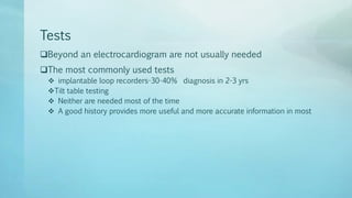
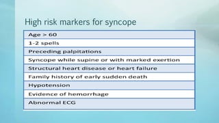
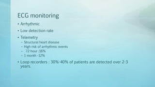
Syncope
Fainting within the first 20 seconds of arising from lying or
sitting almost always have orthostatic hypotension
 1%-2% of emergency
 30%-50% of 1-2% are admitted
 Most common is vasovagal syncope-50%
 orthostatic hypotension -7%
 Cardiac syncope -7%
 Structural heart disease - 3%
 The 30-day mortality - 0.7%,
 30-day adverse outcome-4.5%
Define
• Syncope
– transient
– loss of consciousness
– rapid onset
– short duration,
– spontaneous recovery,
– associated with at least 1 of
– (1) Specific forms of syncope (like vasovagal syncope); and/or
– (2) the absence of features suggesting another cause of loss of consciousness
(like epileptic convulsions or hypoglycemia).
Aim
 Structural substrate ?
 Risk factors?
 Syncope spell detail?
 Induce syncope under controlled conditions(Tilt table)
Tests
Beyond an electrocardiogram are not usually needed
The most commonly used tests
 implantable loop recorders-30-40% diagnosis in 2-3 yrs
Tilt table testing
 Neither are needed most of the time
 A good history provides more useful and more accurate information in most
History
• 4 distinct phases
(1) the context before symptoms;
(2) prodromal symptoms
(3) how the patient felt afterward
(4)Try to get a bystander
• Vasovagal syncope and orthostatic vs from arrhythmic
• Strenuous exertion –Arrhythmias
Physical Examination
• No physical findings are completely diagnostic of syncope or its
causes
– aortic stenosis
– hypertrophic cardiomyopathy
– dilated left ventricle
– carotid sinus supersenistivity,
– orthostatic hypotension.
High risk markers for syncope
Future syncope
Risk score
Diagnostic Tests
• Positive - 1%-4%
12-lead ECG
• Low detection
• Inexpensive
• Detects
– Conduction disease
– repolarization abnormalities
– QT prolongation/abbreviation,
– ischemic heart disease
– ventricular hypertrophy
– Brugada, ARVD
– Cardiomyopathy
–
ECG monitoring
• Arrhythmic
• Low detection rate
• Telemetry
– Structural heart disease
– High risk of arrhythmic events
– 72-hour :16%
– 1 month -12%
• Loop recorders : 30%-40% of patients are detected over 2-3
years.
Echocardiography
• Structural heart disease.
Tilt Table test
• The most common provocative test
• The goal is to trigger clinically reminiscent presyncope or
syncope
• Result of hypotension or bradycardia or both
To avoid syncope during MRI

Syncope

  • 1.
    Syncope Fainting within thefirst 20 seconds of arising from lying or sitting almost always have orthostatic hypotension  1%-2% of emergency  30%-50% of 1-2% are admitted  Most common is vasovagal syncope-50%  orthostatic hypotension -7%  Cardiac syncope -7%  Structural heart disease - 3%  The 30-day mortality - 0.7%,  30-day adverse outcome-4.5%
  • 2.
    Define • Syncope – transient –loss of consciousness – rapid onset – short duration, – spontaneous recovery, – associated with at least 1 of – (1) Specific forms of syncope (like vasovagal syncope); and/or – (2) the absence of features suggesting another cause of loss of consciousness (like epileptic convulsions or hypoglycemia).
  • 3.
    Aim  Structural substrate?  Risk factors?  Syncope spell detail?  Induce syncope under controlled conditions(Tilt table)
  • 4.
    Tests Beyond an electrocardiogramare not usually needed The most commonly used tests  implantable loop recorders-30-40% diagnosis in 2-3 yrs Tilt table testing  Neither are needed most of the time  A good history provides more useful and more accurate information in most
  • 5.
    History • 4 distinctphases (1) the context before symptoms; (2) prodromal symptoms (3) how the patient felt afterward (4)Try to get a bystander • Vasovagal syncope and orthostatic vs from arrhythmic • Strenuous exertion –Arrhythmias
  • 6.
    Physical Examination • Nophysical findings are completely diagnostic of syncope or its causes – aortic stenosis – hypertrophic cardiomyopathy – dilated left ventricle – carotid sinus supersenistivity, – orthostatic hypotension.
  • 7.
    High risk markersfor syncope
  • 8.
  • 9.
  • 10.
  • 11.
    12-lead ECG • Lowdetection • Inexpensive • Detects – Conduction disease – repolarization abnormalities – QT prolongation/abbreviation, – ischemic heart disease – ventricular hypertrophy – Brugada, ARVD – Cardiomyopathy –
  • 12.
    ECG monitoring • Arrhythmic •Low detection rate • Telemetry – Structural heart disease – High risk of arrhythmic events – 72-hour :16% – 1 month -12% • Loop recorders : 30%-40% of patients are detected over 2-3 years.
  • 13.
  • 14.
    Tilt Table test •The most common provocative test • The goal is to trigger clinically reminiscent presyncope or syncope • Result of hypotension or bradycardia or both
  • 15.
    To avoid syncopeduring MRI