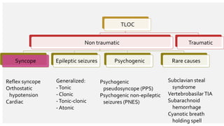
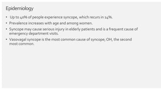
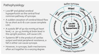
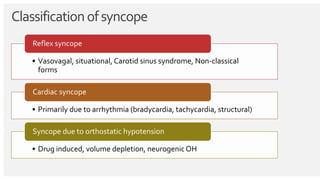
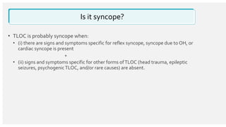
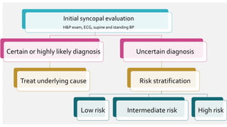
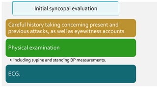
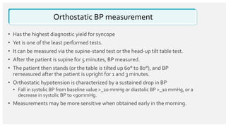
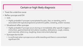
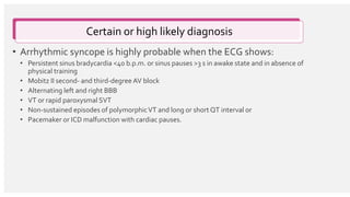
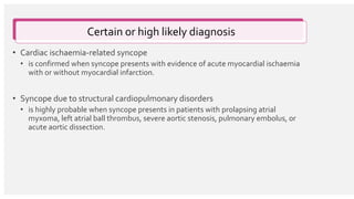
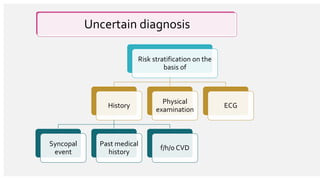
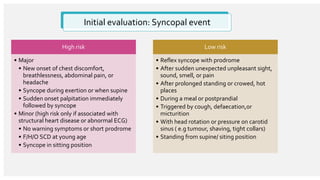
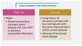
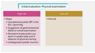
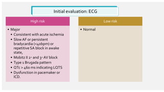
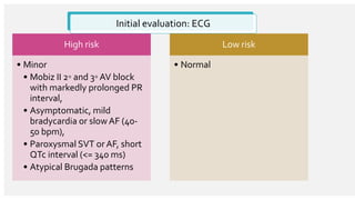
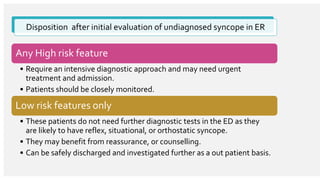
Syncope
Insp Dr Mahadev Deuja
Department of Emergency
Outlines
• Introduction
• Epidemiology
• Pathophysiology
• Classification
• Initial evaluation of patient in emergency
• Risk stratification of undiagnosed syncopal attack
• Disposition after initial evaluation of undiagnosed syncope in ER
Introduction
• Syncope :TLOC due to cerebral hypoperfusion
• Rapid onset
• Short duration
• Spontaneous complete recovery.
TLOC
Non traumatic
Syncope Epileptic seizures Psychogenic Rare causes
Traumatic
Reflex syncope
Orthostatic
hypotension
Cardiac
Generalized:
-Tonic
- Clonic
-Tonic-clonic
- Atonic
Psychogenic
pseudosyncope (PPS)
Psychogenic non-epileptic
seizures (PNES)
Subclavian steal
syndrome
VertebrobasilarTIA
Subarachnoid
hemorrhage
Cyanotic breath
holding spell
Epidemiology
• Up to 40% of people experience syncope, which recurs in 14%.
• Prevalence increases with age and among women.
• Syncope may cause serious injury in elderly patients and is a frequent cause of
emergency department visits.
• Vasovagal syncope is the most common cause of syncope; OH, the second
most common.
Pathophysiology
• Low BP and global cerebral
hypoperfusion as the central final
common pathway of syncope.
• A sudden cessation of cerebral blood flow
for as short as 6–8 s can cause complete
LOC.
• A systolic BP of 50–60 mmHg at heart
level, i.e. 30–45 mmHg at brain level in
the upright position, will cause LOC.
• Systemic BP is the product of cardiac
output and total peripheral resistance; a
fall in either can cause syncope.
• However, in syncope, both mechanisms
often act together to a varying degree.
Classificationofsyncope
• Vasovagal, situational, Carotid sinus syndrome, Non-classical
forms
Reflex syncope
• Primarily due to arrhythmia (bradycardia, tachycardia, structural)
Cardiac syncope
• Drug induced, volume depletion, neurogenic OH
Syncope due to orthostatic hypotension
Is it syncope?
• TLOC is probably syncope when:
• (i) there are signs and symptoms specific for reflex syncope, syncope due to OH, or
cardiac syncope is present
+
• (ii) signs and symptoms specific for other forms ofTLOC (head trauma, epileptic
seizures, psychogenicTLOC, and/or rare causes) are absent.
Initial syncopal evaluation
H&P exam, ECG, supine and standing BP
Certain or highly likely diagnosis
Treat underlying cause
Uncertain diagnosis
Risk stratification
Low risk Intermediate risk High risk
Initial syncopal evaluation
Careful history taking concerning present and
previous attacks, as well as eyewitness accounts
Physical examination
• Including supine and standing BP measurements.
ECG.
Orthostatic BP measurement
• Has the highest diagnostic yield for syncope
• Yet is one of the least performed tests.
• It can be measured via the supine-stand test or the head-up tilt table test.
• After the patient is supine for 5 minutes, BP measured.
• The patient then stands (or the table is tilted up 60° to 80°), and BP
remeasured after the patient is upright for 1 and 3 minutes.
• Orthostatic hypotension is characterized by a sustained drop in BP
• Fall in systolic BP from baseline value >_20 mmHg or diastolic BP >_10 mmHg, or a
decrease in systolic BP to <90mmHg.
• Measurements may be more sensitive when obtained early in the morning.
Certain or high likely diagnosis
• Treat the underline cause
• Reflex syncope and OH
• VVS
• Highly probable if syncope is precipitated by pain, fear, or standing, and is
associated with typical progressive prodrome (pallor, sweating, and/or nausea).
• Situational reflex syncope
• Highly probable if syncope occurs during or immediately after specific triggers –
micturition, gastrointestinal stimulation (swallow, defecation), cough, sneeze,
post-exercise, others (e.g. laughing, brass instrument playing)
• Syncope due to OH
• Is confirmed when syncope occurs while standing and there is concomitant
significant OH.
Certain or high likely diagnosis
• Arrhythmic syncope is highly probable when the ECG shows:
• Persistent sinus bradycardia <40 b.p.m. or sinus pauses >3 s in awake state and in absence of
physical training
• Mobitz II second- and third-degree AV block
• Alternating left and right BBB
• VT or rapid paroxysmal SVT
• Non-sustained episodes of polymorphicVT and long or short QT interval or
• Pacemaker or ICD malfunction with cardiac pauses.
• Cardiac ischaemia-related syncope
• is confirmed when syncope presents with evidence of acute myocardial ischaemia
with or without myocardial infarction.
• Syncope due to structural cardiopulmonary disorders
• is highly probable when syncope presents in patients with prolapsing atrial
myxoma, left atrial ball thrombus, severe aortic stenosis, pulmonary embolus, or
acute aortic dissection.
Certain or high likely diagnosis
Uncertain diagnosis
Risk stratification on the
basis of
History
Syncopal
event
Past medical
history
f/h/o CVD
Physical
examination
ECG
Uncertain diagnosis
• After thorough history, physical examination, ECG and lab investigation, about
half of the people remain undiagnosed syncope in ED.
Initial evaluation: Syncopal event
High risk
• Major
• New onset of chest discomfort,
breathlessness, abdominal pain, or
headache
• Syncope during exertion or when supine
• Sudden onset palpitation immediately
followed by syncope
• Minor (high risk only if associated with
structural heart disease or abnormal ECG)
• No warning symptoms or short prodrome
• F/H/O SCD at young age
• Syncope in sitting position
Low risk
• Reflex syncope with prodrome
• After sudden unexpected unpleasant sight,
sound, smell, or pain
• After prolonged standing or crowed, hot
places
• During a meal or postprandial
• Triggered by cough, defaecation,or
micturition
• With head rotation or pressure on carotid
sinus ( e.g tumour, shaving, tight collars)
• Standing from supine/ siting position
Initial evaluation: Past medical history
High risk
• Major
• Severe structural or
coronary artery
disease ( HF, low LVEF
or previous
myocardial infarction)
Low risk
• Long history of
recurrent syncope with
low risk features with
the same characteristics
of the current episode
• Absence of structural
heart disease
Initial evaluation: Physical examination
High risk
• Major
• Unexplained systolic BP in the
ED < 90mmHg
• Suggestion of gastrointestinal
bleed on rectal examination
• Persistent bradycardia (<40
bpm) in awake state and in
absence of physical training
• Undiagnosed systolic murmur
Low risk
• Normal
Initial evaluation: ECG
High risk
• Major
• Consistent with acute ischemia
• Slow AF or persistent
bradycardia (<40bpm) or
repetitive SA block in awake
state,
• Mobitz II 2◦ and 3◦ AV block
• Type 1 Brugada pattern
• QTc > 460 ms indicating LQTS
• Dysfunction in pacemaker or
ICD.
Low risk
• Normal
Initial evaluation: ECG
High risk
• Minor
• Mobiz II 2◦ and 3◦ AV block
with markedly prolonged PR
interval,
• Asymptomatic, mild
bradycardia or slowAF (40-
50 bpm),
• Paroxysmal SVT or AF, short
QTc interval (<= 340 ms)
• Atypical Brugada patterns
Low risk
• Normal
Disposition after initial evaluation of undiagnosed syncope in ER
Any High risk feature
• Require an intensive diagnostic approach and may need urgent
treatment and admission.
• Patients should be closely monitored.
Low risk features only
• These patients do not need further diagnostic tests in the ED as they
are likely to have reflex, situational, or orthostatic syncope.
• They may benefit from reassurance, or counselling.
• Can be safely discharged and investigated further as a out patient basis.
Additional examinations
• May be performed when needed
• Immediate ECG monitoring
• When there is a suspicion of arrhythmic syncope.
• Echocardiogram
• When there is previous known heart disease, data suggestive of structural heart
disease, or syncope secondary to cardiovascular cause.
• Carotid sinus massage (CSM) in patients aged >40 years.
• Head-up tilt testing when there is suspicion of syncope due to OH or reflex
syncope.
• Blood tests when clinically indicated, e.g
• Haematocrit or haemoglobin when haemorrhage is suspected
• Oxygen saturation and blood gas analysis when hypoxia is suspected
• Troponin when cardiac ischaemia-related syncope is suspected, or
• D-dimer when pulmonary embolism is suspected, etc.
• MRI, CT head, carotid artery imaging, and electroencephalography are not
recommended as part of the routine evaluation for syncope.
Additional examinations
• Syncope is a common presentation
in the emergency setting
• Diagnosing cardiac syncope is
especially important because of
high mortality risk
• Initial evaluation of all syncope
patient should include detail
history, physical examination,
supine and standing BP and ECG.
• Don’t miss life threatening causes
• hemorrhage (eg, gastrointestinal,
subarachnoid), pulmonary embolism,
and cardiac syncope from arrhythmia
or acute coronary syndrome.
Summary
Syncope:
• If you can’t reach obvious cause do
risk stratification
• Patients with low-risk features only,
likely to have reflex, situational, or
orthostatic syncope, be discharged
directly from the ED
• Patients with high-risk features
should not be discharged but receive
an intensive diagnostic evaluation in
a syncope unit or ED observation
unit, or hospitalized. Summary
Syncope:
Medical term for
fainting
Mcq’s
• Which of the following is considered a major high-risk feature associated with
syncope?
(According to the 2018 ESC syncope guidelines)
a) Syncope during exertion or when supine
b) Syncope triggered by cough, defecation, or micturition
c) Syncope after a sudden unexpected sight, sound, smell, or pain
d) Syncope with head rotation or pressure on carotid sinus
• Which of the following is considered a major high-risk ECG finding for
syncope?
• (According to the 2018 ESC syncope guidelines)
a) Mobitz I 2◦ AV block and 1 ◦ AV block with markedly prolonged PR interval
b) Pre-excited QRS complex
c) Short QTc interval (≤ 340 ms)
d) Type I Brugada pattern
Refrences
• Michele Brignole, Angel Moya, Frederik J de Lange, Jean-Claude Deharo, Perry
M Elliott, Alessandra Fanciulli, Artur Fedorowski, Raffaello Furlan, Rose Anne
Kenny, Alfonso Martín,Vincent Probst, Matthew J Reed, Ciara P Rice, Richard
Sutton, Andrea Ungar, J Gert van Dijk, ESC Scientific DocumentGroup; 2018
ESC Guidelines for the diagnosis and management of
syncope, European Heart Journal,Volume 39, Issue 21, 1 June 2018, Pages
1883–1948, https://doi.org/10.1093/eurheartj/ehy037
• emedicine.com. Accessed January 10, 2019].
page 30
ThankYou

Syncope

  • 1.
    Syncope Insp Dr MahadevDeuja Department of Emergency
  • 2.
    Outlines • Introduction • Epidemiology •Pathophysiology • Classification • Initial evaluation of patient in emergency • Risk stratification of undiagnosed syncopal attack • Disposition after initial evaluation of undiagnosed syncope in ER
  • 3.
    Introduction • Syncope :TLOCdue to cerebral hypoperfusion • Rapid onset • Short duration • Spontaneous complete recovery.
  • 4.
    TLOC Non traumatic Syncope Epilepticseizures Psychogenic Rare causes Traumatic Reflex syncope Orthostatic hypotension Cardiac Generalized: -Tonic - Clonic -Tonic-clonic - Atonic Psychogenic pseudosyncope (PPS) Psychogenic non-epileptic seizures (PNES) Subclavian steal syndrome VertebrobasilarTIA Subarachnoid hemorrhage Cyanotic breath holding spell
  • 5.
    Epidemiology • Up to40% of people experience syncope, which recurs in 14%. • Prevalence increases with age and among women. • Syncope may cause serious injury in elderly patients and is a frequent cause of emergency department visits. • Vasovagal syncope is the most common cause of syncope; OH, the second most common.
  • 6.
    Pathophysiology • Low BPand global cerebral hypoperfusion as the central final common pathway of syncope. • A sudden cessation of cerebral blood flow for as short as 6–8 s can cause complete LOC. • A systolic BP of 50–60 mmHg at heart level, i.e. 30–45 mmHg at brain level in the upright position, will cause LOC. • Systemic BP is the product of cardiac output and total peripheral resistance; a fall in either can cause syncope. • However, in syncope, both mechanisms often act together to a varying degree.
  • 7.
    Classificationofsyncope • Vasovagal, situational,Carotid sinus syndrome, Non-classical forms Reflex syncope • Primarily due to arrhythmia (bradycardia, tachycardia, structural) Cardiac syncope • Drug induced, volume depletion, neurogenic OH Syncope due to orthostatic hypotension
  • 8.
    Is it syncope? •TLOC is probably syncope when: • (i) there are signs and symptoms specific for reflex syncope, syncope due to OH, or cardiac syncope is present + • (ii) signs and symptoms specific for other forms ofTLOC (head trauma, epileptic seizures, psychogenicTLOC, and/or rare causes) are absent.
  • 9.
    Initial syncopal evaluation H&Pexam, ECG, supine and standing BP Certain or highly likely diagnosis Treat underlying cause Uncertain diagnosis Risk stratification Low risk Intermediate risk High risk
  • 10.
    Initial syncopal evaluation Carefulhistory taking concerning present and previous attacks, as well as eyewitness accounts Physical examination • Including supine and standing BP measurements. ECG.
  • 11.
    Orthostatic BP measurement •Has the highest diagnostic yield for syncope • Yet is one of the least performed tests. • It can be measured via the supine-stand test or the head-up tilt table test. • After the patient is supine for 5 minutes, BP measured. • The patient then stands (or the table is tilted up 60° to 80°), and BP remeasured after the patient is upright for 1 and 3 minutes. • Orthostatic hypotension is characterized by a sustained drop in BP • Fall in systolic BP from baseline value >_20 mmHg or diastolic BP >_10 mmHg, or a decrease in systolic BP to <90mmHg. • Measurements may be more sensitive when obtained early in the morning.
  • 12.
    Certain or highlikely diagnosis • Treat the underline cause • Reflex syncope and OH • VVS • Highly probable if syncope is precipitated by pain, fear, or standing, and is associated with typical progressive prodrome (pallor, sweating, and/or nausea). • Situational reflex syncope • Highly probable if syncope occurs during or immediately after specific triggers – micturition, gastrointestinal stimulation (swallow, defecation), cough, sneeze, post-exercise, others (e.g. laughing, brass instrument playing) • Syncope due to OH • Is confirmed when syncope occurs while standing and there is concomitant significant OH.
  • 13.
    Certain or highlikely diagnosis • Arrhythmic syncope is highly probable when the ECG shows: • Persistent sinus bradycardia <40 b.p.m. or sinus pauses >3 s in awake state and in absence of physical training • Mobitz II second- and third-degree AV block • Alternating left and right BBB • VT or rapid paroxysmal SVT • Non-sustained episodes of polymorphicVT and long or short QT interval or • Pacemaker or ICD malfunction with cardiac pauses.
  • 14.
    • Cardiac ischaemia-relatedsyncope • is confirmed when syncope presents with evidence of acute myocardial ischaemia with or without myocardial infarction. • Syncope due to structural cardiopulmonary disorders • is highly probable when syncope presents in patients with prolapsing atrial myxoma, left atrial ball thrombus, severe aortic stenosis, pulmonary embolus, or acute aortic dissection. Certain or high likely diagnosis
  • 15.
    Uncertain diagnosis Risk stratificationon the basis of History Syncopal event Past medical history f/h/o CVD Physical examination ECG
  • 16.
    Uncertain diagnosis • Afterthorough history, physical examination, ECG and lab investigation, about half of the people remain undiagnosed syncope in ED.
  • 17.
    Initial evaluation: Syncopalevent High risk • Major • New onset of chest discomfort, breathlessness, abdominal pain, or headache • Syncope during exertion or when supine • Sudden onset palpitation immediately followed by syncope • Minor (high risk only if associated with structural heart disease or abnormal ECG) • No warning symptoms or short prodrome • F/H/O SCD at young age • Syncope in sitting position Low risk • Reflex syncope with prodrome • After sudden unexpected unpleasant sight, sound, smell, or pain • After prolonged standing or crowed, hot places • During a meal or postprandial • Triggered by cough, defaecation,or micturition • With head rotation or pressure on carotid sinus ( e.g tumour, shaving, tight collars) • Standing from supine/ siting position
  • 18.
    Initial evaluation: Pastmedical history High risk • Major • Severe structural or coronary artery disease ( HF, low LVEF or previous myocardial infarction) Low risk • Long history of recurrent syncope with low risk features with the same characteristics of the current episode • Absence of structural heart disease
  • 19.
    Initial evaluation: Physicalexamination High risk • Major • Unexplained systolic BP in the ED < 90mmHg • Suggestion of gastrointestinal bleed on rectal examination • Persistent bradycardia (<40 bpm) in awake state and in absence of physical training • Undiagnosed systolic murmur Low risk • Normal
  • 20.
    Initial evaluation: ECG Highrisk • Major • Consistent with acute ischemia • Slow AF or persistent bradycardia (<40bpm) or repetitive SA block in awake state, • Mobitz II 2◦ and 3◦ AV block • Type 1 Brugada pattern • QTc > 460 ms indicating LQTS • Dysfunction in pacemaker or ICD. Low risk • Normal
  • 21.
    Initial evaluation: ECG Highrisk • Minor • Mobiz II 2◦ and 3◦ AV block with markedly prolonged PR interval, • Asymptomatic, mild bradycardia or slowAF (40- 50 bpm), • Paroxysmal SVT or AF, short QTc interval (<= 340 ms) • Atypical Brugada patterns Low risk • Normal
  • 22.
    Disposition after initialevaluation of undiagnosed syncope in ER Any High risk feature • Require an intensive diagnostic approach and may need urgent treatment and admission. • Patients should be closely monitored. Low risk features only • These patients do not need further diagnostic tests in the ED as they are likely to have reflex, situational, or orthostatic syncope. • They may benefit from reassurance, or counselling. • Can be safely discharged and investigated further as a out patient basis.
  • 23.
    Additional examinations • Maybe performed when needed • Immediate ECG monitoring • When there is a suspicion of arrhythmic syncope. • Echocardiogram • When there is previous known heart disease, data suggestive of structural heart disease, or syncope secondary to cardiovascular cause. • Carotid sinus massage (CSM) in patients aged >40 years. • Head-up tilt testing when there is suspicion of syncope due to OH or reflex syncope.
  • 24.
    • Blood testswhen clinically indicated, e.g • Haematocrit or haemoglobin when haemorrhage is suspected • Oxygen saturation and blood gas analysis when hypoxia is suspected • Troponin when cardiac ischaemia-related syncope is suspected, or • D-dimer when pulmonary embolism is suspected, etc. • MRI, CT head, carotid artery imaging, and electroencephalography are not recommended as part of the routine evaluation for syncope. Additional examinations
  • 25.
    • Syncope isa common presentation in the emergency setting • Diagnosing cardiac syncope is especially important because of high mortality risk • Initial evaluation of all syncope patient should include detail history, physical examination, supine and standing BP and ECG. • Don’t miss life threatening causes • hemorrhage (eg, gastrointestinal, subarachnoid), pulmonary embolism, and cardiac syncope from arrhythmia or acute coronary syndrome. Summary Syncope:
  • 26.
    • If youcan’t reach obvious cause do risk stratification • Patients with low-risk features only, likely to have reflex, situational, or orthostatic syncope, be discharged directly from the ED • Patients with high-risk features should not be discharged but receive an intensive diagnostic evaluation in a syncope unit or ED observation unit, or hospitalized. Summary Syncope: Medical term for fainting
  • 27.
    Mcq’s • Which ofthe following is considered a major high-risk feature associated with syncope? (According to the 2018 ESC syncope guidelines) a) Syncope during exertion or when supine b) Syncope triggered by cough, defecation, or micturition c) Syncope after a sudden unexpected sight, sound, smell, or pain d) Syncope with head rotation or pressure on carotid sinus
  • 28.
    • Which ofthe following is considered a major high-risk ECG finding for syncope? • (According to the 2018 ESC syncope guidelines) a) Mobitz I 2◦ AV block and 1 ◦ AV block with markedly prolonged PR interval b) Pre-excited QRS complex c) Short QTc interval (≤ 340 ms) d) Type I Brugada pattern
  • 29.
    Refrences • Michele Brignole,Angel Moya, Frederik J de Lange, Jean-Claude Deharo, Perry M Elliott, Alessandra Fanciulli, Artur Fedorowski, Raffaello Furlan, Rose Anne Kenny, Alfonso Martín,Vincent Probst, Matthew J Reed, Ciara P Rice, Richard Sutton, Andrea Ungar, J Gert van Dijk, ESC Scientific DocumentGroup; 2018 ESC Guidelines for the diagnosis and management of syncope, European Heart Journal,Volume 39, Issue 21, 1 June 2018, Pages 1883–1948, https://doi.org/10.1093/eurheartj/ehy037 • emedicine.com. Accessed January 10, 2019].
  • 30.