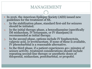
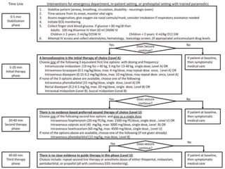
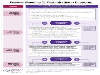
Dr. Kamran Khan
PG Medicine
Year 1
STATUS EPILEPTICUS
DEFINITIONS
 A condition characterized by epileptic seizures that
are sufficiently prolonged or repeated at sufficiently
brief intervals so as to produce and unvarying and
enduring epileptic condition. (WHO 1973)
 A seizure that persists for a sufficient length of
time or is repeated frequently enough that recovery
between attack doesn’t occur. (ILAE Commission
2001)
 Status epilepticus is a condition resulting either from
the failure of the mechanisms responsible for seizure
termination or from the initiation of mechanisms,
which lead to abnormally, prolonged seizures (after
time point t1). It is a condition, which can have long-
term consequences (after time point t2), including
neuronal death, neuronal injury, and alteration of
neuronal networks, depending on the type and
duration of seizures.
(ILAE 2015)
CLASSIFICATION OF STATUS EPILEPTICUS
 Generalized convulsive status epilepticus
 Tonic Clonic Status epilepticus
 Clonic Status epilepticus
 Tonic Status epilepticus
 Myoclonic Status epilepticus
 Generalized non convulsive Status epilepticus
 Absence Status epilepticus
CLASSIFICATION OF STATUS EPILEPTICUS
 Simple partial (focal) Status epilepticus
 Complex partial Status epilepticus
PATHOPHYSIOLOGY
 On a neurochemical level, seizures are sustained by
excess excitation and reduced inhibition.
 Glutamate is the most common excitatory
neurotransmitter and the NMDA (N-methyl-D-
aspartate) receptor subtype is involved.
 Gamma-aminobutyric acid (GABA) is the most
common inhibitory neurotransmitter. Failure of
inhibitory processes is increasingly thought to be the
major mechanism leading to status epilepticus.
 However, changes in gene expression that are
induced by SE result in alterations in the number or
subunit composition of ion channels, receptors, cell
metabolism, and neuronal connectivity.
 Neuronal death probably results from the inability to
handle large increases in intracellular calcium
brought about by prolonged exposure to excitatory
neurotransmitters.
 More prolong the status, the poorer the prognosis
 Neuronal Death occurs after 30 mins of continuous
seizures.
 High mortality related to
 Rhabdomyolysis
 Aspiration
 Metabolic/lactic acidosis
 Respiratory failure
 Neurogenic pulmonary edema
 Myocardial injury
ETIOLOGY
 In adults, the most common precipitant was
cerebrovascular disease (25%), whereas this factor
caused only 3% of pediatric cases.
 SE vary significantly with age. DeLorenzo et al
reported that in patients younger than 16 years, the
most common cause was fever and/or infection
(36%); in contrast, this accounted for only 5% of SE
in adults.
 Only 25% of people who experience seizures or status
epilepticus have epilepsy.
 Stroke (remote or acute)
 Hypoxic injury
 Tumor
 Subarachnoid hemorrhage
 Head trauma
 Drugs eg, cocaine, theophylline, isoniazid (INH) may
cause seizures and is unique in having a specific
antidote, pyridoxine (vitamin B-6),
Flouroquinolones, Flumazenil
 Alcohol withdrawal
 Electrolyte abnormalities
(eg, hyponatremia, hypernatremia,hypoglycemia,
hyperglycemia, hypocalcemia,hepatic
encephalopathy, Uremia)
 Neoplasms
 CNS infections (eg, meningitis, brain abscess,
encephalitis)
 Toxins, notably sympathomimetics
PRESENTATION
 The event usually begins with a series of generalized
tonic, clonic, or tonic-clonic seizures that often are
dramatic.
 If the condition is not treated or is treated inadequately,
SE persists, and the motor manifestations become less
dramatic than before. Eventually, only subtle movements
(eg, nystagmoid jerks of the eyes or twitching of the
shoulder) may be seen—that is, subtle SE. If SE
continues, all motor activity may stop, though EEG
seizures persist (ie, electrical generalized convulsive SE).

 Among patients with established epilepsy,
noncompliance with medications is the rule rather
than the exception. In roughly one third of cases,
status epilepticus is the initial presentation of a
seizure disorder.
SIMPLE PARTIAL STATUS EPILEPTICUS
 By clinical history, nonmotor simple partial SE
involves subjective sensory disturbances, including
the following:
 Focal or unilateral paresthesias or numbness
 Focal visual changes, usually characterized by
flashing lights
 Focal visual obscuration or focal colorful
hallucinations
 Olfactory or gustatory hallucinations
 Atypical rising abdominal sensations
COMPLEX PARTIAL STATUS EPILEPTICUS
 Complex partial SE often begins with a history of
recurrent or prolonged simple partial seizures, or it
may follow or precede a generalized convulsive
seizure. Patients are often confused and have
variable responsiveness. Memory of the event is
usually impaired. Behavior may fluctuate or be
bizarre.
 Many patients have clinical automatisms, as with
typical complex partial seizures, including repetitive
lip-smacking, fumbling, or swallowing movements.
Subtle nystagmus may be observed.
EXAMINATION FINDINGS
 Needle track marks might suggest SE secondary to
the use of illicit, or street, drugs
 Papilledema, a sign of increased intracranial
pressure, suggests a possible mass lesion or brain
infection
 Associated injuries that may be present in patients
with seizures include tongue lacerations (typically
lateral), shoulder dislocations, head trauma, and
facial trauma.
DIFFERENTIAL DIAGNOSES
 Encephalitis
 Heatstroke
 Hypernatremia in Emergency Medicine
 Hyperosmolar Hyperglycemic Nonketotic Coma
 Hypocalcemia in Emergency Medicine
 Hypoglycemia
 Hyponatremia
 Medication-Induced Dystonic Reactions
 Neuroleptic Malignant Syndrome
 Neurological Manifestations of Uremic Encephalopathy
 Withdrawal Syndromes
LABORATORY STUDIES
 Laboratory studies that should be obtained on an
emergency basis include the following:
 Electrolytes
 Calcium
 Magnesium
 Glucose
 Complete blood count
 Renal function tests
 Toxicologic screening
 Anticonvulsant levels
 Liver function tests
 EEG
 ABGs
 CT Scan
 MRI
 Chest Radiography
 Lumbar Puncture
MANAGEMENT
 In 2016, the American Epilepsy Society (AES) issued new
guidelines for the treatment of SE.
1. In the stabilization phase, standard first-aid for seizures
should be initiated.
2. In the initial therapy phase, a benzodiazepine (specifically
IM midazolam, IV lorazepam, or IV diazepam) is
recommended as initial therapy.
3. In the second phase, options include IV fosphenytoin,
valproic acid, or levetiracetam. If none of these is available,
IV phenobarbital is a reasonable alternative.
4. In the third phase, if a patient experiences 40+ minutes of
seizure activity, treatment considerations should include
repeating second-line therapy or anesthetic doses of
thiopental, midazolam, pentobarbital, or propofol.
Status Epilepticus
Status Epilepticus
Status Epilepticus

Status Epilepticus

  • 1.
    Dr. Kamran Khan PGMedicine Year 1 STATUS EPILEPTICUS
  • 2.
    DEFINITIONS  A conditioncharacterized by epileptic seizures that are sufficiently prolonged or repeated at sufficiently brief intervals so as to produce and unvarying and enduring epileptic condition. (WHO 1973)  A seizure that persists for a sufficient length of time or is repeated frequently enough that recovery between attack doesn’t occur. (ILAE Commission 2001)
  • 3.
     Status epilepticusis a condition resulting either from the failure of the mechanisms responsible for seizure termination or from the initiation of mechanisms, which lead to abnormally, prolonged seizures (after time point t1). It is a condition, which can have long- term consequences (after time point t2), including neuronal death, neuronal injury, and alteration of neuronal networks, depending on the type and duration of seizures. (ILAE 2015)
  • 4.
    CLASSIFICATION OF STATUSEPILEPTICUS  Generalized convulsive status epilepticus  Tonic Clonic Status epilepticus  Clonic Status epilepticus  Tonic Status epilepticus  Myoclonic Status epilepticus  Generalized non convulsive Status epilepticus  Absence Status epilepticus
  • 5.
    CLASSIFICATION OF STATUSEPILEPTICUS  Simple partial (focal) Status epilepticus  Complex partial Status epilepticus
  • 6.
    PATHOPHYSIOLOGY  On aneurochemical level, seizures are sustained by excess excitation and reduced inhibition.  Glutamate is the most common excitatory neurotransmitter and the NMDA (N-methyl-D- aspartate) receptor subtype is involved.
  • 7.
     Gamma-aminobutyric acid(GABA) is the most common inhibitory neurotransmitter. Failure of inhibitory processes is increasingly thought to be the major mechanism leading to status epilepticus.  However, changes in gene expression that are induced by SE result in alterations in the number or subunit composition of ion channels, receptors, cell metabolism, and neuronal connectivity.
  • 8.
     Neuronal deathprobably results from the inability to handle large increases in intracellular calcium brought about by prolonged exposure to excitatory neurotransmitters.  More prolong the status, the poorer the prognosis
  • 9.
     Neuronal Deathoccurs after 30 mins of continuous seizures.  High mortality related to  Rhabdomyolysis  Aspiration  Metabolic/lactic acidosis  Respiratory failure  Neurogenic pulmonary edema  Myocardial injury
  • 10.
    ETIOLOGY  In adults,the most common precipitant was cerebrovascular disease (25%), whereas this factor caused only 3% of pediatric cases.  SE vary significantly with age. DeLorenzo et al reported that in patients younger than 16 years, the most common cause was fever and/or infection (36%); in contrast, this accounted for only 5% of SE in adults.  Only 25% of people who experience seizures or status epilepticus have epilepsy.
  • 11.
     Stroke (remoteor acute)  Hypoxic injury  Tumor  Subarachnoid hemorrhage  Head trauma  Drugs eg, cocaine, theophylline, isoniazid (INH) may cause seizures and is unique in having a specific antidote, pyridoxine (vitamin B-6), Flouroquinolones, Flumazenil
  • 12.
     Alcohol withdrawal Electrolyte abnormalities (eg, hyponatremia, hypernatremia,hypoglycemia, hyperglycemia, hypocalcemia,hepatic encephalopathy, Uremia)  Neoplasms  CNS infections (eg, meningitis, brain abscess, encephalitis)  Toxins, notably sympathomimetics
  • 13.
    PRESENTATION  The eventusually begins with a series of generalized tonic, clonic, or tonic-clonic seizures that often are dramatic.  If the condition is not treated or is treated inadequately, SE persists, and the motor manifestations become less dramatic than before. Eventually, only subtle movements (eg, nystagmoid jerks of the eyes or twitching of the shoulder) may be seen—that is, subtle SE. If SE continues, all motor activity may stop, though EEG seizures persist (ie, electrical generalized convulsive SE). 
  • 14.
     Among patientswith established epilepsy, noncompliance with medications is the rule rather than the exception. In roughly one third of cases, status epilepticus is the initial presentation of a seizure disorder.
  • 15.
    SIMPLE PARTIAL STATUSEPILEPTICUS  By clinical history, nonmotor simple partial SE involves subjective sensory disturbances, including the following:  Focal or unilateral paresthesias or numbness  Focal visual changes, usually characterized by flashing lights  Focal visual obscuration or focal colorful hallucinations  Olfactory or gustatory hallucinations  Atypical rising abdominal sensations
  • 16.
    COMPLEX PARTIAL STATUSEPILEPTICUS  Complex partial SE often begins with a history of recurrent or prolonged simple partial seizures, or it may follow or precede a generalized convulsive seizure. Patients are often confused and have variable responsiveness. Memory of the event is usually impaired. Behavior may fluctuate or be bizarre.  Many patients have clinical automatisms, as with typical complex partial seizures, including repetitive lip-smacking, fumbling, or swallowing movements. Subtle nystagmus may be observed.
  • 17.
    EXAMINATION FINDINGS  Needletrack marks might suggest SE secondary to the use of illicit, or street, drugs  Papilledema, a sign of increased intracranial pressure, suggests a possible mass lesion or brain infection  Associated injuries that may be present in patients with seizures include tongue lacerations (typically lateral), shoulder dislocations, head trauma, and facial trauma.
  • 18.
    DIFFERENTIAL DIAGNOSES  Encephalitis Heatstroke  Hypernatremia in Emergency Medicine  Hyperosmolar Hyperglycemic Nonketotic Coma  Hypocalcemia in Emergency Medicine  Hypoglycemia  Hyponatremia  Medication-Induced Dystonic Reactions  Neuroleptic Malignant Syndrome  Neurological Manifestations of Uremic Encephalopathy  Withdrawal Syndromes
  • 19.
    LABORATORY STUDIES  Laboratorystudies that should be obtained on an emergency basis include the following:  Electrolytes  Calcium  Magnesium  Glucose  Complete blood count  Renal function tests  Toxicologic screening  Anticonvulsant levels  Liver function tests
  • 20.
     EEG  ABGs CT Scan  MRI  Chest Radiography  Lumbar Puncture
  • 21.
    MANAGEMENT  In 2016,the American Epilepsy Society (AES) issued new guidelines for the treatment of SE. 1. In the stabilization phase, standard first-aid for seizures should be initiated. 2. In the initial therapy phase, a benzodiazepine (specifically IM midazolam, IV lorazepam, or IV diazepam) is recommended as initial therapy. 3. In the second phase, options include IV fosphenytoin, valproic acid, or levetiracetam. If none of these is available, IV phenobarbital is a reasonable alternative. 4. In the third phase, if a patient experiences 40+ minutes of seizure activity, treatment considerations should include repeating second-line therapy or anesthetic doses of thiopental, midazolam, pentobarbital, or propofol.