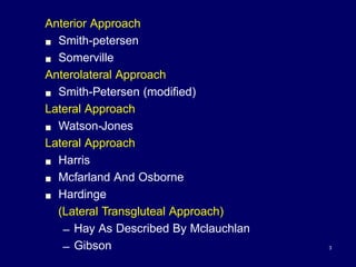
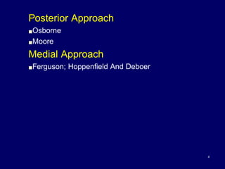
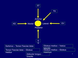
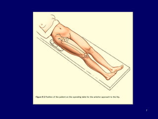
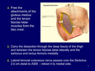
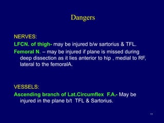
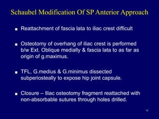
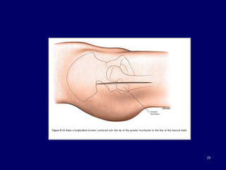
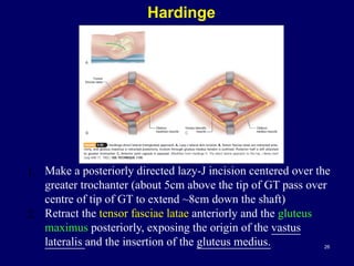
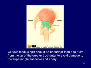
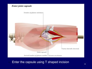
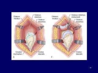
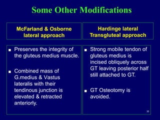
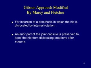
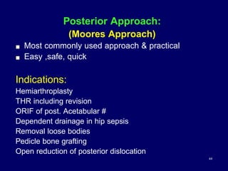
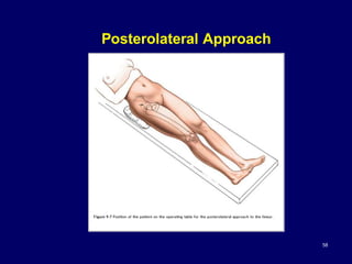
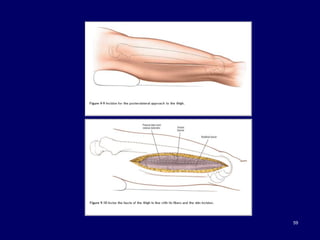
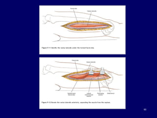
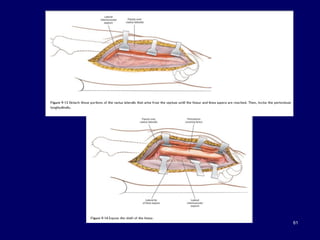
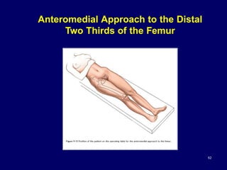
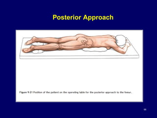
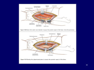
The document describes various surgical approaches to the hip, including anterior, anterolateral, lateral, posterior, and medial approaches. The anterior approach, such as the Smith-Petersen approach, provides access to the front of the hip joint. The lateral approach, including the Hardinge and Watson-Jones approaches, avoids the need for trochanteric osteotomy. The posterior, or Moore's, approach is the most commonly used and provides exposure for procedures like hip replacements. Each approach has specific indications and technical steps described. Potential dangers to nerves and blood vessels are also outlined.
OSTEOLYSIS AND LOOSENING OF total hip arthroplasty IMPLANTS.pptx by dr vasu ...Vasu Srivastava
Aseptic Loosening of implants is caused by osteolysis. It is most significant factor limiting longevity of THA. Revision for loosening is 4x higher than next leading cause (dislocation at 13.6%), and its particularly problematic in younger patients [2].
Osteolysis is bone resorption caused by the body’s response to particulate debris generated as the THA implant wears out. Motion between any two components of the prosthesis (ie the femoral head and the acetabuluar liner, the head-neck junction of the femoral stem, or the liner and shell of the acetabulum) generates debris that floats around the joint. This debris stimulates a host response. Particles of metal, poly, or cement can all cause osteolysis, albeit different types of reaction. Osteolysis is important because it leads to implant loosening and/or periprosthetic fractures.
While osteolysis is the primary cause of loosening, infection must be part of the differential diagnosis.
Historical Perspective: Osteolysis was first described by Harris in 1976 and it was attributed to “cement disease” [3], because it was observed around the femoral component, and this was what started the drive for cementless implants. Yet after significant R&D, and development of cementless implants, osteolysis was still seen around the implants [4], and the histology was similar between cemented [5] and cementless implants [6]. Surgeons then looked for another cause of osteolysis and recognized that it was produced by wear particles.
STAGES OF OSTEOLYSIS
1) Debris production (ie poly wear) is the initial stage (we talk about metal debris in a separate section because it behaves totally differently, see section). Particulate debris in THA is produced by Abrasive and Adhesive wear (whereas the TKA produces delaminating wear: small fissures form within the poly).
▪ Adhesive wear is two surfaces bonding together causing the softer material to “peel” off as a thin film onto the harder surface during motion.
Volumetric wear is a specific type of adhesive wear, and it occurs as the femoral head articulates with the cup liner, and the amount of wear is proportional to the femoral head radius squared (therefore larger femoral head = more wear..this is why the initial Charnley implants, which used conventional poly, used a size 22 femoral head). Linear wear is caused by focused stress on a isolated part of the poly due to abnormal loading.
▪ Abrasive wear occurs when a harder surface (which is never completely smooth) cuts or ploughs through a softer surface, like a cheese grater. Both cause particle formation. Most wear occurs superiorly in the cup (or at the rim in cases of impingement).
The conventional PE wear from articulating with a Cobalt-chrome head is 0.10 mm/year. The ultramolecular weight poly (UMWPE, also known as highly-crosslinked poly) wear is about 0.02 mm/year. What is the difference between conventional and UMWPE?
The majority of elderly patients who receive a hip replacement retain the prosthesis for 15 to 20 years, and sometimes for life. However, some patients may need one or more revisions of a hip replacement, particularly if the initial hip replacement surgery is performed at a young age and the patient chooses to have a very active physical lifestyle.
this ppt provides a comprehensive review & exam oriented details
compiled from journals & old edition textbooks. because ITB contracture has become a rare presentation. & new edition books doesnt speak about it much...
Health Education on prevention of hypertensionRadhika kulvi
Hypertension is a chronic condition of concern due to its role in the causation of coronary heart diseases. Hypertension is a worldwide epidemic and important risk factor for coronary artery disease, stroke and renal diseases. Blood pressure is the force exerted by the blood against the walls of the blood vessels and is sufficient to maintain tissue perfusion during activity and rest. Hypertension is sustained elevation of BP. In adults, HTN exists when systolic blood pressure is equal to or greater than 140mmHg or diastolic BP is equal to or greater than 90mmHg. The
Explore our infographic on 'Essential Metrics for Palliative Care Management' which highlights key performance indicators crucial for enhancing the quality and efficiency of palliative care services.
This visual guide breaks down important metrics across four categories: Patient-Centered Metrics, Care Efficiency Metrics, Quality of Life Metrics, and Staff Metrics. Each section is designed to help healthcare professionals monitor and improve care delivery for patients facing serious illnesses. Understand how to implement these metrics in your palliative care practices for better outcomes and higher satisfaction levels.
CHAPTER 1 SEMESTER V - ROLE OF PEADIATRIC NURSE.pdfSachin Sharma
Pediatric nurses play a vital role in the health and well-being of children. Their responsibilities are wide-ranging, and their objectives can be categorized into several key areas:
1. Direct Patient Care:
Objective: Provide comprehensive and compassionate care to infants, children, and adolescents in various healthcare settings (hospitals, clinics, etc.).
This includes tasks like:
Monitoring vital signs and physical condition.
Administering medications and treatments.
Performing procedures as directed by doctors.
Assisting with daily living activities (bathing, feeding).
Providing emotional support and pain management.
2. Health Promotion and Education:
Objective: Promote healthy behaviors and educate children, families, and communities about preventive healthcare.
This includes tasks like:
Administering vaccinations.
Providing education on nutrition, hygiene, and development.
Offering breastfeeding and childbirth support.
Counseling families on safety and injury prevention.
3. Collaboration and Advocacy:
Objective: Collaborate effectively with doctors, social workers, therapists, and other healthcare professionals to ensure coordinated care for children.
Objective: Advocate for the rights and best interests of their patients, especially when children cannot speak for themselves.
This includes tasks like:
Communicating effectively with healthcare teams.
Identifying and addressing potential risks to child welfare.
Educating families about their child's condition and treatment options.
4. Professional Development and Research:
Objective: Stay up-to-date on the latest advancements in pediatric healthcare through continuing education and research.
Objective: Contribute to improving the quality of care for children by participating in research initiatives.
This includes tasks like:
Attending workshops and conferences on pediatric nursing.
Participating in clinical trials related to child health.
Implementing evidence-based practices into their daily routines.
By fulfilling these objectives, pediatric nurses play a crucial role in ensuring the optimal health and well-being of children throughout all stages of their development.
Deep Leg Vein Thrombosis (DVT): Meaning, Causes, Symptoms, Treatment, and Mor...The Lifesciences Magazine
Deep Leg Vein Thrombosis occurs when a blood clot forms in one or more of the deep veins in the legs. These clots can impede blood flow, leading to severe complications.
Medical Technology Tackles New Health Care Demand - Research Report - March 2...pchutichetpong
M Capital Group (“MCG”) predicts that with, against, despite, and even without the global pandemic, the medical technology (MedTech) industry shows signs of continuous healthy growth, driven by smaller, faster, and cheaper devices, growing demand for home-based applications, technological innovation, strategic acquisitions, investments, and SPAC listings. MCG predicts that this should reflects itself in annual growth of over 6%, well beyond 2028.
According to Chris Mouchabhani, Managing Partner at M Capital Group, “Despite all economic scenarios that one may consider, beyond overall economic shocks, medical technology should remain one of the most promising and robust sectors over the short to medium term and well beyond 2028.”
There is a movement towards home-based care for the elderly, next generation scanning and MRI devices, wearable technology, artificial intelligence incorporation, and online connectivity. Experts also see a focus on predictive, preventive, personalized, participatory, and precision medicine, with rising levels of integration of home care and technological innovation.
The average cost of treatment has been rising across the board, creating additional financial burdens to governments, healthcare providers and insurance companies. According to MCG, cost-per-inpatient-stay in the United States alone rose on average annually by over 13% between 2014 to 2021, leading MedTech to focus research efforts on optimized medical equipment at lower price points, whilst emphasizing portability and ease of use. Namely, 46% of the 1,008 medical technology companies in the 2021 MedTech Innovator (“MTI”) database are focusing on prevention, wellness, detection, or diagnosis, signaling a clear push for preventive care to also tackle costs.
In addition, there has also been a lasting impact on consumer and medical demand for home care, supported by the pandemic. Lockdowns, closure of care facilities, and healthcare systems subjected to capacity pressure, accelerated demand away from traditional inpatient care. Now, outpatient care solutions are driving industry production, with nearly 70% of recent diagnostics start-up companies producing products in areas such as ambulatory clinics, at-home care, and self-administered diagnostics.
Telehealth Psychology Building Trust with Clients.pptxThe Harvest Clinic
Telehealth psychology is a digital approach that offers psychological services and mental health care to clients remotely, using technologies like video conferencing, phone calls, text messaging, and mobile apps for communication.
India Clinical Trials Market: Industry Size and Growth Trends [2030] Analyzed...Kumar Satyam
According to TechSci Research report, "India Clinical Trials Market- By Region, Competition, Forecast & Opportunities, 2030F," the India Clinical Trials Market was valued at USD 2.05 billion in 2024 and is projected to grow at a compound annual growth rate (CAGR) of 8.64% through 2030. The market is driven by a variety of factors, making India an attractive destination for pharmaceutical companies and researchers. India's vast and diverse patient population, cost-effective operational environment, and a large pool of skilled medical professionals contribute significantly to the market's growth. Additionally, increasing government support in streamlining regulations and the growing prevalence of lifestyle diseases further propel the clinical trials market.
Growing Prevalence of Lifestyle Diseases
The rising incidence of lifestyle diseases such as diabetes, cardiovascular diseases, and cancer is a major trend driving the clinical trials market in India. These conditions necessitate the development and testing of new treatment methods, creating a robust demand for clinical trials. The increasing burden of these diseases highlights the need for innovative therapies and underscores the importance of India as a key player in global clinical research.
Defecation
Normal defecation begins with movement in the left colon, moving stool toward the anus. When stool reaches the rectum, the distention causes relaxation of the internal sphincter and an awareness of the need to defecate. At the time of defecation, the external sphincter relaxes, and abdominal muscles contract, increasing intrarectal pressure and forcing the stool out
The Valsalva maneuver exerts pressure to expel faeces through a voluntary contraction of the abdominal muscles while maintaining forced expiration against a closed airway. Patients with cardiovascular disease, glaucoma, increased intracranial pressure, or a new surgical wound are at greater risk for cardiac dysrhythmias and elevated blood pressure with the Valsalva maneuver and need to avoid straining to pass the stool.
Normal defecation is painless, resulting in passage of soft, formed stool
CONSTIPATION
Constipation is a symptom, not a disease. Improper diet, reduced fluid intake, lack of exercise, and certain medications can cause constipation. For example, patients receiving opiates for pain after surgery often require a stool softener or laxative to prevent constipation. The signs of constipation include infrequent bowel movements (less than every 3 days), difficulty passing stools, excessive straining, inability to defecate at will, and hard feaces
IMPACTION
Fecal impaction results from unrelieved constipation. It is a collection of hardened feces wedged in the rectum that a person cannot expel. In cases of severe impaction the mass extends up into the sigmoid colon.
DIARRHEA
Diarrhea is an increase in the number of stools and the passage of liquid, unformed feces. It is associated with disorders affecting digestion, absorption, and secretion in the GI tract. Intestinal contents pass through the small and large intestine too quickly to allow for the usual absorption of fluid and nutrients. Irritation within the colon results in increased mucus secretion. As a result, feces become watery, and the patient is unable to control the urge to defecate. Normally an anal bag is safe and effective in long-term treatment of patients with fecal incontinence at home, in hospice, or in the hospital. Fecal incontinence is expensive and a potentially dangerous condition in terms of contamination and risk of skin ulceration
HEMORRHOIDS
Hemorrhoids are dilated, engorged veins in the lining of the rectum. They are either external or internal.
FLATULENCE
As gas accumulates in the lumen of the intestines, the bowel wall stretches and distends (flatulence). It is a common cause of abdominal fullness, pain, and cramping. Normally intestinal gas escapes through the mouth (belching) or the anus (passing of flatus)
FECAL INCONTINENCE
Fecal incontinence is the inability to control passage of feces and gas from the anus. Incontinence harms a patient’s body image
PREPARATION AND GIVING OF LAXATIVESACCORDING TO POTTER AND PERRY,
An enema is the instillation of a solution into the rectum and sig
The dimensions of healthcare quality refer to various attributes or aspects that define the standard of healthcare services. These dimensions are used to evaluate, measure, and improve the quality of care provided to patients. A comprehensive understanding of these dimensions ensures that healthcare systems can address various aspects of patient care effectively and holistically. Dimensions of Healthcare Quality and Performance of care include the following; Appropriateness, Availability, Competence, Continuity, Effectiveness, Efficiency, Efficacy, Prevention, Respect and Care, Safety as well as Timeliness.
6. Anterior Iliofemoral Approach
(Smith Peterson)
! Gives safe access to hip & ilium
Indications:
1. Open reduction of congenital dislocations when
dislocated femoral head is anterosuperior to
acetabulum
2. Synovial biopsies
3. Intra articular fusions
4. THR
5. Hemiarthroplasty
6. Excision of tumors (pelvis)
7. Pelvic osteotomies using upper part of approach
6
8. 1. Begin the
incision at the
middle of the iliac
crest
2. Carry it anteriorly
to ASIS and
distally and
slightly laterally
10 to 12 cm
8
3. Divide the superficial and deep fasciae
9. 9
4. Free the
attachments of the
gluteus medius
and the tensor
fasciae latae
muscles from the
iliac crest.
5. Carry the dissection through the deep fascia of the thigh
and between the tensor fasciae latae laterally and the
sartorius and rectus femoris medially
6. Lateral femoral cutaneous nerve passes over the Sartorius
2.5 cm distal to ASIS ; retract it to medial side.
10. 10
7. Expose and incise
the capsule
transversely and
reveal the femoral
head and proximal
margin of
acetabulum.
8. Capsule may be sectioned along its attachment to the
acetabular labrum (cotyloid ligament) to give the required
exposure.
9. If necessary, the ligamentum teres may be divided with a
curved knife or with scissors.
11. Dangers
NERVES:
LFCN. of thigh- may be injured b/w sartorius & TFL.
Femoral N. – may be injured if plane is missed during
deep dissection as it lies anterior to hip , medial to RF,
lateral to the femoralA.
VESSELS:
Ascending branch of Lat.Circumflex F.A.- May be
injured in the plane b/t TFL & Sartorius.
11
12. ! Reattachment of fascia lata to iliac crest difficult
! Osteotomy of overhang of iliac crest is performed
b/w Ext. Oblique medially & fascia lata to as far as
origin of g.maximus.
! TFL, G.medius & G.minimus dissected
subperiosteally to expose hip joint capsule.
! Closure – Iliac osteotomy fragment reattached with
non-absorbable sutures through holes drilled.
Schaubel Modification Of SP Anterior Approach
12
13. 13
Somerville
•Transverse “bikini” incision for irreducible congenital
dislocation of the hip
•sequential steps must be performed:
1. psoas tenotomy,
2. Complete medial capsulotomy including the
transverse acetabular ligament,
3. excision of hypertrophied ligamentum teres,
4. reduction of the femoral head into acetabulum.
14. 1. Make a straight skin incision, beginning anteriorly
inferiorly and medial to ASIS and coursing obliquely
superiorly and posteriorly to the middle of iliac crest
2. Reflect the abductor muscles subperiosteally from
the iliac wing distally to the capsule of the joint.
14
15. 15
3. Increase exposure of the capsule by separating
the tensor fasciae latae from the sartorius for
about 2.5 cm inferior to the anterior superior spine.
16. Anterolateral Approach:
( Watson-Jones )
! Most commonly used for THR
! Abductor mechanism released either by trochanteric
osteotomy / by cutting the ant.part of GL.medius & the
whole Gl. minimus of the G.T
Indications:
1. THR
2. ORIF of fracture NOF
3. Hemiarthroplasty
4. Synovial biopsy
5. Biopsy Femoral N.
16
22. ! Deep surgical dissection consists in detaching part or all
of the abductor mechanism and dissecting up the
femoral neck superficial to the capsule.
! Incise the capsule of the joint longitudinally along the
anterosuperior surface of the femoral neck.
22
23. ! Nerve
! Femoral N - Placing retractors into substance of
iliopsoas Or overexuberant retraction can
damage it.
! Vessels
! Femoral Artery & Vein – damaged by
acetabular retractors that penetrate iliopsoas
substance.
! Profunda Femoris Artery
Dangers
23
24. Lateral Approach To Hip
! Excellent approach to hip replacement.
! No need for trochanteric osteotomy.
! Early mobilisation of pt possible as the Gl.medius is
preserved.
! But not a wider approach as anterolateral approach.
Position:
Supine with GT at the edge of the table.
24
26. Hardinge
26
1. Make a posteriorly directed lazy-J incision centered over the
greater trochanter (about 5cm above the tip of GT pass over
centre of tip of GT to extend ~8cm down the shaft)
2. Retract the tensor fasciae latae anteriorly and the gluteus
maximus posteriorly, exposing the origin of the vastus
lateralis and the insertion of the gluteus medius.
27. ! Incise the tendon of the gluteus medius obliquely
across the greater trochanter, leaving the posterior half
still attached to the trochanter
! Split the GL. Medius starting in the middle of GT. 27
28. 28
Gluteus medius split should be no farther than 4 to 5 cm
from the tip of the greater trochanter to avoid damage to
the superior gluteal nerve and artery
29. ! Elevate the tendinous insertions of the anterior
portions of the gluteus minimus and vastus lateralis
muscles.
! Abduction of the thigh exposes the anterior capsule
of the hip joint.
! Incise the capsule as desired.
! During closure, repair the tendon of the gluteus
medius with nonabsorbable braided sutures
29
30. • Enter the capsule using T shaped incision 30
31. Dangers:
Nerves:
! Sup.GL.N. damage at the upper end of incision above
GT.
! Prevented by stay suture in the GL. Med
! Femoral N. damaged by inadvertly placed retraction
! Prevented by placing retractor strictly on the bone.
Vessels:
! Fem. Vessels by retractor
31
32. Harris Approach
! Lateral approach for extensive exposure of the hip.
! Permits hip dislocation ant & post.
! But requires GT osteotomy.
So risks are - Trochanteric non-union,
Trochanteric bursitis,
Heterotopic ossification
32
35. Some Other Modifications
McFarland & Osborne
lateral approach
! Preserves the integrity of
the gluteus medius muscle.
! Combined mass of
G.medius & Vastus
lateralis with their
tendinous junction is
elevated & retracted
anteriorly.
Hardinge lateral
Transgluteal approach
! Strong mobile tendon of
gluteus medius is
incised obliquely across
GT leaving posterior half
still attached to GT.
! GT Osteotomy is
avoided.
35
38. GIBSON MODIFIED Kocher and Langenbeck
incision making it more anterior but still angled.
! Iliotibial band is incised along with its fibres, gluteus
medius & minimus are divided at their insertions
leaving enough tendon attached.
! So, closure is easy & post-op rehabilitation is rapid
Posterolateral approach
38
39. 39
1. Begin the proximal limb of the incision at a point 6 to
8 cm anterior to the posterior superior iliac spine and
just distal to the iliac crest 15 to 18 cm, overlying the
anterior border of the gluteus maximus muscle.
2. Incise the iliotibial band in line with its fibers
3. Separate the posterior border of the gluteus medius
muscle from the adjacent piriformis tendon by blunt
dissection.
40. 40
4. Divide the gluteus medius and
minimus muscles at their
insertions, but leave enough of
their tendons attached to
greater trochanter.
5. Incise the capsule superiorly in
the axis of the femoral neck
from the acetabulum to the
intertrochanteric line
41. 41
6. The hip now can be dislocated by flexing the hip and knee
and abducting and externally rotating the thigh
7. To preserve the insertion of the abductor muscles,
osteotomize the trochanter and later reattach it with two
wire loops, 6.5-mm lag screws, or cable grip.
8. Wire loops are passed through the insertion of the muscles
proximal to the trochanter and through a hole drilled in the
femoral shaft 4 cm distal to the osteotomy.
43. Gibson Approach Modified
By Marcy and Fletcher
! For insertion of a prosthesis in which the hip is
dislocated by internal rotation.
! Anterior part of the joint capsule is preserved to
keep the hip from dislocating anteriorly after
surgery.
43
44. Posterior Approach:
(Moores Approach)
! Most commonly used approach & practical
! Easy ,safe, quick
Indications:
Hemiarthroplasty
THR including revision
ORIF of post. Acetabular #
Dependent drainage in hip sepsis
Removal loose bodies
Pedicle bone grafting
Open reduction of posterior dislocation
44
47. Position:
True lateral with affected limb above
Landmark: GT
Incision:
! 10-15cm curved centered on posterior aspect of GT
! Begin proximally 6-8cms posterosuperior to posterior aspect of
GT
! Continue to GT
! Curve the incision in line with fibers of Gluteus Maximus
! Continue along shaft of femur.
Incision is identical to Kocher-Langenbeck Approach ,
except localized posterior to GT
47
48. 48
Retract GL.Maximus & deep
fascia to expose posterolateral
aspect of hip & sciatic N.
Internally rotate the hip to move
sciatic n. Away from the field.
Short external rotator muscles
have been freed from femur and
retracted medially to expose
joint capsule
50. ! capsule has been opened, and hip joint has been
dislocated by flexing, adducting, and internally rotating
thigh.
50
51. MEDIAL APPROACH
(LUDOLFFS APPROACH)
INDICATIONS:
! Open reduction of congenital dislocation of hip.
! Biopsy & RX of tumors of the inf.portion of femoral
neck & medial aspect of proximal shaft.
! Psoas release
! Obturator neurectomy.
! By making short transverse/longitudinal incision-
used for adductor release
51
52. POSITION:
Supine with affected hip flexed , abducted & externally rotated.
Sole of foot lies along the medial side of opp. Knee.
LANDMARKS:
Adductor longus traced to its origin
Pubic tubercle
GT
52
53. INCISION:
Longitudinal incision on the medial thigh starting 3cm below
pubic tubercle that runs down over adductor longus
Length depends on amount of femur to be exposed
53