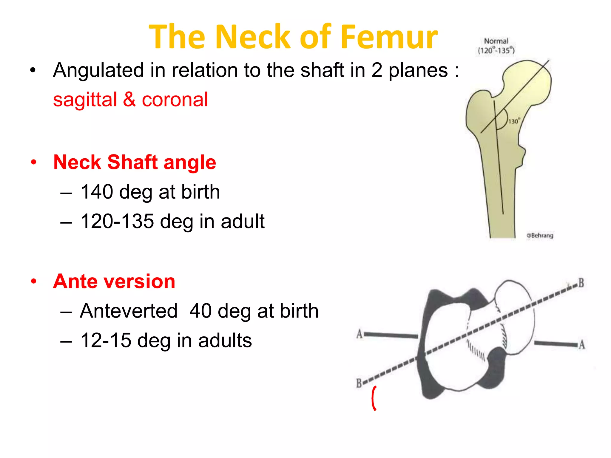
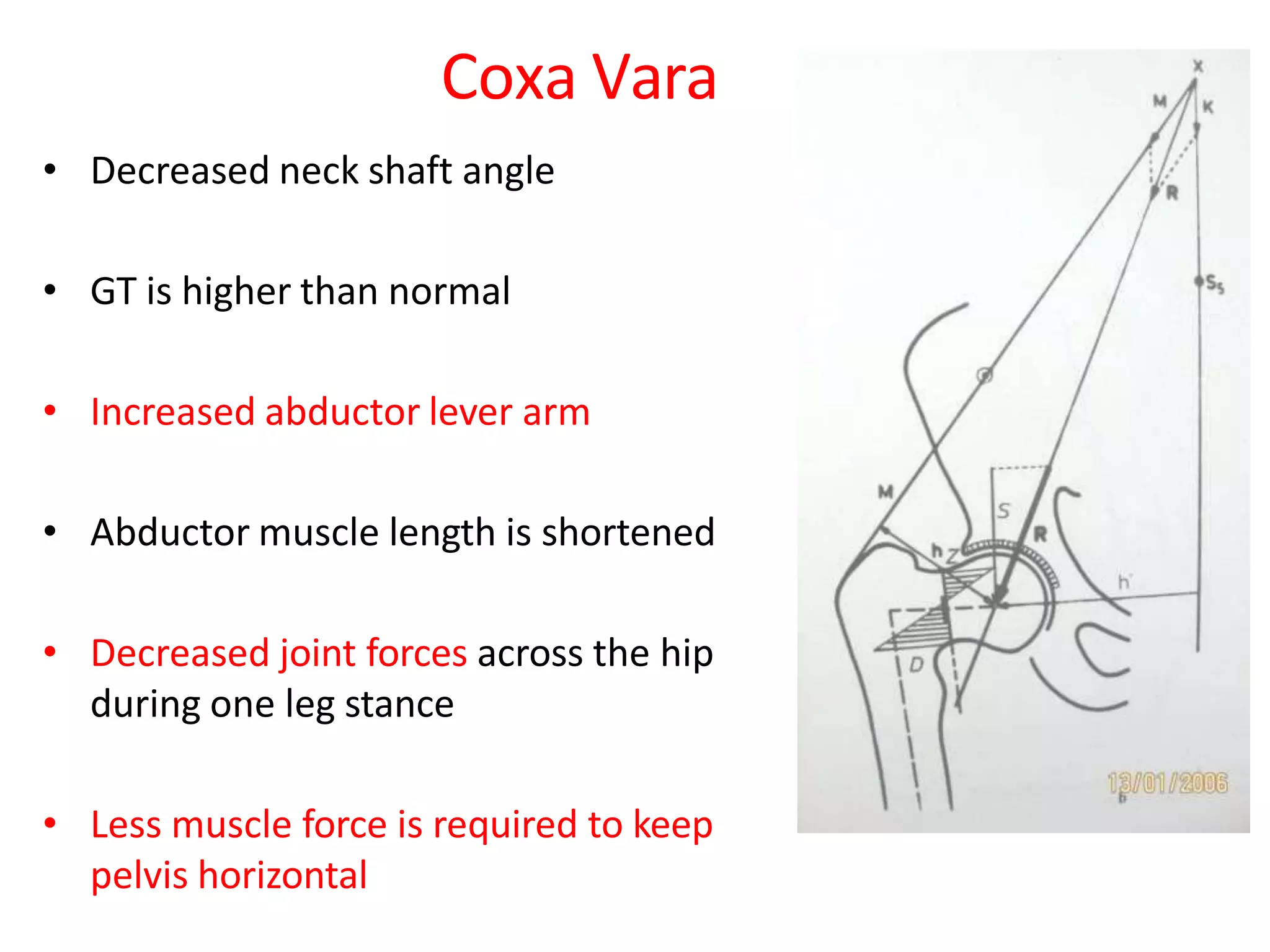
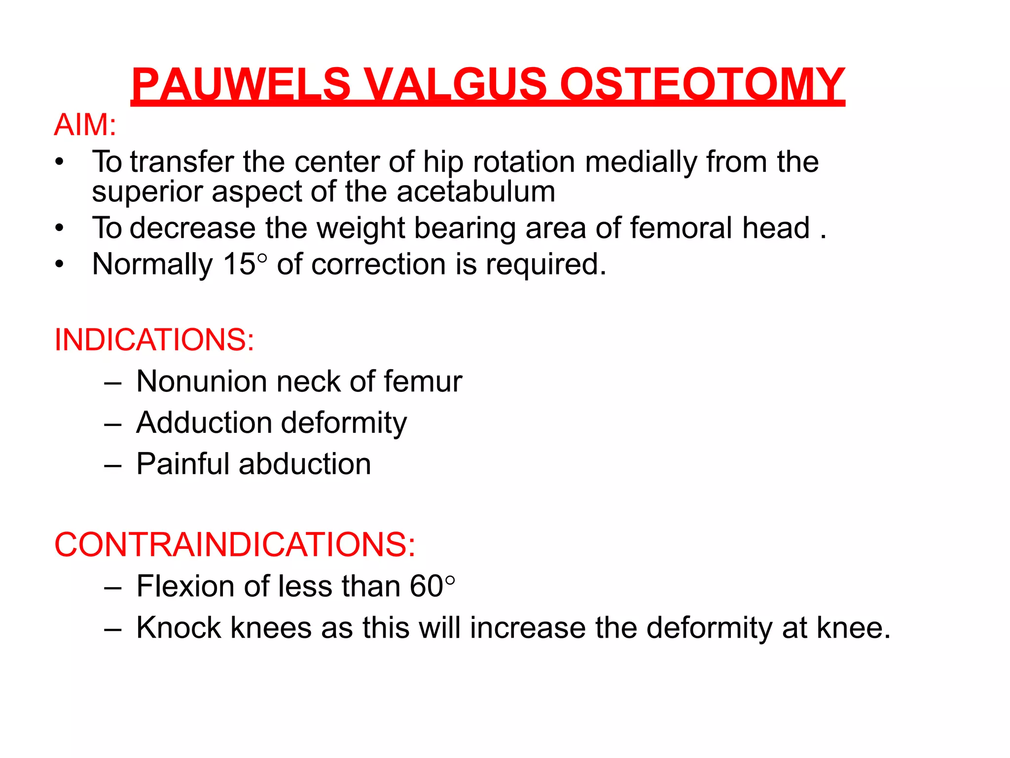
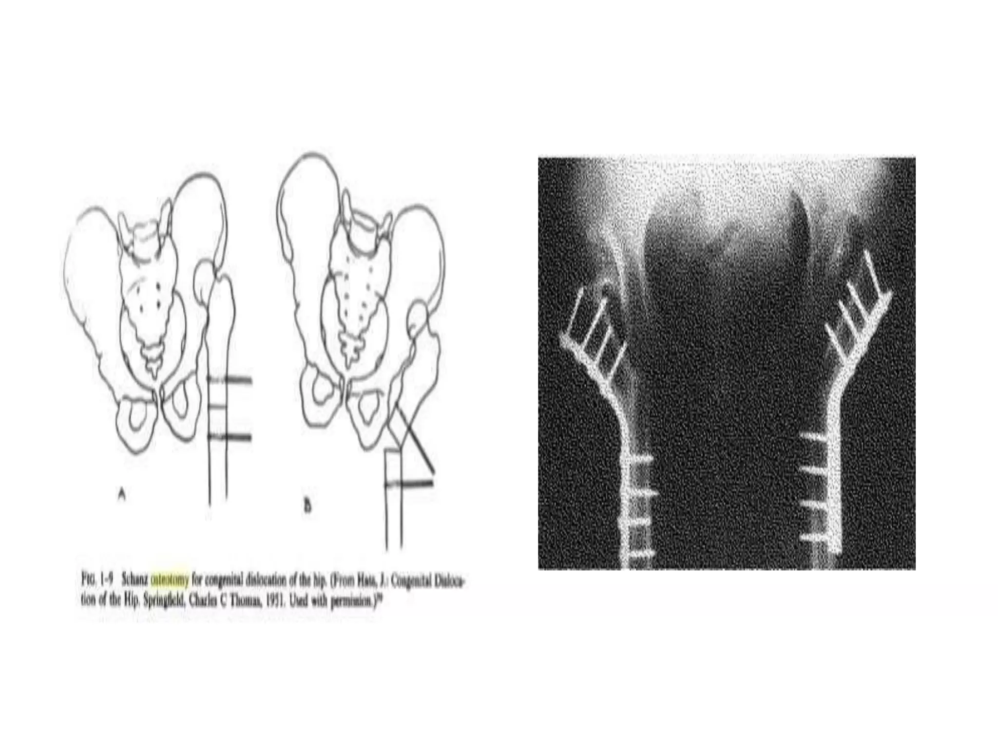
2. • Surgical corrective procedure used to
- obtain a correct biomechanical alignment
of the extremity
- achieve equivocal load transmission
• + / - removal of a portion of bone.
3. Objectives
• Improve coverage of head
• Containment of head
• Moves normal articular cartilage into weight
bearing zone
• Improves motion
• Relieves pain
• To correct leg length inequality by shortening
/ lengthening
4. The Neck of Femur
• Angulated in relation to the shaft in 2 planes :
sagittal & coronal
• Neck Shaft angle
– 140 deg at birth
– 120-135 deg in adult
• Ante version
– Anteverted 40 deg at birth
– 12-15 deg in adults
5. Acetabular Direction
• long axis of acetabulum
points
– forwards : 15-200
ante version
– 450 inferior inclination ante version
7. defined as force generated within a joint in response to forces
acting on the joint
in the hip, it is the result of the need to balance the moment
arms of the body weight and abductor tension
maintains a level pelvis
Joint reaction force
- 3W in single leg stance
-5W in walking
-10W while running
Joint reaction force
8.
9.
10. Biomechanics in neck deformities :
Coxa valga
• Increased neck shaft angle
• GT is at lower level
• Shortened abductor lever arm
• Body wt arm remains same
• Increased joint forces in hip during one leg
stance
• More muscle force required to keep
pelvis horizontal
12. Coxa Vara
• Decreased neck shaft angle
• GT is higher than normal
• Increased abductor lever arm
• Abductor muscle length is shortened
• Decreased joint forces across the hip
during one leg stance
• Less muscle force is required to keep
pelvis horizontal
14. Proximal femoral Osteotomies are classified
Proximal Femoral Osteotomies
According to Anatomical location
High cervical
1. Intertrochanteric
2. Subtrochanteric
3. Greater Trochanteric
Based on displacement of distal
fragment
• Torsional/derotation
• Trans positional
• Angulation
– adductional/varus
– abductional/valgus
– flexion/ extension osteotomies
15. • Based on Indications
– To obtain pain relief
• Osteoarthritis.
– Pauwels varus osteotomy.
– Pauwels valgus osteotomy.
– McMurrays osteotomy
– In proximal femur fractures
• ununited fractures of femoral neck.
– McMurry’s osteotomy.
– Pauwels valgus osteotomy
– Dickson's high geometric osteotomy.
– SchanzAngulation Osteotomy.
• unstable intertrochanteric fractures.
– Dimon Hughston Osteotomy.
– Sarmiento’s Osteotomy
16. PROXIMAL FEMORAL OSTEOTOMIES
Based on the displacement of distal fragment
1.TRANSPOSITIONAL OSTEOTOMY:
Longitudinal axis of distal fragment
remains parallel to the longitudinal
terminal axis of proximal fragment.
Used in : Fracture neck of femur,
OA.
Eg: McMurray osteotomy,
Pauwel’s osteotomy
17. 2.ANGULATION OSTEOTOMY :
Longitudinal axis of distal fragment
forms an angle with that of proximal
fragment .
- Sagittal plane : Extension osteotomy for FFD
-Coronal plane : varus osteotomy
valgus osteotomy
18. Based on INDICATION – Osteoarthritis of hip
• AIM OF OSTEOTOMY :
1. RELIEF OF PAIN: Mechanical : reducing the ratio
between abductor and body weight lever arm,
relaxing capsule.
Haemodynamic: Also by decreasing
the intra osseous pressure.
2.CORRECTION OF DEFORMITY: flexion, adduction,
external rotation.
3.REVERSAL OF DEGENERATIVE PROCESS: helped
by increase in joint space.
19. • Osteotomies in Osteoarthritis of hip :
–Pauwels varus osteotomy.
–Pauwels valgus osteotomy.
–McMurrays osteotomy
20. McMurray’s Displacement Osteotomy
INDICATIONS:
1. Nonunion of femoral neck
2. Advanced osteoarthritis .
PREOPERATIVE PLANNING :
Determination of the size of the bone wedge to be
removed, the position of the seating chisel which will
determine the size and angle of the blade plate to be
used.
AIM :
– Line of weight bearing is shifted medially
– Shearing force at the nonunion is decreased, because the
fracture surface has become more horizontal
21. • Oblique osteotomy made in the shaft of the femur -
• Its lower border on the outer margin being slightly below
the level of lesser trochanter
• Terminates on the inner side b/w lesser trochanter and
neck
• Shaft of femur is displaced inwards by abduction of the
limb & digital pressure on the upper and outer aspect of
lower fragment – complete inward displacement
22.
23. Postoperative care
• Mobilize the patient as soon as symptoms
permit.
• Maintain touch-down weight bearing until union
occurs.
• Active and assisted range of motion exercises
for the hip and knee.
• Once union occurs, unrestricted rehabilitation is
possible.
24. Pauwels Varus Osteotomy
AIM :
• To elevate the greater trochanter and move it
laterally, while moving the abductor and
psoas muscles medially,
• To Restore joint congruity
• Decrease the force acting on the edge of the
acetabulum moves to the middle of weight
bearing surface.
INDICATIONS:
– Antalgic abductor limb
– Abduction deformity
– Painful adduction
– Neck shaft angle > 135° .
25.
26.
27. CONTRAINDICATIONS:
– Fixed external rotation of > 25°
– Flexion of 70° or less.
DISADVANTAGES:
• Shortens the limb to some degrees.
• Creates a trendelenberg gait.
• Increases the prominence of greater trochanter.
• Overloading of the medial compartment of knee.
28.
29. PAUWELS VALGUS OSTEOTOMY
AIM:
• To transfer the center of hip rotation medially from the
superior aspect of the acetabulum
• To decrease the weight bearing area of femoral head .
• Normally 15° of correction is required.
INDICATIONS:
– Nonunion neck of femur
– Adduction deformity
– Painful abduction
CONTRAINDICATIONS:
– Flexion of less than 60°
– Knock knees as this will increase the deformity at knee.
30. • After insertion of guide wire & chisel 2cm proximal to
osteotomy site similar to explained before :-
31. Osteotomies in Nonunion neck of femur
DICKSON HIGH GEOMETRIC OSTEOTOMY
• Principle - the line of vertical SHEAR force is
converted to a horizontal (impacting force).
• In this distal fragment is abducted to 60° after
making osteotomy just below the greater
trochanter & fixed with plate.
• High rate of union
• Lengthens limb
• Improves abductor strength
32.
33. SCHANZ ANGULATION OSTEOTOMY
AIM :
To turn the shaft from the
adducted to abducted
position, so that the
shearing stress of weight
bearing and muscle
retraction becomes an
impaction force.
INDICATIONS:
• Nonunion fracture neck of
femur
• Congenital dislocation of
hip
34. • The femur is cut transversely at ischial tuberosity level
& the proximal fragment is adducted until it rests
against the side wall of the pelvis.
• This lengthens the distance of the gluteus medius and
provides a fulcrum so that adequate leverage of the
muscle is obtained.
• A plate is prepared and angulated sufficiently
• At operation, the bone is sectioned and the plate is
attached to proximal fragment.
• Then, the distal fragment is abducted, extended and
approximated to the distal half of the plate, which is
then attached.
35.
36. LEGG CALVE PERTHES DISEASE:
PATHOLOGY:
• Self limited disease of avascular necrosis of
ossification center of the capital epiphysis,
resulting in variable degree of deformity of femoral
head.
AIM:
• To prevent or minimize residual deformity of
femoral head by creating the biomechanical
environment which is not detrimental to normal
growth and remodeling of epiphysis.
• This is achieved by containing the femoral head
within the acetabulum.
37. VARUS DE-ROTATION OSTEOTOMY
AIM :
• By reducing the ante-version and neck shaft angle to
obtain maximum coverage of the femoral head.
• This osteotomy is done before 4 years of age, as after
this age, there are less chances of Acetabular
remodeling.
DISADVANTAGES:
1. Excessive varus angulation that may not correct with
growth
2. Further shortening of already shortened extremity
3. Possibility of a gluteus lurch produced by decreasing
the length of the lever arm of the gluteus musculature.
38. TECHNIQUE:
• With patient supine make lateral
incision from greater trochanter
distally 8 to 12cm exposing lateral
aspect of femur.
• Mark the level of osteotomy at the
level of lesser trochanter or
slightly distal.
39. • Insert the guide pin
and do reaming of
the femoral head.
• Insert the barrel guide
into the back of the
implanted lag screw.
• Make the osteotomy cut
& tilt the head into varus .
40. • Using the side plate and screws firmly join the
proximal and distal fragments.
• Spica cast is worn for 8-12 weeks and internal
fixation can be removed after 1-2 years.
41. SLIPPED CAPITAL FEMORAL EPIPHYSIS
• In this condition, the epiphysis is displaced inferiorly
causing adduction and external rotation deformity of
the limb.
AIM:
Osteotomy is performed here to reposition the
femoral head (epiphysis) concentrically within the
acetabulum.
INDICATIONS:
– Chronic slip with moderate to severe
displacement.
– Malunited slip
42. TWO BASIC TYPES:
• Closing wedge osteotomy of neck: Usually associated with
serious complications of AVN and chondrolysis, therefore
these osteotomies are not recommended. These are of four
types.
a. The technique of Fish
b. Technique of Dunn just distal to slip
c. Base of neck technique by Kramer et al d.
d. Technique of Abraham et al
• Compensatory osteotomies through the Trochantric region:
These osteotomies produce a deformity in the opposite
direction. It includes
a. Ball and socket osteotomy
b.Biplane intertrochanteric osteotomy
(Southwick)
43. 1. CUNEIFORM OSTEOTOMY
OF FEMORAL NECK (FISH):
• Fish recommended this in
moderate to severe slips of
more than 30°.
• Capsule is incised & femoral
neck is exposed.
• Locate the physis.
• Determine the size of wedge
to be removed by noting the
degree of slip.
44. • Adjacent to the
epiphyseal plate, a wedge
shaped piece of bone is
superiorly with
removed with its base
directed anteriorly and
apex
psotero-inferiorly.
• Take care that osteotome
does not penetrate the
intact posterior periosteum,
damaging retinacular
vessels.
45. • Reduce the epiphysis
by flexion, abduction
and internal rotation of
limb, taking care to put
much tension on the
posterior periosteum,
capsule and vessels.
After reduction fix the epiphysis to neck with 2-3 pins six
inches long threaded on one half of their length. Do not
penetrate articular cartilage.
46. CUNIEFORM OSTEOTOMY OF FEMORAL NECK
(DUNN):
• Dunn described an osteotomy for severe
chronic slips in children with open physis.
• This procedure should not be done if the
physis is closed.
• Anterosuperior wedge of the most superior part
of the femoral neck is removed
47. TECHNIQUE :
• Through a lateral approach
• A. Greater trochanter is detached.
• B. Synovium is elevated from anterior and
postero-lateral surface of femoral neck with
periosteum elevator.
48. • C. Head is freed of all fibrocartilage and callus.
• D. Osteotomy line on upper end of femoral neck
is made for excision of trapezoid segment. (
anterosuperior wedge )
49. • E. Head of femur is replaced on femoral neck
and three threaded Steinmann pins are used for
fixation of shaft, head, and neck of femur.
• F. Two cancellous screws are used to fix greater
trochanter in normal position.
50. Osteotomies at base of neck – KRAMER,BARMADA
• Intracapsular base of neck osteotomy – for
chronic slips with > 20 ° of deformity
• Extracapsular base of neck osteotomy – for
moderate to severe chronic slips with > 30 ° of
head shaft angle
51.
52. Intertrochanteric Osteotomy - SOUTHWICK
• BIPLANE osteotomy
• Anterolaterally based wedge
• At the level of lesser trochanter
• Indications :
- for chronic / healed slips with head shaft deformities
between 30 – 70 °
• Corrects extension / varus deformity with flexion /
extension of the distal fragment , and internal rotation as
needed
54. OSTEOTOMY TO CORRECT UNSTABLE
INTERTROCHANTERIC FRACTURES
• Dimon and Hughston :
– Medial displacement osteotomy to stabilise unstable 4
part IT fracture
– In 4part # adductors tend to displace fracture into varus
secondary to lack of medial cortical opposition
– Chang et al - anatomical reduction allow greater load
shearing by bone than medial displacement osteotomy.
(CORR 1987 Dec;(225):141-6)
55. • Technique:
• If GT remains attached to femur ,a transverse osteotomy needs to
be made at a level 2cm below LT
• GT fragment is reflected superiorly
• Steinman pin inserted into the superior third of femoral head
• Key the calcar spike (proximal fragment )into the medially displaced
distal fragment
• Guide wire is placed into the lower half of femoral head
• The wire position will ensure a more valgus orientation of femoral
neck,once the screw and side plate have been applied
• Determine and insert the appropriate compression screw
• Abduct the thigh to bring the reduction into valgus
• Apply the side plate – 135deg, short barrel
• Release traction and apply the compression screw
• Reattach the GT fragment with wires
57. SARMIENTO OSTEOTOMY
• Involves creating an oblique osteotomy of the distal
fragment(valgus osteotomy) to obtain stability in unstable
IT #
• Changes # plane from verticle to near horizontal
• Creates contact b/w medial and posterior cortex of
proximal and distal fragments
• Goal – to obtain medial stability
• Adv – valgus realignment of proximal fragment makes up
for less of length at osteotomy site so that limb lengths
are equal
58. Technique:
• A 45° oblique osteotomy of distal fragment begins just
below flare of GT & crosses distally and medially to exit
about 1cm distal to the apex of #
• Guide wire and then implant are inserted at 90° to plane
of # of proximal fragment
• With more vertical alignment of # ,insert guide pin so that
it ends up more inferiorly in the femoral head ( other wise
,the osteotomy will be placed in varus)
• Insert 135 sliding screw in usual manner
• # is reduced and impacted
• Medial cortex opposition and hence stability are restored