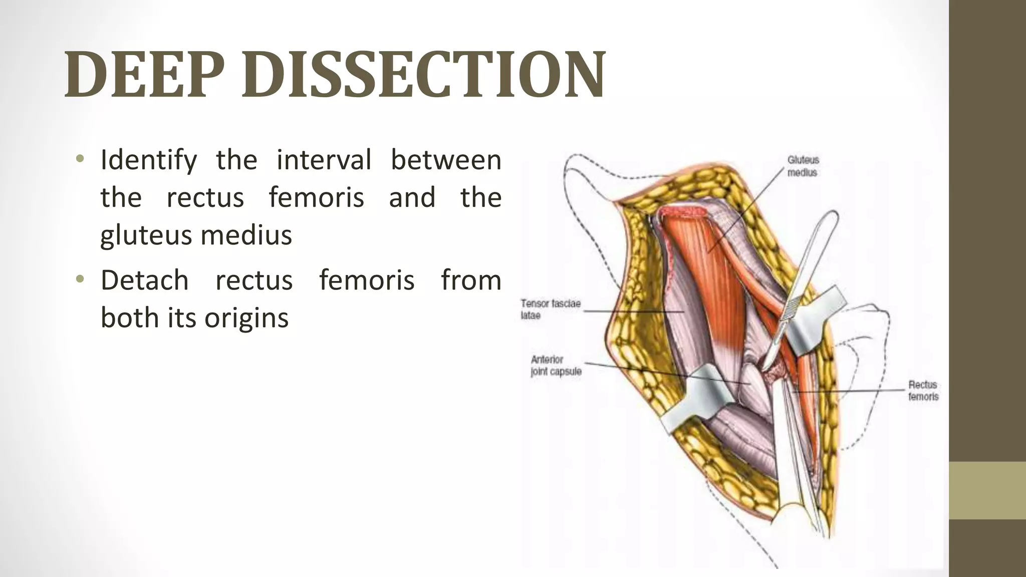
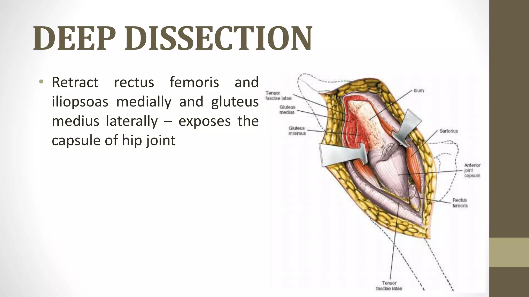
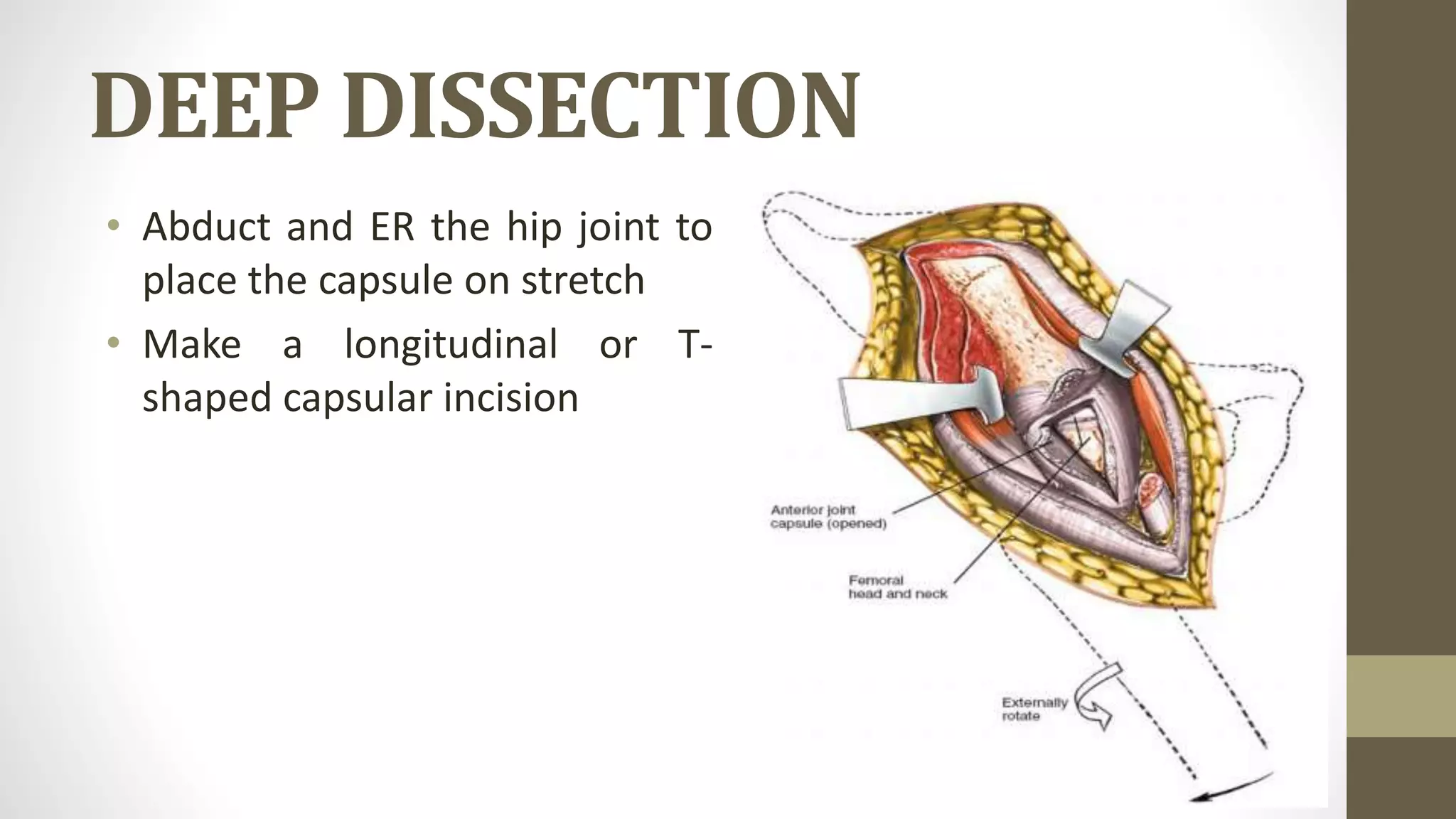
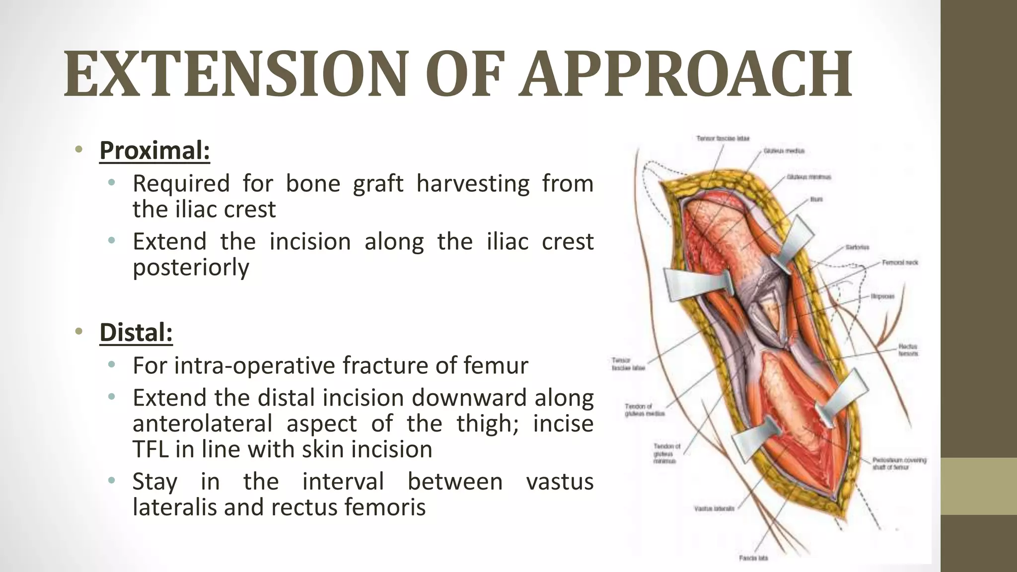
ANTERIOR
APPROACH TO THE
HIP
Presented by: Dr. Sachin. M.
2nd year PGT, Dept. of Orthopaedics, SMCH
Moderator: Dr. A. K. Sipani
Prof & HOD, Dept. of Orthopaedics, SMCH
INTRODUCTION
• Also known as Smith – Peterson approach
• Provides access to the hip joint and the ilium
• INDICATIONS:
• Open reduction of CDH
• THA, Hemiarthroplasty
• Synovial biopsies, irrigation and debridement of the hip joint
• Intra-articular fusions
• Excision of the tumors, especially of the pelvis
• Pelvic osteotomies
POSITION AND PREPARATION
• Anaesthesia:
• Spinal block or general
anaesthesia
• Position of the patient:
• Supine
• Use sandbag in cases of
pelvic osteotomies
INTERNERVOUS PLANE
• Superficial:
• Sartorius – femoral nv.
• Tensor fasciae latae – superior
gluteal nv.
INTERNERVOUS PLANE
• Deep:
• Rectus femoris – femoral
nv.
• Gluteus medius – superior
gluteal nv.
LANDMARKS
• Iliac crest: subcutaneous and easily palpable
• ASIS: move anteriorly along the iliac crest to feel a bony
prominence
• Easily palpable in thin patients
INCISION
• 8 to 10cm incision along the
anterior half of the iliac crest
up to ASIS
• Curve the incision inferiorly,
extend vertically (8 to 10cm)
directing towards the lateral
side of the patella
SUPERFICIAL DISSECTION
• Identify the interval between
sartorius and TFL
• Dissect through the
subcutaneous fat (avoid LFCN)
SUPERFICIAL DISSECTION
• Incise fascia on the medial
border of TFL
• Detach TFL from its iliac origin
to develop internervous plane
SUPERFICIAL DISSECTION
• Ligate the ascending branch of lateral
femoral circumflex artery
DEEP DISSECTION
• Identify the interval between
the rectus femoris and the
gluteus medius
• Detach rectus femoris from
both its origins
DEEP DISSECTION
• Retract rectus femoris and
iliopsoas medially and gluteus
medius laterally – exposes the
capsule of hip joint
DEEP DISSECTION
• Abduct and ER the hip joint to
place the capsule on stretch
• Make a longitudinal or T-
shaped capsular incision
EXTENSION OF APPROACH
• Proximal:
• Required for bone graft harvesting from
the iliac crest
• Extend the incision along the iliac crest
posteriorly
• Distal:
• For intra-operative fracture of femur
• Extend the distal incision downward along
anterolateral aspect of the thigh; incise
TFL in line with skin incision
• Stay in the interval between vastus
lateralis and rectus femoris
DANGERS
• Lateral femoral cutaneous nerve:
• Passes under the inguinal ligament
• Variations are common – courses
either medial or lateral to ASIS
• Avoid injuring the nerve while
incising the fascia between sartorius
and TFL
• Femoral nerve:
• Stays in the femoral triangle
• Safe as far as we stay lateral to the
sartorius muscle
DANGERS
• Ascending branch of lateral
femoral circumflex artery
• Usually found in the interval
between sartorius and TFL
• Ligate to avoid excessive
haemorrhage
Anterior approach to the hip - Dr. Sachin M

Anterior approach to the hip - Dr. Sachin M

  • 1.
    ANTERIOR APPROACH TO THE HIP Presentedby: Dr. Sachin. M. 2nd year PGT, Dept. of Orthopaedics, SMCH Moderator: Dr. A. K. Sipani Prof & HOD, Dept. of Orthopaedics, SMCH
  • 2.
    INTRODUCTION • Also knownas Smith – Peterson approach • Provides access to the hip joint and the ilium • INDICATIONS: • Open reduction of CDH • THA, Hemiarthroplasty • Synovial biopsies, irrigation and debridement of the hip joint • Intra-articular fusions • Excision of the tumors, especially of the pelvis • Pelvic osteotomies
  • 3.
    POSITION AND PREPARATION •Anaesthesia: • Spinal block or general anaesthesia • Position of the patient: • Supine • Use sandbag in cases of pelvic osteotomies
  • 4.
    INTERNERVOUS PLANE • Superficial: •Sartorius – femoral nv. • Tensor fasciae latae – superior gluteal nv.
  • 5.
    INTERNERVOUS PLANE • Deep: •Rectus femoris – femoral nv. • Gluteus medius – superior gluteal nv.
  • 6.
    LANDMARKS • Iliac crest:subcutaneous and easily palpable • ASIS: move anteriorly along the iliac crest to feel a bony prominence • Easily palpable in thin patients
  • 7.
    INCISION • 8 to10cm incision along the anterior half of the iliac crest up to ASIS • Curve the incision inferiorly, extend vertically (8 to 10cm) directing towards the lateral side of the patella
  • 8.
    SUPERFICIAL DISSECTION • Identifythe interval between sartorius and TFL • Dissect through the subcutaneous fat (avoid LFCN)
  • 9.
    SUPERFICIAL DISSECTION • Incisefascia on the medial border of TFL • Detach TFL from its iliac origin to develop internervous plane
  • 10.
    SUPERFICIAL DISSECTION • Ligatethe ascending branch of lateral femoral circumflex artery
  • 11.
    DEEP DISSECTION • Identifythe interval between the rectus femoris and the gluteus medius • Detach rectus femoris from both its origins
  • 12.
    DEEP DISSECTION • Retractrectus femoris and iliopsoas medially and gluteus medius laterally – exposes the capsule of hip joint
  • 13.
    DEEP DISSECTION • Abductand ER the hip joint to place the capsule on stretch • Make a longitudinal or T- shaped capsular incision
  • 14.
    EXTENSION OF APPROACH •Proximal: • Required for bone graft harvesting from the iliac crest • Extend the incision along the iliac crest posteriorly • Distal: • For intra-operative fracture of femur • Extend the distal incision downward along anterolateral aspect of the thigh; incise TFL in line with skin incision • Stay in the interval between vastus lateralis and rectus femoris
  • 15.
    DANGERS • Lateral femoralcutaneous nerve: • Passes under the inguinal ligament • Variations are common – courses either medial or lateral to ASIS • Avoid injuring the nerve while incising the fascia between sartorius and TFL • Femoral nerve: • Stays in the femoral triangle • Safe as far as we stay lateral to the sartorius muscle
  • 16.
    DANGERS • Ascending branchof lateral femoral circumflex artery • Usually found in the interval between sartorius and TFL • Ligate to avoid excessive haemorrhage