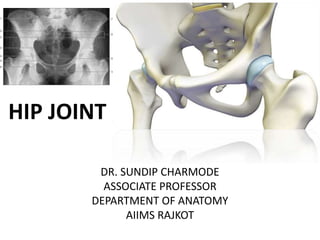
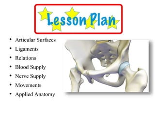
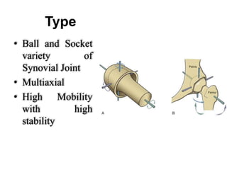
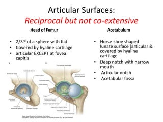
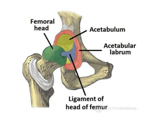
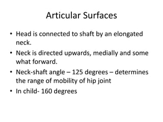
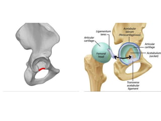
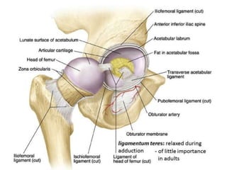
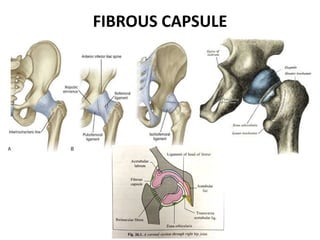
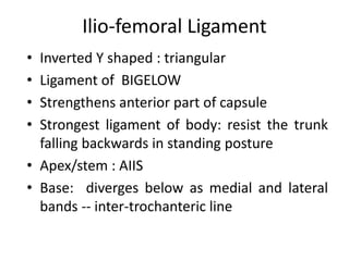
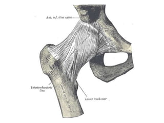
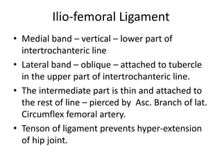
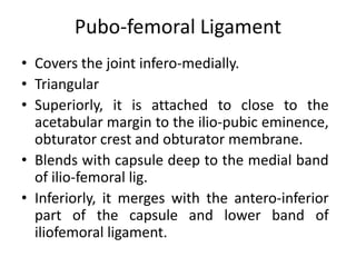
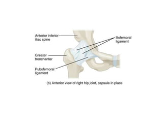
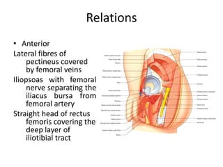
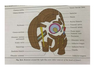
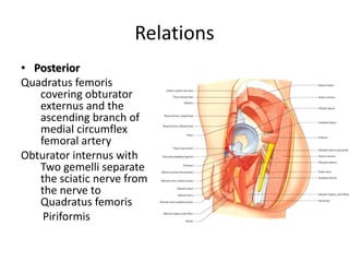
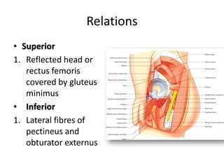
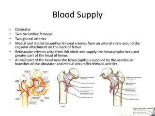
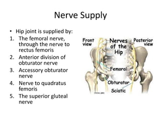
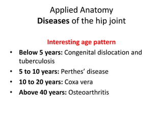
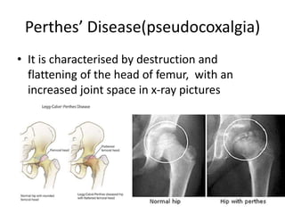
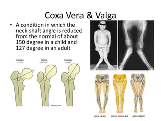
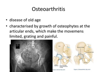
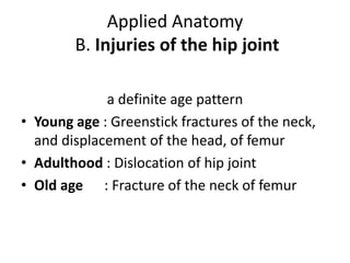
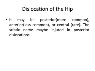
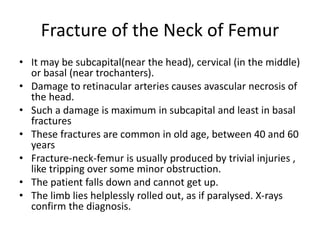
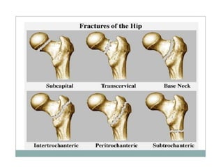
HIP JOINT
DR. SUNDIP CHARMODE
ASSOCIATE PROFESSOR
DEPARTMENT OF ANATOMY
AIIMS RAJKOT
• Type of Joint
• Articular Surfaces
• Ligaments
• Relations
• Blood Supply
• Nerve Supply
• Movements
• Applied Anatomy
Type
• Ball and Socket
variety of
Synovial Joint
• Multiaxial
• High Mobility
with high
stability
Articular Surfaces:
Reciprocal but not co-extensive
Head of Femur
• 2/3rd of a sphere with flat
• Covered by hyaline cartilage
• articular EXCEPT at fovea
capitis
Acetabulum
• Horse-shoe shaped
lunate surface (articular &
covered by hyaline
cartilage
• Deep notch with narrow
mouth
• Articular notch
• Acetabular fossa
Articular Surfaces
• Head is connected to shaft by an elongated
neck.
• Neck is directed upwards, medially and some
what forward.
• Neck-shaft angle – 125 degrees – determines
the range of mobility of hip joint
• In child- 160 degrees
ARTICULAR SURFACES: SOCKET
• Cup shaped depression, acetabulum, where
ilium, ischium and pubis meet separated by a
tri-radiate Y shaped cartilage.
• Acetabular notch:
• Acetabular labrum:
• Transverse acetabular ligament and base of
ligament of head of femur.
• Acetabular fossa is non articular.
• Articular surface – lunate surface
ARTICULAR SURFACES: SOCKET
• Acetabular fossa is filled with cushion of fibro-
fatty tissue covered by synovial membrane.
• Intracapsular but extra-synovial
• Haversian fat is in liquid condition at room
temperature.
• The gap between the bony notch and
Transverse ligament transmit Acetabular
branches of obturator and medial circumflex
femoral vessels.
LIGAMENTS
• Fibrous capsule
• Synovial membrane
• Ilio-femoral
• Pubo-femoral
• Ischio-femoral
• Ligament of head of
femur
• Acetabular labrum
• Transverse acetabular
ligament
FIBROUS CAPSULE
• Attachment
• On hip bone: to the acetabular margin about
5-6 mm above the labrum. Blends with
anterior surface of labrum in front and
transverse acetabular ligament below.
• To the bone above and behind the
acetabulum.
• On femur: to the inter-trochanteric line in
front and 1 cm medial to inter-trochanteric
crest behind
FIBROUS CAPSULE
FIBROUS CAPSULE
• Thick antero-superiorly : part
subjected to max tension in
standing
• Thin and loosely attached
postero-inferiorly, is stretched
in abduction.
• 2 type of fibres
outer longitudinal : many of
them reflect to neck of femur
K/s retinacula (contain blood
vessels supplying head and
neck of femur)
inner circular K/s Zona
Orbicularis
SYNOVIAL MEMBRANE
• Lines the inner surface of
fibrous capsule,
• Intra-capsular portion of
neck of femur,
• Both surfaces of
acetabular labrum,
• Transverse ligament
• Fat in acetabular fossa
• Round ligament of head
of femur
SYNOVIAL MEMBRANE
• The SM invests the ligament of head of femur
as a flattened cone and is attached to the
femoral head at the peripheral margin of the
fovea.
• Towards the transverse ligament, the layers
diverge. Inferior layer covers the fibrous
capsule and superior layer covers the
acetabular fat.
• Oval gap between Pubo-femoral and ilio-
femoral ligaments.
Joint Cavity
• Communicates with
the sub-tendinous
bursa beneath the
tendon of psoas
major, through a
circular opening in
the capsule between
pubo-femoral and
vertical band of ilio-
femoral ligaments.
Acetabular Labrum
• Cotyloid ligament
• Fibro-cartilagenous rim attached to the margins
of acetabulum, triangula rim cross-section.
• Narrows the mouthy of acetabulum which helps
in holding the head of femur in position
Acetabular Labrum
Transverse Ligament of Acetabulum
• A part of the labrum
which bridges the
acetabular notch, but it
has no cartilage cells
• The notch thus
converted into a
foramen transmits
vessels (acetabular) and
nerves to the joint.
• It blends with base of
ligament of head of
femur and lower part of
fibrous capsule.
Ligament Of the Head of Femur
• Round ligament or ligamentum teres femoris
• Flat, triangular ligament/ fibrous band, covered
by S Membrane.
• Apex : fovea capitis
• Base: Two ends of the the acetabular notch and
blends with transverse acetabular ligament.
• Very thin/even absent
• Transmits arteries to the head of femur from
the acetabular branches of the obturator and
medical circumflex femoral arteries.
Ligament Of the Head of Femur
• The ligamentum teres is stretched in
adduction of semi-flexed hip and is relaxed in
abduction.
• The ligament does not increase the stability
of the joint.
Ilio-femoral Ligament
• Inverted Y shaped : triangular
• Ligament of BIGELOW
• Strengthens anterior part of capsule
• Strongest ligament of body: resist the trunk
falling backwards in standing posture
• Apex/stem : AIIS
• Base: diverges below as medial and lateral
bands -- inter-trochanteric line
Ilio-femoral Ligament
• Medial band – vertical – lower part of
intertrochanteric line
• Lateral band – oblique – attached to tubercle
in the upper part of intertrochanteric line.
• The intermediate part is thin and attached to
the rest of line – pierced by Asc. Branch of lat.
Circumflex femoral artery.
• Tenson of ligament prevents hyper-extension
of hip joint.
Pubo-femoral Ligament
• Covers the joint infero-medially.
• Triangular
• Superiorly, it is attached to close to the
acetabular margin to the ilio-pubic eminence,
obturator crest and obturator membrane.
• Blends with capsule deep to the medial band
of ilio-femoral lig.
• Inferiorly, it merges with the antero-inferior
part of the capsule and lower band of
iliofemoral ligament.
Pubo-femoral Ligament
• A gap between pubo-femoral and medial
band of ilio-femoral ligaments.
• Psoas bursa communicates with joint cavity
Ischifemoral Ligament
• Comparatively weak, and
covers the joint
posteriorly.
• Fibers pass spirally
upwards and laterally
behind the neck of femur.
• Most fibers continuous
with zona orbicularis
Relations
• Anterior
Lateral fibres of
pectineus covered
by femoral veins
Iliopsoas with femoral
nerve separating the
iliacus bursa from
femoral artery
Straight head of rectus
femoris covering the
deep layer of
iliotibial tract
Relations
• Posterior
Quadratus femoris
covering obturator
externus and the
ascending branch of
medial circumflex
femoral artery
Obturator internus with
Two gemelli separate
the sciatic nerve from
the nerve to
Quadratus femoris
Piriformis
Relations
• Superior
1. Reflected head or
rectus femoris
covered by gluteus
minimus
• Inferior
1. Lateral fibres of
pectineus and
obturator externus
Blood Supply
• Obturator
• Two circumflex femoral
• Two gluteal arteries
• Medial and lateral circumflex femoral arteries form an arterial circle around the
capsular attachment on the neck of femur
• Retinacular arteries arise from this circle and supply the intracapsular neck and
greater part of the head of femur.
• A small part of the head near the fovea capitis is supplied by the acetabular
branches of the obturator and medial circumflex femoral arteries
Nerve Supply
• Hip joint is supplied by:
1. The femoral nerve,
through the nerve to
rectus femoris
2. Anterior division of
obturator nerve
3. Accessory obturator
nerve
4. Nerve to quadratus
femoris
5. The superior gluteal
nerve
Movements
• Flexion and extension occur around a
transverse axis around the neck of femur
which pass through the centre of head of
femoral head.
• Flexion with extended knee – 90-120 and 120
degrees
• Loosely packed and relaxed capsule
Movements
• Extension – 10-20 degrees and is limited by ilio-
femoral ligament. Closely packed capsule.
• Adduction and abduction occur around a antero-
posterior axis, passing through the centre of head
of femur.
• Medial and lateral rotations occur around a
vertical axis. Upper end - through the centre of
head of femur and lower end of axis - lateral
condyle of femur when foot is on ground.
Movements
• Lower end of axis – through any part of foot
when foot is above the ground.
• Circumduction is a combination of the
foregoing movements
Muscles producing Movement
1. Flexion-Ilio-Psoas major. Pectineus, RF, Sartorius.
Add. Longus helps in initial phase.
2. Extension-Gluteus maximus and hamstrings
3. Adduction-Adductors longus, brevis and
magnus. Medial part of pectineus and gracilis.
4. Abduction-Glutei medius and minimus
5. Medial Rotation- Tensor fasciae latae and the
anterior fibres of glutei medius and minimus
6. Lateral rotation-Two obturators, two gemelli
and quadratus femoris
Applied Anatomy
Diseases of the hip joint
Interesting age pattern
• Below 5 years: Congenital dislocation and
tuberculosis
• 5 to 10 years: Perthes’ disease
• 10 to 20 years: Coxa vera
• Above 40 years: Osteoarthritis
Congenital Dislocation
• More common in the hip than any other joint of the body
• The head of the femur slips upwards onto the gluteal
surface of the ilium because the upper margin of
acetabulum is developmentally deficient
• This causes lurching gait, and the Trendelenburg’s test is
positive.
Tuberculosis
• Osseous destruction & marrow edema
involving the bones forming hip articulation
along with synovial collection & reduced joint
space.
Perthes’ Disease(pseudocoxalgia)
• It is characterised by destruction and
flattening of the head of femur, with an
increased joint space in x-ray pictures
Coxa Vera & Valga
• A condition in which the
neck-shaft angle is reduced
from the normal of about
150 degree in a child and
127 degree in an adult
Osteoarthritis
• disease of old age
• characterised by growth of osteophytes at the
articular ends, which make the movemens
limited, grating and painful.
Applied Anatomy
B. Injuries of the hip joint
a definite age pattern
• Young age : Greenstick fractures of the neck,
and displacement of the head, of femur
• Adulthood : Dislocation of hip joint
• Old age : Fracture of the neck of femur
Dislocation of the Hip
• It may be posterior(more common),
anterior(less common), or central (rare). The
sciatic nerve maybe injured in posterior
dislocations.
Fracture of the Neck of Femur
• It may be subcapital(near the head), cervical (in the middle)
or basal (near trochanters).
• Damage to retinacular arteries causes avascular necrosis of
the head.
• Such a damage is maximum in subcapital and least in basal
fractures
• These fractures are common in old age, between 40 and 60
years
• Fracture-neck-femur is usually produced by trivial injuries ,
like tripping over some minor obstruction.
• The patient falls down and cannot get up.
• The limb lies helplessly rolled out, as if paralysed. X-rays
confirm the diagnosis.
Trochanteric Fracture
 Intertrochanteric(between the Trochanters)
 Peritrochanteric(along the Trochanters) or
 Subtrochanteric(below the trochanters).
• These fractures occur in strong, adult subjects,
and are produced by severe, violent injuries.
Applied Anatomy
Shortening of the lower limb
• By fractures/dislocation and tuberculosis
• The length of the lower limb is measured from
anterior superior iliac spine to medical
malleolus
Displacement of the greater Trochanter
(in fractures and dislocations)
Shenton’s line
Nelaton’s Line /Bryant’s triangle
Applied Anatomy
Disease of the hip, like tuberculosis, may cause
referred pain in the knee because of the
common nerve supply of the two joints.

Hip Joint.pptx

  • 1.
    HIP JOINT DR. SUNDIPCHARMODE ASSOCIATE PROFESSOR DEPARTMENT OF ANATOMY AIIMS RAJKOT
  • 2.
    • Type ofJoint • Articular Surfaces • Ligaments • Relations • Blood Supply • Nerve Supply • Movements • Applied Anatomy
  • 3.
    Type • Ball andSocket variety of Synovial Joint • Multiaxial • High Mobility with high stability
  • 4.
    Articular Surfaces: Reciprocal butnot co-extensive Head of Femur • 2/3rd of a sphere with flat • Covered by hyaline cartilage • articular EXCEPT at fovea capitis Acetabulum • Horse-shoe shaped lunate surface (articular & covered by hyaline cartilage • Deep notch with narrow mouth • Articular notch • Acetabular fossa
  • 6.
    Articular Surfaces • Headis connected to shaft by an elongated neck. • Neck is directed upwards, medially and some what forward. • Neck-shaft angle – 125 degrees – determines the range of mobility of hip joint • In child- 160 degrees
  • 7.
    ARTICULAR SURFACES: SOCKET •Cup shaped depression, acetabulum, where ilium, ischium and pubis meet separated by a tri-radiate Y shaped cartilage. • Acetabular notch: • Acetabular labrum: • Transverse acetabular ligament and base of ligament of head of femur. • Acetabular fossa is non articular. • Articular surface – lunate surface
  • 10.
    ARTICULAR SURFACES: SOCKET •Acetabular fossa is filled with cushion of fibro- fatty tissue covered by synovial membrane. • Intracapsular but extra-synovial • Haversian fat is in liquid condition at room temperature. • The gap between the bony notch and Transverse ligament transmit Acetabular branches of obturator and medial circumflex femoral vessels.
  • 11.
    LIGAMENTS • Fibrous capsule •Synovial membrane • Ilio-femoral • Pubo-femoral • Ischio-femoral • Ligament of head of femur • Acetabular labrum • Transverse acetabular ligament
  • 12.
    FIBROUS CAPSULE • Attachment •On hip bone: to the acetabular margin about 5-6 mm above the labrum. Blends with anterior surface of labrum in front and transverse acetabular ligament below. • To the bone above and behind the acetabulum. • On femur: to the inter-trochanteric line in front and 1 cm medial to inter-trochanteric crest behind
  • 14.
  • 15.
    FIBROUS CAPSULE • Thickantero-superiorly : part subjected to max tension in standing • Thin and loosely attached postero-inferiorly, is stretched in abduction. • 2 type of fibres outer longitudinal : many of them reflect to neck of femur K/s retinacula (contain blood vessels supplying head and neck of femur) inner circular K/s Zona Orbicularis
  • 16.
    SYNOVIAL MEMBRANE • Linesthe inner surface of fibrous capsule, • Intra-capsular portion of neck of femur, • Both surfaces of acetabular labrum, • Transverse ligament • Fat in acetabular fossa • Round ligament of head of femur
  • 17.
    SYNOVIAL MEMBRANE • TheSM invests the ligament of head of femur as a flattened cone and is attached to the femoral head at the peripheral margin of the fovea. • Towards the transverse ligament, the layers diverge. Inferior layer covers the fibrous capsule and superior layer covers the acetabular fat. • Oval gap between Pubo-femoral and ilio- femoral ligaments.
  • 18.
    Joint Cavity • Communicateswith the sub-tendinous bursa beneath the tendon of psoas major, through a circular opening in the capsule between pubo-femoral and vertical band of ilio- femoral ligaments.
  • 19.
    Acetabular Labrum • Cotyloidligament • Fibro-cartilagenous rim attached to the margins of acetabulum, triangula rim cross-section. • Narrows the mouthy of acetabulum which helps in holding the head of femur in position
  • 20.
  • 21.
    Transverse Ligament ofAcetabulum • A part of the labrum which bridges the acetabular notch, but it has no cartilage cells • The notch thus converted into a foramen transmits vessels (acetabular) and nerves to the joint. • It blends with base of ligament of head of femur and lower part of fibrous capsule.
  • 23.
    Ligament Of theHead of Femur • Round ligament or ligamentum teres femoris • Flat, triangular ligament/ fibrous band, covered by S Membrane. • Apex : fovea capitis • Base: Two ends of the the acetabular notch and blends with transverse acetabular ligament. • Very thin/even absent • Transmits arteries to the head of femur from the acetabular branches of the obturator and medical circumflex femoral arteries.
  • 24.
    Ligament Of theHead of Femur
  • 26.
    Ligament Of theHead of Femur • The ligamentum teres is stretched in adduction of semi-flexed hip and is relaxed in abduction. • The ligament does not increase the stability of the joint.
  • 27.
    Ilio-femoral Ligament • InvertedY shaped : triangular • Ligament of BIGELOW • Strengthens anterior part of capsule • Strongest ligament of body: resist the trunk falling backwards in standing posture • Apex/stem : AIIS • Base: diverges below as medial and lateral bands -- inter-trochanteric line
  • 30.
    Ilio-femoral Ligament • Medialband – vertical – lower part of intertrochanteric line • Lateral band – oblique – attached to tubercle in the upper part of intertrochanteric line. • The intermediate part is thin and attached to the rest of line – pierced by Asc. Branch of lat. Circumflex femoral artery. • Tenson of ligament prevents hyper-extension of hip joint.
  • 31.
    Pubo-femoral Ligament • Coversthe joint infero-medially. • Triangular • Superiorly, it is attached to close to the acetabular margin to the ilio-pubic eminence, obturator crest and obturator membrane. • Blends with capsule deep to the medial band of ilio-femoral lig. • Inferiorly, it merges with the antero-inferior part of the capsule and lower band of iliofemoral ligament.
  • 33.
    Pubo-femoral Ligament • Agap between pubo-femoral and medial band of ilio-femoral ligaments. • Psoas bursa communicates with joint cavity
  • 35.
    Ischifemoral Ligament • Comparativelyweak, and covers the joint posteriorly. • Fibers pass spirally upwards and laterally behind the neck of femur. • Most fibers continuous with zona orbicularis
  • 37.
    Relations • Anterior Lateral fibresof pectineus covered by femoral veins Iliopsoas with femoral nerve separating the iliacus bursa from femoral artery Straight head of rectus femoris covering the deep layer of iliotibial tract
  • 39.
    Relations • Posterior Quadratus femoris coveringobturator externus and the ascending branch of medial circumflex femoral artery Obturator internus with Two gemelli separate the sciatic nerve from the nerve to Quadratus femoris Piriformis
  • 40.
    Relations • Superior 1. Reflectedhead or rectus femoris covered by gluteus minimus • Inferior 1. Lateral fibres of pectineus and obturator externus
  • 41.
    Blood Supply • Obturator •Two circumflex femoral • Two gluteal arteries • Medial and lateral circumflex femoral arteries form an arterial circle around the capsular attachment on the neck of femur • Retinacular arteries arise from this circle and supply the intracapsular neck and greater part of the head of femur. • A small part of the head near the fovea capitis is supplied by the acetabular branches of the obturator and medial circumflex femoral arteries
  • 42.
    Nerve Supply • Hipjoint is supplied by: 1. The femoral nerve, through the nerve to rectus femoris 2. Anterior division of obturator nerve 3. Accessory obturator nerve 4. Nerve to quadratus femoris 5. The superior gluteal nerve
  • 43.
    Movements • Flexion andextension occur around a transverse axis around the neck of femur which pass through the centre of head of femoral head. • Flexion with extended knee – 90-120 and 120 degrees • Loosely packed and relaxed capsule
  • 44.
    Movements • Extension –10-20 degrees and is limited by ilio- femoral ligament. Closely packed capsule. • Adduction and abduction occur around a antero- posterior axis, passing through the centre of head of femur. • Medial and lateral rotations occur around a vertical axis. Upper end - through the centre of head of femur and lower end of axis - lateral condyle of femur when foot is on ground.
  • 45.
    Movements • Lower endof axis – through any part of foot when foot is above the ground. • Circumduction is a combination of the foregoing movements
  • 46.
    Muscles producing Movement 1.Flexion-Ilio-Psoas major. Pectineus, RF, Sartorius. Add. Longus helps in initial phase. 2. Extension-Gluteus maximus and hamstrings 3. Adduction-Adductors longus, brevis and magnus. Medial part of pectineus and gracilis. 4. Abduction-Glutei medius and minimus 5. Medial Rotation- Tensor fasciae latae and the anterior fibres of glutei medius and minimus 6. Lateral rotation-Two obturators, two gemelli and quadratus femoris
  • 48.
    Applied Anatomy Diseases ofthe hip joint Interesting age pattern • Below 5 years: Congenital dislocation and tuberculosis • 5 to 10 years: Perthes’ disease • 10 to 20 years: Coxa vera • Above 40 years: Osteoarthritis
  • 49.
    Congenital Dislocation • Morecommon in the hip than any other joint of the body • The head of the femur slips upwards onto the gluteal surface of the ilium because the upper margin of acetabulum is developmentally deficient • This causes lurching gait, and the Trendelenburg’s test is positive.
  • 51.
    Tuberculosis • Osseous destruction& marrow edema involving the bones forming hip articulation along with synovial collection & reduced joint space.
  • 52.
    Perthes’ Disease(pseudocoxalgia) • Itis characterised by destruction and flattening of the head of femur, with an increased joint space in x-ray pictures
  • 53.
    Coxa Vera &Valga • A condition in which the neck-shaft angle is reduced from the normal of about 150 degree in a child and 127 degree in an adult
  • 54.
    Osteoarthritis • disease ofold age • characterised by growth of osteophytes at the articular ends, which make the movemens limited, grating and painful.
  • 55.
    Applied Anatomy B. Injuriesof the hip joint a definite age pattern • Young age : Greenstick fractures of the neck, and displacement of the head, of femur • Adulthood : Dislocation of hip joint • Old age : Fracture of the neck of femur
  • 56.
    Dislocation of theHip • It may be posterior(more common), anterior(less common), or central (rare). The sciatic nerve maybe injured in posterior dislocations.
  • 57.
    Fracture of theNeck of Femur • It may be subcapital(near the head), cervical (in the middle) or basal (near trochanters). • Damage to retinacular arteries causes avascular necrosis of the head. • Such a damage is maximum in subcapital and least in basal fractures • These fractures are common in old age, between 40 and 60 years • Fracture-neck-femur is usually produced by trivial injuries , like tripping over some minor obstruction. • The patient falls down and cannot get up. • The limb lies helplessly rolled out, as if paralysed. X-rays confirm the diagnosis.
  • 58.
    Trochanteric Fracture  Intertrochanteric(betweenthe Trochanters)  Peritrochanteric(along the Trochanters) or  Subtrochanteric(below the trochanters). • These fractures occur in strong, adult subjects, and are produced by severe, violent injuries.
  • 60.
    Applied Anatomy Shortening ofthe lower limb • By fractures/dislocation and tuberculosis • The length of the lower limb is measured from anterior superior iliac spine to medical malleolus
  • 61.
    Displacement of thegreater Trochanter (in fractures and dislocations) Shenton’s line Nelaton’s Line /Bryant’s triangle
  • 62.
    Applied Anatomy Disease ofthe hip, like tuberculosis, may cause referred pain in the knee because of the common nerve supply of the two joints.