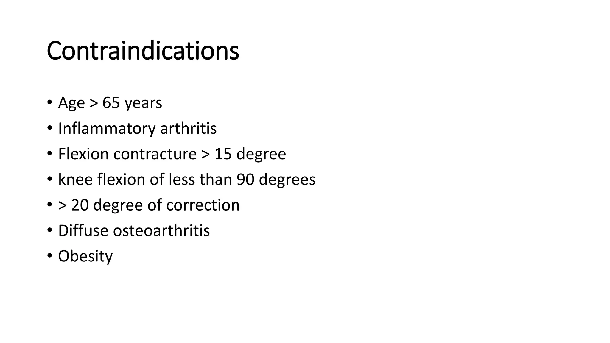
Topic : HIGH TIBIAL OSTEOTOMY
Moderator- Dr. Satish S
Presenter – Dr. Karthik M V
Introduction
• High tibial osteotomy is a well-established procedure for the
treatment of unicompartmental osteoarthritis of the knee.
• Most reports have shown approximately 80% satisfactory results
at 5 years and 60% at 10 years after high tibial osteotomy.
Osteotomy since 20th century
• First introduced by Jackson & Waugh in
1961
• Jackson operated distal to the tibial
tuberosity
• Classic osteotomy of Coventry 1965 was
a closed-wedge valgization type including
a fibula osteotomy, and was performed
proximal to the tibial tuberosity
AXIS
• Each long bone has 2 axis
1. Mechanical axis
2. Anatomic axis
• MECHANICAL AXIS - The mechanical axis
of a bone is defined as the straight line
connecting the joint center points of the
proximal and distal joints.
• Mechanical axis is always a straight line
connecting two joint center points
MECHANICAL AXIS
ANATOMIC AXIS
• ANATOMIC AXIS - The anatomic axis of a long
bone is the mid-diaphyseal line of that bone.
• In straight bones, the anatomic axis follows
the straight middiaphyseal path.
In Tibia Mechanical and
Anatomic axis are
Parallel, but not same.
The tibial anatomic-
mechanical angle (AMA)
is 0°
The Anatomic axis
slightly medial to the
Mechanical axis.
In the femur, the
mechanical and anatomic
axes are different and
converge distally.
The normal femoral AMA
is 7±2°.
Joint Orientation Angles
• It is the angle formed between the joint orientation line
and either the mechanical or anatomic axis.
 m – mechanical axis
 a – anatomic axis
 M – medial
 L – lateral
 A – anterior
 P – posterior
 F – femur
 T – Tibia
Mechanical axis of lower limb (Mikulicz line)
• In frontal plane, the line passing from the
center of the femoral head to the center of
the ankle plafond
• Typically passes immediately medial to the
center of the knee.
• Malalignment occurs when the center of the
knee does not lie close to this line.
Mechanical axis deviation (MAD)
• The distance between the mechanical axis of
the lower limb and the center of the knee in
the frontal plane is the MAD.
• Normally, 1 mm to 15 mm medial tothe knee
joint center.
MECHANICAL AXES DEVIATION
• MAD >15 mm medial to the knee
midpoint- varus malalignment.
• MAD > 10mm lateral to knee joint mid
point- valgus malalignment.
Center of rotation of angulation(CORA)
• It is the intersection of the proximal axis and distal
axis of a deformed bone is called as CORA.
• It is the point about which a deformity may be
rotated to achieve correction.
• Either anatomic or mechanical axis can be used.
Radiographic assessment
 Full length AP view (frontal plane)
 Full length LAT view (sagittal plane).
 The true AP view of the knee is obtained in the knee forward
position (patella centered on the femoral condyles)
 The correct method is to orient the patella forward, irrespective of
the foot position
The knee is kept in full
extension. To see the proximal
femur, the pelvis is rotated
posteriorly 30°- 45° without
rotating the knee on the study
side.
Osteotomy
• An osteotomy is used to separate the deformed bone segments to
allow realignment of the anatomic and mechanical axes.
• The ability of an osteotomy to restore alignment depends on
a. CORA
b. the correction axis (ACA)
c. the location of the osteotomy.
OSTEOTOMY RULE 1
• When the osteotomy line and
ACA pass through any of the
CORAs, realignment occurs
without translation
OSTEOTOMY RULE 2
• When the ACA is through the
CORA but the osteotomy is at
a different level, the axis will
realign by angulation and
translation at the osteotomy
site.
OSTEOTOMY RULE 3
• When the osteotomy and
ACA are at a level above or
below the CORAs, a
translational deformity will
result.
WEDGE OSTEOTOMY
• The type of osteotomy is determined by location of osteotomy
relative to locations of the CORA and the ACA
• The CORA and ACA may lie on concave or convex side of cortex, or
in the middle of the bone.
OPENING WEDGE OSTEOTOMY
• The ACA-CORA lie on the convex cortex of the deformity,
the correction will result in an opening wedge osteotomy.
• All points on the convex side of the ACA-CORA will be
compressed, whereas all points on the concave side will
be distracted.
• In an opening wedge osteotomy, the cortex on the concave
side is distracted to restore the alignment.
• There is a wedge-shaped bone defect with its base on the
concave side
• An opening wedge osteotomy increases the bone length.
Closing Wedge Osteotomy
• The ACA-CORA lie on the concave cortex of the
deformity, the correction will result in an closing
wedge osteotomy.
• In a closing wedge osteotomy, the cortex on the
convex side is compressed, this involves removal of a
bone wedge across the entire bone diameter to
restore alignment.
• A closing wedge osteotomy decreases bone length.
Neutral Wedge Osteotomy
• The ACA-CORA lie in the middle of the bone, the
correction will result in an Neutral wedge osteotomy.
• A bone wedge is removed from the convex side to allow
realignment.
• The cortex on the convex side is compressed and
concave side is distracted.
• Has no effect on bone length.
Fujisawa point
• The best results from HTO were
obtained when the mechanical axis
line of the limb passed through the
30%- 40% lateral plateau region.
We call this the Fujisawa point.
AMOUNT OF CORRECTION
• The aim of the mechanical
axis is at 10–15 % of the scale
in the lateral compartment
for patients who have lost
one third of their medial
compartment cartilage,
• 20–25 % if two thirds of
medial cartilage is lost,
• and at 30–35 % if the medial
cartilage is completely lost
PLANNING ALGORITHM FOR HIGH-TIBIAL
OSTEOTOMY
• Preoperative radiographic
evaluation for high-tibial
osteotomy (HTO) includes
• bilateral weight-bearing AP
views in full extension
• bilateral weight-bearing (PA)
tunnel views in 45° of flexion
(Rosenberg view)
ANGLE OF CORRECTION
Varus malalignment caused due to
ligamentous laxity
MINIACI METHOD
AMOUNT OF WEDGE REQUIRED ?
• if tibia is 57 mm wide, length of wedge=degrees of correction Length
= Diameter of tibia(at osteotomy site) X 0.02 X Angle
• The size of the wedge
removed as roughly 1 degree
of correction for each 1 mm
of length at the base of the
wedge
• In a well-aligned knee, load distribution is not well-balanced but
physiologically 60 % in the medial and 40 % in the lateral
compartment, the medial compartment has been estimated to take
75% of the load during single-leg stance
• When the mechanical axis is moved into 6° of valgus, the load is 40%
medial and 60% lateral.
• Mechanical alignment of the lower limb should be moved into 2°-6°
of mechanical valgus
• Principle - HTO unloads the affected part of knee joint by correcting
malalignment and redistributing stresses hence relieves pain and
restores function.
• Main indication – correct Varus Malalignment in Medial
unicompartmental Osteoarthritis
• Aim – unload Medial compartment by slightly overcorrecting into
valgus – reduce pain, slow degenerative process, delay joint
replacement.
IDEAL CANDIDATE FOR HTO
• Is younger than 65 years (male) respectively 55 years(female)
• Has almost normal range of motion
• Malalignment test < 15 degree
• Is a non-smoker
• Has a certain pain tolerance
• May have ACL or PCL deficiency (can be addressed by the surgery)
• Should preferably have a BMI under 30
Contraindications
• Age > 65 years
• Inflammatory arthritis
• Flexion contracture > 15 degree
• knee flexion of less than 90 degrees
• > 20 degree of correction
• Diffuse osteoarthritis
• Obesity
Four basic types are most commonly used:
1. Lateral closing wedge, valgus producing osteotomy
2. Medial opening wedge osteotomy with bone graft
3. Dome (barrel vault) osteotomy of Macquet
4. Opening wedge osteotomy using external fixators (hemicallotasis)
- Ilizarov
-External fixators
Lateral Closing Wedge Osteotomy
• Coventry described a closing wedge osteotomy made proximal to the
tibial tuberosity.
• Normally, there is valgus alignment of 5 to 8 degrees
• The amount of correction of the arthritic knee needed to achieve a
normal angle is calculated, and an additional 3 to 5 degrees of
overcorrection is added to achieve approximately 10 degrees of
valgus.
• leaving a thin posteromedial lip of
bone on the proximal tibial
fragment.
• osteotomy is closed after removal of
the wedge of bone, this posterior lip
overrides the proximal end of the
distal fragment and gives added
support and stability
• Removing the inferomedial portion of the
fibular head
Or
• disruption of the proximal tibiofibular
syndesmosis
• Care should be taken to not injure the
peroneal nerve.
Procedure
• Curved incision
from the head
of the fibula to
2 cm below
the tibial
tubercle.
Peroneal nerve
protected.
A, Incision. B, Positioning of transverse osteotomy guide. C, Determination of correct position of jig.
D, Placement of oblique osteotomy guide over 3.2-mm smooth pins and performance of osteotomy.
E, Placement of L-shaped osteotomy plate. F, Application of compression clamp
Leave 15–20 mm of tibial plateau to avoid fracture.
G, Application of slow compression. H, Fixation of distal plate.
Advantages
• Most stable
• Early consolidation
• Early mobilisation
• Exploration of knee joint through same approach
Disadvantages
• Limb shortening
• Nerve injury
• LCL laxity
Post op
• Ambulation is begun on the second day after surgery, and 50% weight
bearing is allowed for the first 6 weeks with the use of crutches
• Active ROM from POD 2
Medial Opening Wedge Osteotomy
• Hernigou et al. described a medial opening wedge
tibial osteotomy which they thought is more
precise and allows more exact correction than does
a lateral closing wedge
• Opening wedge osteotomy should be done if the
involved extremity is 2 cm or more shorter than the
contralateral extremity.
• Indicated in pt with MCL laxity
• A lateral hinge fracture with extension into the
lateral tibial plateau is a common complication
Why MOWO is better ?
• Allows precise correction, amount of correction can be changed
intraoperatively
• Proximal Tibiofibular joint need not be disrupted
• Only one saw cut
• No leg shortening
• Avoids dissection of Peroneal nerve
SURGICAL TECHNIQUE
Two k- wire parallel to each other
Tomo fix bone spreader
Chisel to increase osteotomy gap Bone spreader forceps
IMPLANT DESIGN OF THE ANGULAR LOCKING
PLATE FIXATOR TOMOFIX
SURGICAL TECHNIQUE
SURGICAL TECHNIQUE
• a temporary lag screw is inserted into the first
plate hole distal to the osteotomy.
• This compresses the lateral hinge by pulling the
distal osteotomy segment towards the plate
fixator.
• The osteotomy gap is watched constantly while
the lag screw is tightened slowly to avoid
secondary loss of correction.
• In the last step of the operation this screw is
replaced by a bicortical locking screw.
Removal of Lag screw
Replaced by Locking screw
Complications of Medial Opening Wedge Osteotomy
• Delay healing and Non union
• Leg lengthening
• Prolonged weight bearing
• Loss of correction
BARREL VAULT /DOME
OSTEOTOMY
▶ Maquet described barrel vault osteotomy.
▶ In which he believed more accuracy and
adjustability of correction.
▶ It may be considered if a correction angle >20*
▶ Because this osteotomy is inherently stable, so
internal fixation not required.
▶ Maquet use special jig to orient dome
osteotomy properly.
▶ In this procedure Distal tibia can be translated
if needed to change patellar tracking for
patellofemoral degenerative changes.
Opening wedge hemicallotasis
• Schwartsman advocated the use Ilizarov Ex fixator after percutaneous
tibial osteotomy distal to the tibial tuberosity
• adjustments can be made postoperatively on the basis of standing,
weight-bearing radiographs
• placement of the osteotomy below the tibial tubercle minimizes
patellar complications
Advantages
• Improved stability of fixation
• Immediate weight bearing
• Knee motion of 0 to 90 degrees in the circular frame.
Disadvantages
• Pin loosening
• Pin site infection
• Pt compliance
Effects of HTO on cartilage
• HTO alone promote cartilage regeneration
• HTO  degenerated articular cartilage covered by fibrocartilage
• HTO + cartilage restoration techniques
Microfractures
Abrasion arthroplasty
Stem cell therapy
References
• Osteotomies around the Knee Indications—Planning—Surgical
techniques using plate fixators -2009
• Campbell’s operative orthopaedics -14th edition
• Principles of deformity correction – Dror paley
• Turek’s Orthopaedics – 7th edition

HIGH TIBIAL OSTEOTOMY AROUND THE KNEE.pptx

  • 1.
    Topic : HIGHTIBIAL OSTEOTOMY Moderator- Dr. Satish S Presenter – Dr. Karthik M V
  • 2.
    Introduction • High tibialosteotomy is a well-established procedure for the treatment of unicompartmental osteoarthritis of the knee. • Most reports have shown approximately 80% satisfactory results at 5 years and 60% at 10 years after high tibial osteotomy.
  • 3.
    Osteotomy since 20thcentury • First introduced by Jackson & Waugh in 1961 • Jackson operated distal to the tibial tuberosity • Classic osteotomy of Coventry 1965 was a closed-wedge valgization type including a fibula osteotomy, and was performed proximal to the tibial tuberosity
  • 4.
    AXIS • Each longbone has 2 axis 1. Mechanical axis 2. Anatomic axis
  • 5.
    • MECHANICAL AXIS- The mechanical axis of a bone is defined as the straight line connecting the joint center points of the proximal and distal joints. • Mechanical axis is always a straight line connecting two joint center points MECHANICAL AXIS
  • 6.
    ANATOMIC AXIS • ANATOMICAXIS - The anatomic axis of a long bone is the mid-diaphyseal line of that bone. • In straight bones, the anatomic axis follows the straight middiaphyseal path.
  • 7.
    In Tibia Mechanicaland Anatomic axis are Parallel, but not same. The tibial anatomic- mechanical angle (AMA) is 0° The Anatomic axis slightly medial to the Mechanical axis.
  • 8.
    In the femur,the mechanical and anatomic axes are different and converge distally. The normal femoral AMA is 7±2°.
  • 9.
    Joint Orientation Angles •It is the angle formed between the joint orientation line and either the mechanical or anatomic axis.  m – mechanical axis  a – anatomic axis  M – medial  L – lateral  A – anterior  P – posterior  F – femur  T – Tibia
  • 10.
    Mechanical axis oflower limb (Mikulicz line) • In frontal plane, the line passing from the center of the femoral head to the center of the ankle plafond • Typically passes immediately medial to the center of the knee. • Malalignment occurs when the center of the knee does not lie close to this line.
  • 11.
    Mechanical axis deviation(MAD) • The distance between the mechanical axis of the lower limb and the center of the knee in the frontal plane is the MAD. • Normally, 1 mm to 15 mm medial tothe knee joint center.
  • 12.
    MECHANICAL AXES DEVIATION •MAD >15 mm medial to the knee midpoint- varus malalignment. • MAD > 10mm lateral to knee joint mid point- valgus malalignment.
  • 14.
    Center of rotationof angulation(CORA) • It is the intersection of the proximal axis and distal axis of a deformed bone is called as CORA. • It is the point about which a deformity may be rotated to achieve correction. • Either anatomic or mechanical axis can be used.
  • 15.
    Radiographic assessment  Fulllength AP view (frontal plane)  Full length LAT view (sagittal plane).  The true AP view of the knee is obtained in the knee forward position (patella centered on the femoral condyles)  The correct method is to orient the patella forward, irrespective of the foot position
  • 18.
    The knee iskept in full extension. To see the proximal femur, the pelvis is rotated posteriorly 30°- 45° without rotating the knee on the study side.
  • 19.
    Osteotomy • An osteotomyis used to separate the deformed bone segments to allow realignment of the anatomic and mechanical axes. • The ability of an osteotomy to restore alignment depends on a. CORA b. the correction axis (ACA) c. the location of the osteotomy.
  • 20.
    OSTEOTOMY RULE 1 •When the osteotomy line and ACA pass through any of the CORAs, realignment occurs without translation
  • 21.
    OSTEOTOMY RULE 2 •When the ACA is through the CORA but the osteotomy is at a different level, the axis will realign by angulation and translation at the osteotomy site.
  • 22.
    OSTEOTOMY RULE 3 •When the osteotomy and ACA are at a level above or below the CORAs, a translational deformity will result.
  • 23.
    WEDGE OSTEOTOMY • Thetype of osteotomy is determined by location of osteotomy relative to locations of the CORA and the ACA • The CORA and ACA may lie on concave or convex side of cortex, or in the middle of the bone.
  • 24.
    OPENING WEDGE OSTEOTOMY •The ACA-CORA lie on the convex cortex of the deformity, the correction will result in an opening wedge osteotomy. • All points on the convex side of the ACA-CORA will be compressed, whereas all points on the concave side will be distracted. • In an opening wedge osteotomy, the cortex on the concave side is distracted to restore the alignment. • There is a wedge-shaped bone defect with its base on the concave side • An opening wedge osteotomy increases the bone length.
  • 26.
    Closing Wedge Osteotomy •The ACA-CORA lie on the concave cortex of the deformity, the correction will result in an closing wedge osteotomy. • In a closing wedge osteotomy, the cortex on the convex side is compressed, this involves removal of a bone wedge across the entire bone diameter to restore alignment. • A closing wedge osteotomy decreases bone length.
  • 28.
    Neutral Wedge Osteotomy •The ACA-CORA lie in the middle of the bone, the correction will result in an Neutral wedge osteotomy. • A bone wedge is removed from the convex side to allow realignment. • The cortex on the convex side is compressed and concave side is distracted. • Has no effect on bone length.
  • 30.
    Fujisawa point • Thebest results from HTO were obtained when the mechanical axis line of the limb passed through the 30%- 40% lateral plateau region. We call this the Fujisawa point.
  • 31.
    AMOUNT OF CORRECTION •The aim of the mechanical axis is at 10–15 % of the scale in the lateral compartment for patients who have lost one third of their medial compartment cartilage, • 20–25 % if two thirds of medial cartilage is lost, • and at 30–35 % if the medial cartilage is completely lost
  • 32.
    PLANNING ALGORITHM FORHIGH-TIBIAL OSTEOTOMY • Preoperative radiographic evaluation for high-tibial osteotomy (HTO) includes • bilateral weight-bearing AP views in full extension • bilateral weight-bearing (PA) tunnel views in 45° of flexion (Rosenberg view)
  • 33.
    ANGLE OF CORRECTION Varusmalalignment caused due to ligamentous laxity
  • 34.
  • 35.
    AMOUNT OF WEDGEREQUIRED ? • if tibia is 57 mm wide, length of wedge=degrees of correction Length = Diameter of tibia(at osteotomy site) X 0.02 X Angle • The size of the wedge removed as roughly 1 degree of correction for each 1 mm of length at the base of the wedge
  • 36.
    • In awell-aligned knee, load distribution is not well-balanced but physiologically 60 % in the medial and 40 % in the lateral compartment, the medial compartment has been estimated to take 75% of the load during single-leg stance • When the mechanical axis is moved into 6° of valgus, the load is 40% medial and 60% lateral. • Mechanical alignment of the lower limb should be moved into 2°-6° of mechanical valgus
  • 37.
    • Principle -HTO unloads the affected part of knee joint by correcting malalignment and redistributing stresses hence relieves pain and restores function. • Main indication – correct Varus Malalignment in Medial unicompartmental Osteoarthritis • Aim – unload Medial compartment by slightly overcorrecting into valgus – reduce pain, slow degenerative process, delay joint replacement.
  • 38.
    IDEAL CANDIDATE FORHTO • Is younger than 65 years (male) respectively 55 years(female) • Has almost normal range of motion • Malalignment test < 15 degree • Is a non-smoker • Has a certain pain tolerance • May have ACL or PCL deficiency (can be addressed by the surgery) • Should preferably have a BMI under 30
  • 39.
    Contraindications • Age >65 years • Inflammatory arthritis • Flexion contracture > 15 degree • knee flexion of less than 90 degrees • > 20 degree of correction • Diffuse osteoarthritis • Obesity
  • 40.
    Four basic typesare most commonly used: 1. Lateral closing wedge, valgus producing osteotomy 2. Medial opening wedge osteotomy with bone graft 3. Dome (barrel vault) osteotomy of Macquet 4. Opening wedge osteotomy using external fixators (hemicallotasis) - Ilizarov -External fixators
  • 41.
    Lateral Closing WedgeOsteotomy • Coventry described a closing wedge osteotomy made proximal to the tibial tuberosity. • Normally, there is valgus alignment of 5 to 8 degrees • The amount of correction of the arthritic knee needed to achieve a normal angle is calculated, and an additional 3 to 5 degrees of overcorrection is added to achieve approximately 10 degrees of valgus.
  • 42.
    • leaving athin posteromedial lip of bone on the proximal tibial fragment. • osteotomy is closed after removal of the wedge of bone, this posterior lip overrides the proximal end of the distal fragment and gives added support and stability
  • 43.
    • Removing theinferomedial portion of the fibular head Or • disruption of the proximal tibiofibular syndesmosis • Care should be taken to not injure the peroneal nerve.
  • 44.
    Procedure • Curved incision fromthe head of the fibula to 2 cm below the tibial tubercle. Peroneal nerve protected. A, Incision. B, Positioning of transverse osteotomy guide. C, Determination of correct position of jig.
  • 45.
    D, Placement ofoblique osteotomy guide over 3.2-mm smooth pins and performance of osteotomy. E, Placement of L-shaped osteotomy plate. F, Application of compression clamp Leave 15–20 mm of tibial plateau to avoid fracture.
  • 46.
    G, Application ofslow compression. H, Fixation of distal plate.
  • 47.
    Advantages • Most stable •Early consolidation • Early mobilisation • Exploration of knee joint through same approach Disadvantages • Limb shortening • Nerve injury • LCL laxity Post op • Ambulation is begun on the second day after surgery, and 50% weight bearing is allowed for the first 6 weeks with the use of crutches • Active ROM from POD 2
  • 48.
    Medial Opening WedgeOsteotomy • Hernigou et al. described a medial opening wedge tibial osteotomy which they thought is more precise and allows more exact correction than does a lateral closing wedge • Opening wedge osteotomy should be done if the involved extremity is 2 cm or more shorter than the contralateral extremity. • Indicated in pt with MCL laxity • A lateral hinge fracture with extension into the lateral tibial plateau is a common complication
  • 49.
    Why MOWO isbetter ? • Allows precise correction, amount of correction can be changed intraoperatively • Proximal Tibiofibular joint need not be disrupted • Only one saw cut • No leg shortening • Avoids dissection of Peroneal nerve
  • 50.
  • 51.
    Two k- wireparallel to each other
  • 53.
    Tomo fix bonespreader Chisel to increase osteotomy gap Bone spreader forceps
  • 54.
    IMPLANT DESIGN OFTHE ANGULAR LOCKING PLATE FIXATOR TOMOFIX
  • 55.
  • 56.
    SURGICAL TECHNIQUE • atemporary lag screw is inserted into the first plate hole distal to the osteotomy. • This compresses the lateral hinge by pulling the distal osteotomy segment towards the plate fixator. • The osteotomy gap is watched constantly while the lag screw is tightened slowly to avoid secondary loss of correction. • In the last step of the operation this screw is replaced by a bicortical locking screw.
  • 57.
  • 58.
  • 59.
    Complications of MedialOpening Wedge Osteotomy • Delay healing and Non union • Leg lengthening • Prolonged weight bearing • Loss of correction
  • 61.
    BARREL VAULT /DOME OSTEOTOMY ▶Maquet described barrel vault osteotomy. ▶ In which he believed more accuracy and adjustability of correction. ▶ It may be considered if a correction angle >20* ▶ Because this osteotomy is inherently stable, so internal fixation not required. ▶ Maquet use special jig to orient dome osteotomy properly. ▶ In this procedure Distal tibia can be translated if needed to change patellar tracking for patellofemoral degenerative changes.
  • 62.
    Opening wedge hemicallotasis •Schwartsman advocated the use Ilizarov Ex fixator after percutaneous tibial osteotomy distal to the tibial tuberosity • adjustments can be made postoperatively on the basis of standing, weight-bearing radiographs • placement of the osteotomy below the tibial tubercle minimizes patellar complications
  • 63.
    Advantages • Improved stabilityof fixation • Immediate weight bearing • Knee motion of 0 to 90 degrees in the circular frame. Disadvantages • Pin loosening • Pin site infection • Pt compliance
  • 65.
    Effects of HTOon cartilage • HTO alone promote cartilage regeneration • HTO  degenerated articular cartilage covered by fibrocartilage • HTO + cartilage restoration techniques Microfractures Abrasion arthroplasty Stem cell therapy
  • 66.
    References • Osteotomies aroundthe Knee Indications—Planning—Surgical techniques using plate fixators -2009 • Campbell’s operative orthopaedics -14th edition • Principles of deformity correction – Dror paley • Turek’s Orthopaedics – 7th edition

Editor's Notes

  • #4 Diff b/w osteotomy prox and distal to tib tuberosity