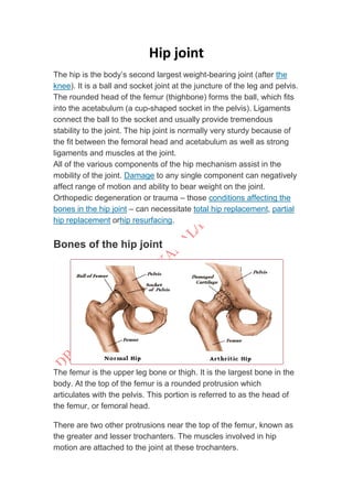
The hip joint is a ball and socket joint that connects the femur to the pelvis. It is the body's largest weight bearing joint. The rounded head of the femur fits into the cup-shaped acetabulum of the pelvis. Strong ligaments and muscles provide stability to the joint. Damage to any of the hip joint components can negatively affect its range of motion and weight bearing ability, and may require hip replacement surgery. The hip allows for flexion, extension, abduction, adduction, internal and external rotation.
MyTradeTV Glazing Digital Magazine July 2014mytradetv
The MyTradeTV Glass and Glazing Digital Magazine is full of Aluminium, Conservatories, Doors, Glazing Events, Glass, Machinery, Recruitment, Windows, uPVC, Double Glazing, Triple Glazing News, Information, Videos, Podcasts.
all information about
it include :-
1- DEFINITION
2- SIGNS AND SYMPTOMS
3- MECHANISM
4- RISK FACTOR
5- COMPLICATIONS
6- PRECAUTION
7- PREVENTION
8- EPIDEMIOLOGY
9- TREATMENT
and finally video about the mechanism of action
if you van not open it
hit this link
https://www.youtube.com/watch?v=ZuUfGeoN_cw
i hope it meet what you want
this slideshow describes about the hip joint anatomy, biomechanics and its pathomechanics along with angles of hip joint. the slide show also briefs about the pelvic femoral rhythm in daily activities
Femoral Head (Superiorly, Medially, Anteriorly).
Acetabulum (Inferiorly, Laterally, Anteriorly).
Horseshoe-shaped (Acetabular Notch).
The deepest portion (Acetabular Fossa).
Labrum Acetabular:
Is a wedged fibrocartilaginous ring inserted into the acetabular rim to increase the acetabular concavity.
Factory Supply Best Quality Pmk Oil CAS 28578–16–7 PMK Powder in Stockrebeccabio
Factory Supply Best Quality Pmk Oil CAS 28578–16–7 PMK Powder in Stock
Telegram: bmksupplier
signal: +85264872720
threema: TUD4A6YC
You can contact me on Telegram or Threema
Communicate promptly and reply
Free of customs clearance, Double Clearance 100% pass delivery to USA, Canada, Spain, Germany, Netherland, Poland, Italy, Sweden, UK, Czech Republic, Australia, Mexico, Russia, Ukraine, Kazakhstan.Door to door service
Hot Selling Organic intermediates
Title: Sense of Smell
Presenter: Dr. Faiza, Assistant Professor of Physiology
Qualifications:
MBBS (Best Graduate, AIMC Lahore)
FCPS Physiology
ICMT, CHPE, DHPE (STMU)
MPH (GC University, Faisalabad)
MBA (Virtual University of Pakistan)
Learning Objectives:
Describe the primary categories of smells and the concept of odor blindness.
Explain the structure and location of the olfactory membrane and mucosa, including the types and roles of cells involved in olfaction.
Describe the pathway and mechanisms of olfactory signal transmission from the olfactory receptors to the brain.
Illustrate the biochemical cascade triggered by odorant binding to olfactory receptors, including the role of G-proteins and second messengers in generating an action potential.
Identify different types of olfactory disorders such as anosmia, hyposmia, hyperosmia, and dysosmia, including their potential causes.
Key Topics:
Olfactory Genes:
3% of the human genome accounts for olfactory genes.
400 genes for odorant receptors.
Olfactory Membrane:
Located in the superior part of the nasal cavity.
Medially: Folds downward along the superior septum.
Laterally: Folds over the superior turbinate and upper surface of the middle turbinate.
Total surface area: 5-10 square centimeters.
Olfactory Mucosa:
Olfactory Cells: Bipolar nerve cells derived from the CNS (100 million), with 4-25 olfactory cilia per cell.
Sustentacular Cells: Produce mucus and maintain ionic and molecular environment.
Basal Cells: Replace worn-out olfactory cells with an average lifespan of 1-2 months.
Bowman’s Gland: Secretes mucus.
Stimulation of Olfactory Cells:
Odorant dissolves in mucus and attaches to receptors on olfactory cilia.
Involves a cascade effect through G-proteins and second messengers, leading to depolarization and action potential generation in the olfactory nerve.
Quality of a Good Odorant:
Small (3-20 Carbon atoms), volatile, water-soluble, and lipid-soluble.
Facilitated by odorant-binding proteins in mucus.
Membrane Potential and Action Potential:
Resting membrane potential: -55mV.
Action potential frequency in the olfactory nerve increases with odorant strength.
Adaptation Towards the Sense of Smell:
Rapid adaptation within the first second, with further slow adaptation.
Psychological adaptation greater than receptor adaptation, involving feedback inhibition from the central nervous system.
Primary Sensations of Smell:
Camphoraceous, Musky, Floral, Pepperminty, Ethereal, Pungent, Putrid.
Odor Detection Threshold:
Examples: Hydrogen sulfide (0.0005 ppm), Methyl-mercaptan (0.002 ppm).
Some toxic substances are odorless at lethal concentrations.
Characteristics of Smell:
Odor blindness for single substances due to lack of appropriate receptor protein.
Behavioral and emotional influences of smell.
Transmission of Olfactory Signals:
From olfactory cells to glomeruli in the olfactory bulb, involving lateral inhibition.
Primitive, less old, and new olfactory systems with different path
MANAGEMENT OF ATRIOVENTRICULAR CONDUCTION BLOCK.pdfJim Jacob Roy
Cardiac conduction defects can occur due to various causes.
Atrioventricular conduction blocks ( AV blocks ) are classified into 3 types.
This document describes the acute management of AV block.
Anti ulcer drugs and their Advance pharmacology ||
Anti-ulcer drugs are medications used to prevent and treat ulcers in the stomach and upper part of the small intestine (duodenal ulcers). These ulcers are often caused by an imbalance between stomach acid and the mucosal lining, which protects the stomach lining.
||Scope: Overview of various classes of anti-ulcer drugs, their mechanisms of action, indications, side effects, and clinical considerations.
Lung Cancer: Artificial Intelligence, Synergetics, Complex System Analysis, S...Oleg Kshivets
RESULTS: Overall life span (LS) was 2252.1±1742.5 days and cumulative 5-year survival (5YS) reached 73.2%, 10 years – 64.8%, 20 years – 42.5%. 513 LCP lived more than 5 years (LS=3124.6±1525.6 days), 148 LCP – more than 10 years (LS=5054.4±1504.1 days).199 LCP died because of LC (LS=562.7±374.5 days). 5YS of LCP after bi/lobectomies was significantly superior in comparison with LCP after pneumonectomies (78.1% vs.63.7%, P=0.00001 by log-rank test). AT significantly improved 5YS (66.3% vs. 34.8%) (P=0.00000 by log-rank test) only for LCP with N1-2. Cox modeling displayed that 5YS of LCP significantly depended on: phase transition (PT) early-invasive LC in terms of synergetics, PT N0—N12, cell ratio factors (ratio between cancer cells- CC and blood cells subpopulations), G1-3, histology, glucose, AT, blood cell circuit, prothrombin index, heparin tolerance, recalcification time (P=0.000-0.038). Neural networks, genetic algorithm selection and bootstrap simulation revealed relationships between 5YS and PT early-invasive LC (rank=1), PT N0—N12 (rank=2), thrombocytes/CC (3), erythrocytes/CC (4), eosinophils/CC (5), healthy cells/CC (6), lymphocytes/CC (7), segmented neutrophils/CC (8), stick neutrophils/CC (9), monocytes/CC (10); leucocytes/CC (11). Correct prediction of 5YS was 100% by neural networks computing (area under ROC curve=1.0; error=0.0).
CONCLUSIONS: 5YS of LCP after radical procedures significantly depended on: 1) PT early-invasive cancer; 2) PT N0--N12; 3) cell ratio factors; 4) blood cell circuit; 5) biochemical factors; 6) hemostasis system; 7) AT; 8) LC characteristics; 9) LC cell dynamics; 10) surgery type: lobectomy/pneumonectomy; 11) anthropometric data. Optimal diagnosis and treatment strategies for LC are: 1) screening and early detection of LC; 2) availability of experienced thoracic surgeons because of complexity of radical procedures; 3) aggressive en block surgery and adequate lymph node dissection for completeness; 4) precise prediction; 5) adjuvant chemoimmunoradiotherapy for LCP with unfavorable prognosis.
These lecture slides, by Dr Sidra Arshad, offer a quick overview of physiological basis of a normal electrocardiogram.
Learning objectives:
1. Define an electrocardiogram (ECG) and electrocardiography
2. Describe how dipoles generated by the heart produce the waveforms of the ECG
3. Describe the components of a normal electrocardiogram of a typical bipolar leads (limb II)
4. Differentiate between intervals and segments
5. Enlist some common indications for obtaining an ECG
Study Resources:
1. Chapter 11, Guyton and Hall Textbook of Medical Physiology, 14th edition
2. Chapter 9, Human Physiology - From Cells to Systems, Lauralee Sherwood, 9th edition
3. Chapter 29, Ganong’s Review of Medical Physiology, 26th edition
4. Electrocardiogram, StatPearls - https://www.ncbi.nlm.nih.gov/books/NBK549803/
5. ECG in Medical Practice by ABM Abdullah, 4th edition
6. ECG Basics, http://www.nataliescasebook.com/tag/e-c-g-basics
Ozempic: Preoperative Management of Patients on GLP-1 Receptor Agonists Saeid Safari
Preoperative Management of Patients on GLP-1 Receptor Agonists like Ozempic and Semiglutide
ASA GUIDELINE
NYSORA Guideline
2 Case Reports of Gastric Ultrasound
The prostate is an exocrine gland of the male mammalian reproductive system
It is a walnut-sized gland that forms part of the male reproductive system and is located in front of the rectum and just below the urinary bladder
Function is to store and secrete a clear, slightly alkaline fluid that constitutes 10-30% of the volume of the seminal fluid that along with the spermatozoa, constitutes semen
A healthy human prostate measures (4cm-vertical, by 3cm-horizontal, 2cm ant-post ).
It surrounds the urethra just below the urinary bladder. It has anterior, median, posterior and two lateral lobes
It’s work is regulated by androgens which are responsible for male sex characteristics
Generalised disease of the prostate due to hormonal derangement which leads to non malignant enlargement of the gland (increase in the number of epithelial cells and stromal tissue)to cause compression of the urethra leading to symptoms (LUTS
New Directions in Targeted Therapeutic Approaches for Older Adults With Mantl...i3 Health
i3 Health is pleased to make the speaker slides from this activity available for use as a non-accredited self-study or teaching resource.
This slide deck presented by Dr. Kami Maddocks, Professor-Clinical in the Division of Hematology and
Associate Division Director for Ambulatory Operations
The Ohio State University Comprehensive Cancer Center, will provide insight into new directions in targeted therapeutic approaches for older adults with mantle cell lymphoma.
STATEMENT OF NEED
Mantle cell lymphoma (MCL) is a rare, aggressive B-cell non-Hodgkin lymphoma (NHL) accounting for 5% to 7% of all lymphomas. Its prognosis ranges from indolent disease that does not require treatment for years to very aggressive disease, which is associated with poor survival (Silkenstedt et al, 2021). Typically, MCL is diagnosed at advanced stage and in older patients who cannot tolerate intensive therapy (NCCN, 2022). Although recent advances have slightly increased remission rates, recurrence and relapse remain very common, leading to a median overall survival between 3 and 6 years (LLS, 2021). Though there are several effective options, progress is still needed towards establishing an accepted frontline approach for MCL (Castellino et al, 2022). Treatment selection and management of MCL are complicated by the heterogeneity of prognosis, advanced age and comorbidities of patients, and lack of an established standard approach for treatment, making it vital that clinicians be familiar with the latest research and advances in this area. In this activity chaired by Michael Wang, MD, Professor in the Department of Lymphoma & Myeloma at MD Anderson Cancer Center, expert faculty will discuss prognostic factors informing treatment, the promising results of recent trials in new therapeutic approaches, and the implications of treatment resistance in therapeutic selection for MCL.
Target Audience
Hematology/oncology fellows, attending faculty, and other health care professionals involved in the treatment of patients with mantle cell lymphoma (MCL).
Learning Objectives
1.) Identify clinical and biological prognostic factors that can guide treatment decision making for older adults with MCL
2.) Evaluate emerging data on targeted therapeutic approaches for treatment-naive and relapsed/refractory MCL and their applicability to older adults
3.) Assess mechanisms of resistance to targeted therapies for MCL and their implications for treatment selection
The hemodynamic and autonomic determinants of elevated blood pressure in obes...
Hip joint
1. KINESOLOGY OF HIP JOINT
PRESENTED BY :
DR.ASER MOHAMED KAMAL
PHYSICAL THERAPIST
2. Hip joint
The hip is the body’s second largest weight-bearing joint (after the
knee). It is a ball and socket joint at the juncture of the leg and pelvis.
The rounded head of the femur (thighbone) forms the ball, which fits
into the acetabulum (a cup-shaped socket in the pelvis). Ligaments
connect the ball to the socket and usually provide tremendous
stability to the joint. The hip joint is normally very sturdy because of
the fit between the femoral head and acetabulum as well as strong
ligaments and muscles at the joint.
All of the various components of the hip mechanism assist in the
mobility of the joint. Damage to any single component can negatively
affect range of motion and ability to bear weight on the joint.
Orthopedic degeneration or trauma – those conditions affecting the
bones in the hip joint – can necessitate total hip replacement, partial
hip replacement orhip resurfacing.
Bones of the hip joint
The femur is the upper leg bone or thigh. It is the largest bone in the
body. At the top of the femur is a rounded protrusion which
articulates with the pelvis. This portion is referred to as the head of
the femur, or femoral head.
There are two other protrusions near the top of the femur, known as
the greater and lesser trochanters. The muscles involved in hip
motion are attached to the joint at these trochanters.
3. The acetabulum is a concave area in the pelvis, into which the
femoral head fits. The pelvis is a girdle of bones, connected at the
front by cartilage pad, called the pubis, and at the back by the lowest
four fused vertebrae (the sacrum). The sacro-iliac joints are located
where the sacrum meets the pelvis.
The bone surfaces of the femoral head and acetabulum have a
smooth durable layer of articular cartilage that cushions the ends of
the bones and allows for smooth movement.
Pelvis
o 1/5 pubis
o 2/5 ischium
o 2/5 ilium
o Pelvis full ossification = 15-25 y.o.
Acetabulum
o Concave socket
o Lateral, inferior, anterior (LIA)
o Roundness ↓ w/ age
o Inferior = acetabular notch
o Central/deepest part = acetabular fossa
o Anteversion = anterior orientation of acetabulum
Men = 18.50
Women = 21.5 0
Pathologic ↑ = ↓ jt. stability, risk for anterior
dislocation of femoral head
Femur
o Circular
o Smaller in women
o Fovea
Inferior to medial pt. of femoral head
Attachment of ligament of femoral
head
4. o Medially, superiorly, anteriorly (SAM)
o Neck = 5cm long
o Angulation
Angle of inclination (medial)
Frontal plane bet. Femoral neck & shaft
Early infancy = 1500
Adult = 1250
Elderly = 1200
↓ in women due to width of female pelvis
Pathologic ↑ = coxa valga
Pathologic ↓ = coxa vara
Angle of torsion
Transverse plane bet. Femoral neck &
condyles
Anterior torsion =relative lateral
rotation
↓ w/age
Newborn = 400
Adult = 150
Anteversion
o Pathologic ↑
o Internal femoral torsion
o ↓ ER
o ↑ IR
Retroversion
o Pathologic ↓
o External femoral torsion
o Frog-leg position
FABER
True physiologic position of hip
o A congruent fit under low load would lead to
incongruence under high load
o Periphery of acetabulum in contact, fossa is non-articular
5. Bony landmarks:
Femur:
1. Greater trochanger – lateral aspect of thigh just
distal to hip joint.
2. Medial and lateral condyles– on distal end of femur
3. Medial and lateral epicondyles (Epicondyles)
Patella:
“Knee cap” – anterior aspect of knee (a sesamoid bone
located in quadriceps tendon).
HIP JOINT
› ROM
o Flex 0-1200
o Hypertext 0-100
o Abd 0-450
o Add across30-400
o ER 0-450
o IR 0-350
› Close-packed
o Full ext, IR, Abd
› Open-packed
o 300
flex
o 300
abd
o Slight ER
› Capsular pattern
o Flex, abd, IR
› End feel
o Flex soft/firm
o Ext firm
o Abd soft/firm
o Add soft/firm
o IR firm
o ER firm
› End Feels
› Normal:
o Flexion & Adduction
Elastic or Tissue Approximation
6. o SLR
Elastic
o Extension & Abduction
Elastic/Firm
› IR & ER
Elastic/Firm
› Tonic labyrinthine & optical righting reflexes
o Head effectively behaves as if it’s fixed in a
vertical position
o Maintains head over BOS
› When hip flexor ms. Is tight, keep LOG w/in BOS
o Open-chain response = displacement of
head from vertical (Fig. A)
o Closed-chain response = maintain head in
upright position (Fig. B)
› Acetabulum of pelvis + head of femur
› Diarthrodial, ball-and-socket jt. w/ 30
freedom:
o flex/ext in sagittal plane
o abd/add in frontal plane
o IR/ER in transverse plane
› 10
function of hip
o To support wt. of head, arms & trunk (HAT)
› Also provides pathway for transmission of forces bet. Pelvis & LEs
› Hip tends to operate in a closed kinematic chain
o Proximal end = head
o Distal end = foot
Hip joint capsule or socket
You may hear your hip
surgeon refer to the capsule
or socket, when describing
the structure of the hip joint.
The joint capsule is a thick
ligamentous structure
surrounding the entire joint.
Inside the capsule, the surfaces of the hip joint are covered by a thin
tissue called the synovial membrane. This membrane nourishes and
lubricates the joint.
7. o capsule has major contribution to stability
o femoral neck = intracapsular
o greater & lesser trochanters = extracapsular
o thickened anterosuperiorly
o thin & loose posteroinferiorly
Ligaments
As noted above, the stability of the hip joint is
directly related to its muscles and ligaments. The
most notable ligaments in the hip joint are:
Iliofemoral ligament, which connects the pelvis
to the femur at the front of the joint. It keeps the
hip from hyper-extension
o Iliofemoral ligament
Y ligament of Bigelow
Origin = AIIS
2 arms fan out to insert =
intertrochanteric line of femur
Strongest ligament of hip
Taut in hyperextension
Superior fibers taut in adduction
Inferior tense during abduction
Pubofemoral ligament, which attaches the most forward part of
the pelvis known as the pubis to the femur
Origin = anterior pubic ramus
Insertion = anterior intertrochanteric fossa
Taut in hip abd & ext
8. Ischiofemoral ligament, which attaches to the
ischium (the lowest part of the pelvis) and between
the two trochanters of the femur.
Origin = posterior acetabular rim,
acetabulum labrum
Insertion = spiral around femoral neck
Spiral fibers taut during ext, loosen in
flex
o Position of stability
Full extension of hip
o Position of vulnerability
Flex & add (such as sitting w/thighs
crossed)
Ligamentum teres
Triangular
Ligament of head of femur
Labrum
The labrum is a circular layer of cartilage which surrounds the outer
part of the acetabulum effectively making the socket deeper to
provide more stability for the joint. Labrum tears are not an
uncommon hip injury.
o Acetabular labrum
Fibrocartilage rimming entire periphery
o Transverse acetabular ligament
Roof of tunnel passage for blood vessels & nerves
entering hip
o Has Center Edge angle (CE) or angle of Wiberg
Men = 380
Women = 350
Smaller CE angle (more vertical) = ↓ coverage of
head of femur, ↑ risk superior dislocation of femoral
head
↑ w/age
9. Arthrokinematics
o Movement of convex femoral head on concave
acetabulum
Femoral head glides opposite motion of distal
femur
Flex = Head spins posterior
Ext = anterior spin
o When wt. bearing
Femur fixed, concave acetabulum moves over
convex femoral head
Acetabulum glides in same direction
Hip Mobilization
Flexion: Femur rolls superior & glides inferiorly on pelvis
Extension: Femur rolls inferior & glides superior on pelvis
Abduction: Femur rolls lateral/superior & glides inferior on
pelvis
Adduction: Femur rolls medial/inferior & glides superior on
pelvis
Internal Rotation: Femur rolls medial & glides lateral on
pelvis
External Rotation: Femur rolls lateral & glides medial on
pelvis
Osteokinematics
o Flexion = 900
w/ knee extended
o Normal gait on level ground requires
300
hip flexion
100
hyperextension
50
abd/add/IR/ER
o Anterior pelvic tilt
Sagittal plane
Hip flexion
ASIS anteriorly & inferiorly, symphisis down
o Posterior pelvic tilt
Hip extension
Symphisis pubis up
Posterior pelvis closer to femur
10. o Lateral pelvic tilt
Frontal plane
One hip joint serves as pivot/axis
Opposite iliac crest elevates (hip hike) or drop
(pelvic drop)
Reference is side farthest from supporting hip
o Pelvic rotation
Transverse plane
Occurs in single-limb support around axis of
supporting hip jt.
Forward rotation
Side opposite supporting hip moves
anteriorly
Backward rotation
Side opposite supporting hip moves
posteriorly
o Lumbar-Pelvic Rhythm
Open-chain
E.g. reaching the floor
Hip flexion up to 900
only
Anterior tilt of pelvis on femurs
Flexion of lumbar spine adds 450
E.g. side-lying abduction
Lateral tilt of pelvis & lumbar spine adds
450
o Closed chain response to motions of pelvis
Keeps one or both feet on the ground
Maintain head upright & vertical
Anterior pelvic tilt during hip flexion = head &
trunk displaced forward + lumbar extension
Posterior pelvic tilt + lumbar flexion to keep head
forward over sacrum
11. pelvic motion co-hip motion
compensatory
lumbar
anterior tilt hip flex lumbar ext
posterior tilt hip ext lumbar flex
lateral tilt (drop) right hip add right lateral flex
lateral tilt (hike) right hip abd left lateral flex
forward rot right hip IR rotation to left
backward rot right hip ER rotation to right
Muscle Groups
The muscles of the hip consist of four main
groups
1. Gluteal group: The gluteals are the
muscles in your buttocks. The gluteal muscles
include the gluteus maximus, gluteus
medius, gluteus minimus, and tensor fasciae
latae. They cover the lateral surface of
the ilium.
12. 2. Adductor group: The adductor brevis, adductor
longus, adductor magnus, pectineus, and gracilis make up
the adductor group.
3. Iliopsoas group: The iliacus and psoas major comprise
the iliopsoas group.
4. Lateral rotator group: This group consists of
the externus and internus obturators, the piriformis,
the superior and inferior gemelli, and the quadratus femoris.
5. Other hip muscles: Additional muscles, such as
the rectus femoris and the sartorius, can cause some
movement in the hip joint. However these muscles primarily
move the knee, and not generally classified as muscles of
the hip.
*The hamstring muscles, which originates mostly from the ischial
tuberosity inserting on the tibia/fibula, has a large moment
assisting with hip extension.
Gluteus maximus
Origin Gluteal surface of ilium, lumbar
fascia, sacrum, sacrotuberous ligament
Insertion Gluteal tuberosity of the
femur and iliotibial tract
Nerve Inferior gluteal nerve (L5, S1 and
S2 nerve roots)
ACTION Extends and laterally rotates
hip. Maintains knee extended via
iliotibial tract
Gluteus Medius
Origin: Outer surface of the ilium, between the iliac crest and
the posterior gluteal line above and the anterior gluteal line
below.
Insertion: Posterolateral surface of the greater trochanter of
the femur.
Action: Abduction of the hip, internal rotation of thigh.
Innervation: Superior gluteal nerve L4, 5, S1).
13. Gluteus Minimus
Origin: Outer surface of the ilium, between the anterior and
inferior gluteal lines, and the edge of the greater sciatic notch.
Insertion: Anterior surface of the greater trochanter of the
femur.
Action: Abduction of the thigh, internal rotation of thigh.
Innervation: Superior gluteal nerve L4, 5, S1).
Tensor Fasciae Latae
Origin: Outer surface of the anterior iliac crest, between the
tubercle of the iliac spine. A thick fascia covers the outer surface of
the muscle, making it appear to be sandwiched between the layers
of fasciae latae.
Insertion: By the iliotibial band anterior surface of the lateral
condyle of the tibia.
Action: Assists with flexion of the thigh at the hip, assists with
adduction of the thigh at the hip
Innervation: Superior gluteal nerve (4 -5, S1)
Adductor Magnus Muscle
Origin:
Anterior: Inferior pubic ramus and the ramus of the ischium
Posterior: Inferolateral aspect of the ischial tuberosity
Insertion:
Anterior: Medial margin of the gluteal tuberosity of the femur,
medial to gluteus maximus.
Posterior: By a broad attachment into the linea aspera and the
proximal part of the medial supracondylar line and by a small
tendon to the adductor tubercle.
Action: Adduction of the thigh at the hip, extension of the
thigh at the hip
Innervation: Posterior division of the obturator nerve (L2 – 4)
Adductor Longus
Origin: Anterior surface of the pubis, in the angle between the
crest and pubic symphysis.
Insertion: Lower two-thirds of the medial lip of the linea aspera
on the posterior surface.
Action: Adduction of the thigh at the hip, assists with internal
rotation of the thigh at the hip, assists with flexion of the thigh at
the hip
14. Innervation: Anterior division of the obturator nerve (L2 -3)
Adductor Brevis
Origin: Inferior ramus and body of the pubis, between gracilis and
obturator externus.
Insertion: Along a line from the lesser trochanter to the linea
aspera, the upper third of the linea aspera, downward along the
upper third of the linea aspera, immediately behind the pectineus
and the upper part of adductor longus
Action: Adduction of the thigh at the hip, assists with internal
rotation of the thigh at the hip.
Innervation: Anterior division of the obturator nerve (L2 -3)
Pectineus
Origin: Pectineal line of the pubis and a narrow area of the superior
pubic ramus below it.
Insertion: A vertical line from the lesser trochanter to the linea
aspera
Action: Assists with flexion of the thigh at the hip, assists with
adduction of the thigh at the hip
Innervation: Anterior division of the femoral nerve (L2 – 3)
Gracilis
Origin: Lower half of the body of the pubis, the inferior
pubic ramus, and the adjoining part of the ischial ramus.
Insertion: Upper part of the medial flare of the tibia, just
below the medial condyle, proximal and slightly anterior to
the attachment of the semitendinosus, and posterior and
somewhat inferior to the attachment of sartorius.
Action: Adduction of the thigh at the hip, assists with
internal rotation of the thigh at the hip, assists with flexion
of the thigh at the hip
Innervation: Anterior division of the obturator nerve (L2
– 3)
15. Iliacus
Origin: Superior two-thirds of the
internal surface of the iliac fossa, the
inner lip of the iliac crest, the ventral
surface of the sacroiliac and iliolumbar
ligaments, and the upper surface of the
lateral part of the sacrum.
Insertion: The lesser trochanter of the
femur after being joined by the tendon
of psoas major. The conjoined tendon
passes under the inguinal ligament to
enter the thigh
Action: Flexion of thigh at hip, assists in extension of the lumbar spine
Innervation: Femoral nerve (L2, 3)
Primary Actions of the Iliacus:
1. Flexion of thigh at the hip
2. Flexion of the pelvis at the hip
Psoas Major
Origin: Anterior surfaces of the transverse processes of T12-L5
vertebrae, the upper two thirds of the iliacus
Insertion: The lesser trochanter of the femur after being joined by the
iliacus
Action: Flexion of thigh at hip, assists in extension of the lumbar spine
Innervation: Lumbar plexus (L2, 3, 4)
Primary Actions of the Psoas Major:
1. Flexion of thigh at the hip
Secondary Actions of the Psoas Major:
2. Assists with extension of the lumbar spine
3. Lateral Flexion of the spine when acting unilaterally
16. Piriformis
Origin: Anterior surfaces of the sacrum by three or four
slips off the portions of bone between the foramina of the
sacrum, the ilium near the posterior inferior iliac spine, the
capsule of the sacro-iliac joint, and occasionally the upper
part of the sacrotuberous ligament.
Insertion: By a rounded tendon to the upper part of the
medial surface of the greater trochanter, occasionally
blending with the common tendon of obturator internus,
gemellus superior, and gemellus inferior.
Action: Assists with lateral rotation and abduction of the thigh
Innervation: Nerve to piriformis (S1, 2)
Sartorius
Origin: Inferior portion of the anterior
superior iliac spine
Insertion: Upper medial surface of tibial
shaft at the tibial flare
Action: Assists with hip flexion, knee flexion,
medial rotation of the knee, lateral rotation
of the hip
Innervation: Anterior division of the femoral
nerve (L3- 4)