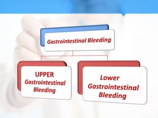
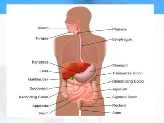
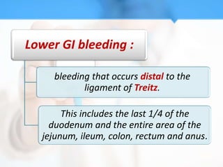
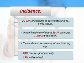
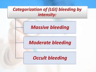
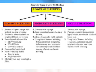
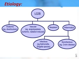
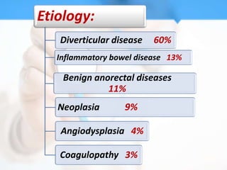
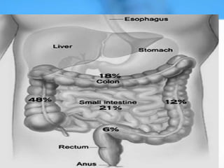
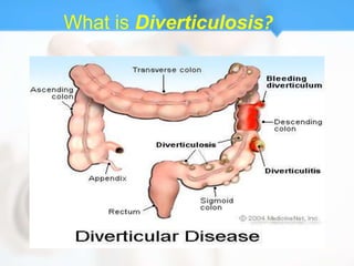
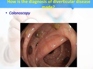
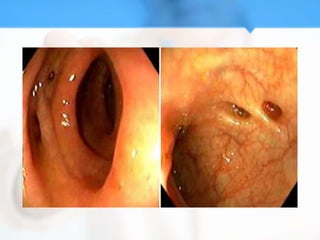
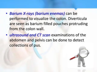
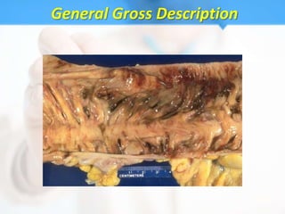
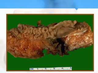
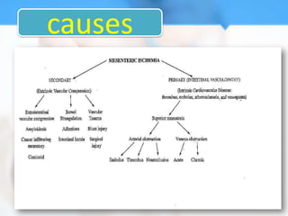
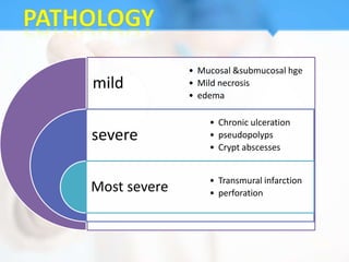
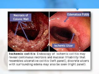
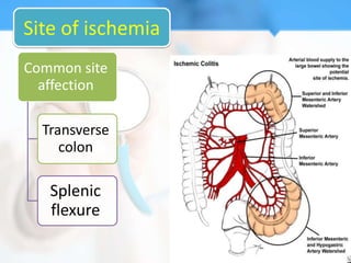
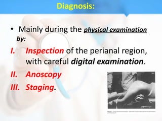
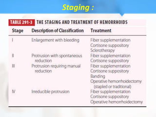
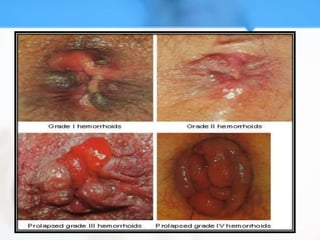
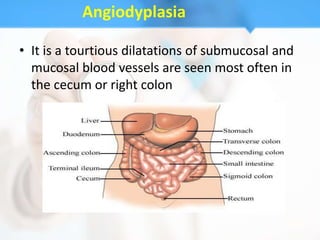
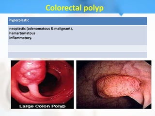
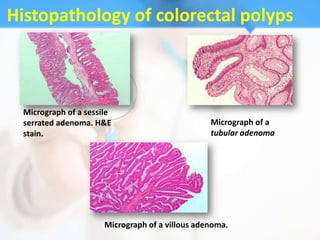
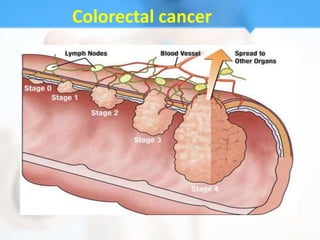
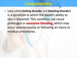
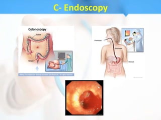
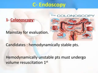
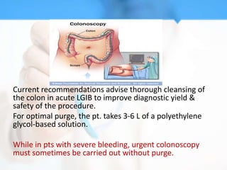
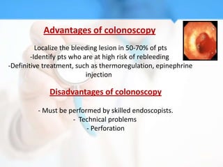
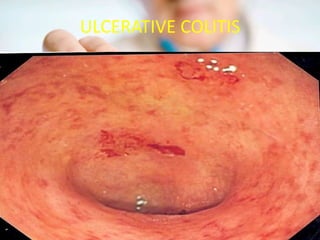
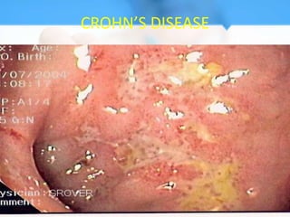
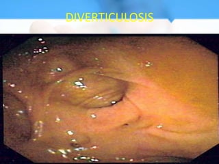
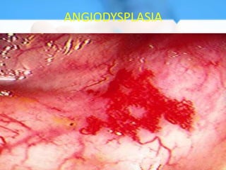
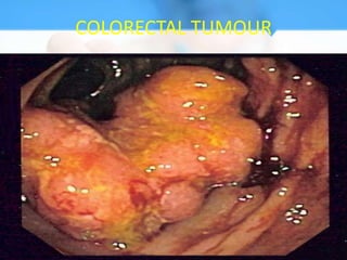
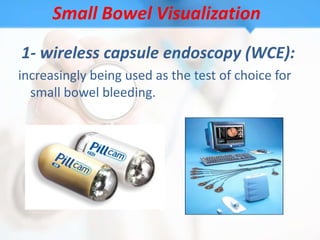
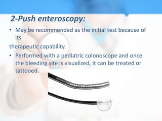
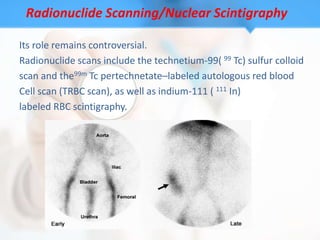
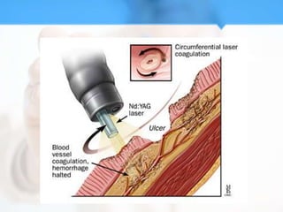
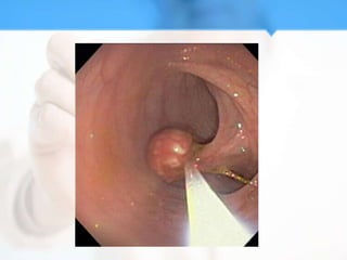
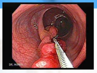
Lower Gastrointestinal Bleeding
 Prof.dr/ Wafaa Hussin Mekki
     Students/ from 285 to 296
•Introduction
•Ayman shahtot (285)
Upper GI bleeding:

  bleeding that occurs proximal to the ligament
                    of Treitz.


    The upper GI tract includes the esophagus,
   stomach, and first part of the small intestine.
Lower GI bleeding :

     bleeding that occurs distal to the
            ligament of Treitz.

       This includes the last 1/4 of the
    duodenum and the entire area of the
  jejunum, ileum, colon, rectum and anus.
Presentation:
It is usually suspected when patients complain of
hematochezia.


Although, the distinctions based upon stool color
are not absolute since melena can be seen
with LGI bleeding from the right colon (or
small intestine), and hematochezia can be
seen with massive upper GI bleeding.
Incidence:
- 20-33% of episodes of gastrointestinal (GI)
               hemorrhage.


- annual incidence of about 20-27 cases per
            100,000 population.

- The incidence rises steeply with advancing
                     age.

- 80% resolve spontaneously.
- 25% will re-bleed.
Categorization of (LGI) bleeding by
            intensity:

         Massive bleeding

        Moderate bleeding

          Occult bleeding
Etiology:
Etiology:
   Diverticular disease   60%
   Inflammatory bowel disease 13%

    Benign anorectal diseases
              11%
   Neoplasia       9%

   Angiodysplasia 4%

   Coagulopathy 3%
Dirverticular diseases
    Aya adel (290)
   Aya abdo (293)
What is Diverticulosis?
Most common site
• Diverticula can occur throughout the colon
  but are most common near the end of the left
  colon referred to as the sigmoid colon.
Risk factors
• increasing age
• constipation
• a diet that is low in dietary fiber
• high intake of meat and red meat
• connective tissue disorders (such as Mara fan
  syndrome) that may cause weakness in the
  colon wall
• hereditary or genetic predisposition
How do diverticula form?
The muscular wall of the colon grows thicker with age,
although the cause of this thickening is unclear. It may reflect
the increasing pressures required by the colon to eliminate
feces. For example, a diet low in fiber can lead to small, hard
stools which are difficult to pass and which require increased
pressure to pass. The lack of fiber and small stools also may
allow segments of the colon to close off from the rest of the
colon when the colonic muscle in the segment contracts. The
pressure in these closed-off segments may become high since
the increased pressure cannot dissipate to the rest of the
colon. Over time, high pressures in the colon push the inner
intestinal lining outward (herniation) through weak areas in
the muscular walls. These pouches or sacs that develop are
called diverticula.
What are the symptoms of diverticular disease?

  • Most patients with diverticulosis have few or no
    symptoms.
  • The most common symptoms of diverticular
    disease include:
   abdominal cramping,
   constipation, and
   diarrhea.
  These symptoms are related to difficulty in passing
    stool through the left colon, which is narrowed by
    diverticular disease.
Complications of diverticulosis
             293
• The most common complication is diverticulitis
  which is a condition in which diverticuli in the
  colon rupture. Which results in infection in the
  tissues that surround the colon
What are the complications of diverticulitis?
 • Bleeding:caused by a small blood vessel in a
   diverticulum that weakens and then bursts
Abscess, Perforation, and Peritonitis


• Diverticulitis may lead to infection, which often
  clears up after a few days of treatment with
  antibiotics. If the infection gets worse, an abscess
  may form in the wall of the colon.
• Infected diverticula may develop perforations.
• Sometimes the perforations leak pus out of the colon
  and form a large abscess in the abdominal cavity, a
  condition called peritonitis.
Fistula

• When diverticulitis-related infection spreads outside
  the colon, the colonic tissue may stick to nearby
  tissues. The organs usually involved are the bladder,
  small intestine, and skin.
• The most common type of fistula occurs between the
  bladder and the colon.
• This type of fistula affects men more often than
  women. It can result in a severe, long-lasting
  infection of the urinary tract. The problem can be
  corrected with surgery to remove the fistula and the
  affected part of the colon.
Intestinal Obstruction
• Scarring caused by infection may lead to
  partial or total blockage of the intestine, called
  intestinal obstruction
How is the diagnosis of diverticular disease
                 made?
• Colonoscopy
• Barium X-rays (barium enemas) can be
  performed to visualize the colon. Diverticula
  are seen as barium filled pouches protruding
  from the colon wall.
• ultrasound and CT scan examinations of the
  abdomen and pelvis can be done to detect
  collections of pus.
General Gross Description
General Micro Description
• Inflammatory bowel disease. 287
        • Enas shams (287)
Is a form of inflammatory bowel disease,
characterized by inflammation with ulcer
formation in the lining of colon.
Site of involvment

                                     Whole
                                     colon
                        Sigmoid&
                        descending
                        colon
           rectum


Inflammation of the distal
terminal ileum may occur
pathology
macroscopic

   Diffuse inflammation
    limited to mucosa
  -Extensive ulceration
  -Mucosa appear as
  inflammatory polyps
pathology
microscopic
  Crypt abscesses
  (the characteristic)
     Progress to
     ulceration
Clinical picture

Bleeding per rectum.
- Diarrhea (bloody diarrhea) with mucous.
- Tenesmus, when the disease is confined to the
   rectum.
-Abdominal discomfort.
- Remissions & exacerbations.
EXTRA INTESTINAL MANIFESTATIONS
ISCHEMIC COLITIS
a medical condition in which inflammation and
injury of the large intestine result from
inadequate blood supply.
causes
• Mucosal &submucosal hge
mild          • Mild necrosis
              • edema

                  • Chronic ulceration
severe            • pseudopolyps
                  • Crypt abscesses


                  • Transmural infarction
Most severe       • perforation
Site of ischemia
Common site
  affection

  Transverse
     colon

   Splenic
   flexure
Clinical picture
( depend on severity of ischemia)
1-abdominal pain
2-rectal bleeding
3-diarrhea
4-fever.
Hemorrhoids
Enal Mahmoud Fahmy(288)
Incidence and Epidemiology:
• Hemorrhoids affect >1 million in western
  civilization per year.

• The prevalence is less in the underdeveloped
  countries and not selective for age or sex.

• Common among pregnant women.
Predisposing factors:
• Constipation.

• Sitting for long periods of time.

• Obesity.

• Heavy Lifting.
Anatomy and Pathophysiology:
        •
Clinical Presentation :
• Patients commonly present to a physician for two
  main reasons:
• Bleeding.

• Protrusion.

• Pain.

• Anal Itching.
Diagnosis:

• Mainly during the physical examination
     by:
I.   Inspection of the perianal region,
     with careful digital examination.
II. Anoscopy
III. Staging.
Staging :
•
Angiodyplasia
 Neoplasm
Aya shawky (289)
Angiodyplasia

• It is a tourtious dilatations of submucosal and
  mucosal blood vessels are seen most often in
  the cecum or right colon
Incidence:

Age                      •     >70
 Sex                                   equal


                         Bleeding from angiodysplasia is usually self-
Mortality/Morbidity       limited, but it can be chronic, recurrent, or
                                even acute and life threatening.

        Site:

77% of angiodysplasias are located in the cecum and ascending colon,

  15% are located in the jejunum and ileum, and the remainder is
           distributed throughout the alimentary tract.
Pathophysiology of angiodysplasia
              most prominent theory


• Repeated episodes of colonic distention
  associated with transient increase in pressure
  and size


• This results in multiple episodes of incresing
  wall tension with obstruction of submucosal
  outflow
Neoplasm
• Neoplastic bleeding can be from a polyp or
  carcinoma.
• Colon cancer is the predominant cause of
  neoplastic bleeding and is responsible for around
  10% of rectal bleeding in patients older than 50
  years.
• The bleeding is usually low-grade and recurrent,
  occurring as a result of mucosal ulceration or
  erosion. Though neoplastic bleeding can present
  as bright red blood per rectum, it is unusual for it
  to cause massive colonic bleeding.
Lower Gastrointestinal Bleeding in
      Children and Adolescents
Intussusception


Polyps and polyposis syndromes
Juvenile polyps and polyposis
Peutz-Jeghers syndrome
Familial adenomatous polyposis (FAP)
Colorectal polyp
hyperplastic

neoplastic (adenomatous & malignant),
hamartomatous
inflammatory.
Histopathology of colorectal polyps


  Micrograph of a sessile
  serrated adenoma. H&E                           Micrograph of a
  stain.                                          tubular adenoma


                       Micrograph of a villous adenoma.
Colorectal cancer
Coagulopathy
Ayman Fawzy Salmona 286
Coagulopathy
• (also called clotting disorder and bleeding disorder)
  is a condition in which the blood’s ability to
  clot is impaired. This condition can cause
  prolonged or excessive bleeding, which may
  occur spontaneously or following an injury or
  medical procedures.
Hypocoagulability

• is an unusual susceptibility to bleeding, that is,
  an increased bleeding diathesis, due to an
  abnormality in coagulation.
Causes
• Acquired
• Autoimmune
• Genetic
Symptoms
               Symptom                    Disorders
                             Wiskott-Aldrich syndrome, where they
                              may resemble a few bruises
Petechiae (red spots)     
                          
                              Acute leukemia
                              Chronic leukemia
                             Vitamin K deficiency

                             Acute leukemia
Purpura and ecchymoses    
                          
                              Chronic leukemia
                              Vitamin K deficiency

                             Wiskott-Aldrich syndrome, especially
Blood in stool            
                              in infancy
                              Acute leukemia

                             Wiskott-Aldrich syndrome
Bleeding gingiva (gums)      Acute leukemia
                             Chronic leukemia
Prolonged nose bleeds        Wiskott-Aldrich syndrome
Complications
                   Complication                                Disorders
Soft tissue bleeding, e.g. deep-muscle bleeding,      Hemophilia
leading to swelling, numbness or pain of a limb.      Von Willebrand disease

Joint damage, potentially with severe pain and
                                                      Hemophilia
even destruction of the joint and development         Von Willebrand disease
of arthritis
Retinal bleeding                                      Acute leukemia
Transfusion transmitted infection, from blood
                                                      Hemophilia
transfusions that are given as treatment.

Adverse reactions to clotting factor treatment.       Hemophilia

Anemia                                                Von Willebrand disease
                                                      Von Willebrand disease Acute
Exsanguination (bleeding to death)                     leukemia
                                                      Vitamin K deficiency
Cerebral hemorrhage                                   Wiskott-Aldrich syndrome
Other causes of LGIB
Infection
• (HIV) is an infrequent cause of LGIB.
• HIV-related opportunistic infections and
   associated etiologies, including:
1. (CMV) colitis,
2. idiopathic colon ulcers,
3. Kaposi sarcoma, and lymphoma
Drug-induced
• Mainly by NSAID and aspirin use, and it is
  more common in the elderly.
• The 2008 Scottish Intercollegiate Guidelines
  Network (SIGN) guideline on the
  management of acute upper and lower
  gastrointestinal bleeding warns that oral
  anticoagulants or corticosteroids should be
  used with caution in patients at risk of GIB,
  especially in those who take NSAIDs or aspirin
Diagnosis
Aya Atef El-Sawy
     291
DIAGNOSIS
• Despite improvement in diagnostic imaging &
  procedures, 10-20% of pts with Lower GIT
  bleeding have no demonstrable bleeding
  source.

• Therefore, this complex problem requires
  systematic evaluation.
A- Initial Evaluation
Patient's history:
-aspirin, vascular disease, past bleeding
  episodes, liver cirrhosis, IBD, coagulopathy.
-duration, frequency, stool colour.

Digital rectal examination

Physical examination to assess the severity of
bleeding:
-HR , BP, postural changes.
B- Laboratory Tests

          CBC

       ESR/ CRP

  Coagulation profile

  Liver function tests

  Renal function tests
C- Endoscopy
C- Endoscopy

1- Colonoscopy:

Mainstay for evaluation.

Candidates : hemodynamically stable pts.

Hemodynamically unstable pts must undergo
volume resuscitation 1st
Current recommendations advise thorough cleansing of
the colon in acute LGIB to improve diagnostic yield &
safety of the procedure.
For optimal purge, the pt. takes 3-6 L of a polyethylene
glycol-based solution.

While in pts with severe bleeding, urgent colonoscopy
must sometimes be carried out without purge.
In cases of suspected perforation or obstruction,
plain abdominal radiography should be
performed before colonoscopy to rule out
these complications.
Advantages of colonoscopy
       -Localize the bleeding lesion in 50-70% of pts
        -Identify pts who are at high risk of rebleeding
-Definitive treatment, such as thermoregulation, epinephrine
                           injection

             Disadvantages of colonoscopy

         - Must be performed by skilled endoscopists.
                    - Technical problems
                         - Perforation
ULCERATIVE COLITIS
CROHN’S DISEASE
DIVERTICULOSIS
ANGIODYSPLASIA
COLORECTAL TUMOUR
2- Esophagogastroduodenoscopy:


It is performed if NG tube aspirate is positive for blood.

About 10% of pts presenting with LGIB have bleeding
originating from the upper GIT.
Aya Abd El-Nasser
      291
Small Bowel Visualization
1- wireless capsule endoscopy (WCE):
increasingly being used as the test of choice for
  small bowel bleeding.
2-Push enteroscopy:
• May be recommended as the initial test because of
  its
therapeutic capability.
• Performed with a pediatric colonoscope and once
  the bleeding site is visualized, it can be treated or
  tattooed.
Radionuclide Scanning/Nuclear Scintigraphy

Its role remains controversial.
Radionuclide scans include the technetium-99( 99 Tc) sulfur colloid
scan and the99m Tc pertechnetate–labeled autologous red blood
Cell scan (TRBC scan), as well as indium-111 ( 111 In)
labeled RBC scintigraphy.
• Nuclear scintigraphy is a sensitive diagnostic tool (86%)
and can detect hemorrhage at rates as low as 0.1
mL/min, as opposed to angiography, which detects
bleeding at rates of 1-1.5 mL/min. This technique is
more sensitive than angiography, but it suffers from a
low specificity compared with endoscopy or
angiography due to its limited resolution.

• Radionuclide scans frequently are performed before
angiography, because the scans detect bleeding at a slower
rate than what can be detected with angiography, thereby
potentially eliminating the need for an invasive procedure.
Advantages:
Noninvasive.
High sensitivity.
Disadvantages:
High false localization rate (3% to 59% ) ,
this often is due to the overlapping segments of bowel
and the migration of tagged RBCs in the large bowel.

 Another disadvantage of radionuclide scans is that the
scans must be performed during active bleeding.
Angiography
• Performed after colonoscopy has failed to identify a
  bleeding site ,can detect bleeding at a rate of more
  than 0.5 mL/min.

• In a patient with active GI bleeding, the radiologist
  first cannulates the superior mesenteric artery,
  because most of the hemodynamically significant
  bleeding originates in the right colon. The
  extravasation of contrast material indicates a
  positive study finding. If the findings from the study
  are negative, the inferior mesenteric artery is
  cannulated, followed by the celiac artery.
Once the bleeding point is identified,
angiography offers potential treatment options,
such as selective vasopressin drip and embolization.
The advantages of angiography include:
1- Accurate localization of the bleeding;
2- Therapeutic utility that includes the use of vasopressin
   infusion or embolization;
3-Does not require preparation of the bowel.

The disadvantages of angiography include:
1- It has a sensitivity of approximately 30-47%;
2- It can only be performed during active bleeding;
3- It has a complication rate of about 9%. Such
   complications include thrombosis, embolization, and
   renal failure.
Barium Enema
Justified only for elective evaluation of unexplained
LGIB.

Barium enema examination is not used in the acute
hemorrhage phase, because it makes subsequent
diagnostic evaluations, including angiography and
colonoscopy impossible.
Abdominal Radiography /CT

• Plain abdominal radiography and/or CT might be carried
  out, depending on the clinical presentation and
  suspected etiology (such as ischemic or inflammatory
  colitis, or in cases where bowel obstruction or
  perforation are suspected).
Histologic Findings

Most colonic diverticula are false pulsion diverticula and
composed only of mucosa and submucosa herniated
through the colonic wall musculature.


Colonic angiodysplasias are vascular ectasias commonly
located on the right side of the colon. Microscopically,
vascular ectasia consists of dilated thin-walled venules
and capillaries localized in the submucosa of the colonic
wall.
Management
Aya Mohamed Khater
       294
• Resuscitation and initial assessment.


• Localization of the bleeding site.


• Therapeutic intervention to stop bleeding at
  the site.
1) Resuscitation and Initial Assessment:

• IV access and administration of normal saline.

• Rapid assessment of vital signs, including heart rate,
  blood pressure, pulse pressure, and urine output.
• Routine laboratory studies (CBC, electrolyte levels,
  and coagulation studies), blood should be typed and
  cross-matched.
• The patient's blood loss and hemodynamic status
  should be evaluated, and in cases of severe bleeding,
  the patient may require invasive hemodynamic
  monitoring .
• Patients in shock should receive fluid volume
  replacement without delay.
Colloid or crystalloid solutions may be used to
        achieve volume restoration before
           administering blood products.
   Red cell transfusion should be considered
    after loss of 30% of the circulating volume.
2) Localization of the Bleeding Site
• In patients who are hemo-dynamically stable with mild to
  moderate bleeding or in patients who have had a massive bleed
  that has stabilized, colonoscopy should be performed initially.
  Once the bleeding site is localized, therapeutic options include
  coagulation and injection with vasoconstrictors or sclerosing
  agents.
• In cases of diverticular bleeding, bipolar probe coagulation,
  epinephrine injection, and metallic clips may be used.
• If recurrent bleeding is present, the affected bowel segment
  can be resected.
• In cases of angiodysplasia, thermal therapy, such as electro-
  coagulation or argon plasma coagulation, is generally
  successful.
3) Therapeutic intervention to stop bleeding at the site.

 Colonoscopy
• Colonoscopy is useful in radiation
  therapy–induced gastrointestinal (GI)
  bleeding and in the treatment of colonic
  polyp lesions.
• Endoscopic treatment of radiation-
  induced bleeding includes topical
  application of formalin, Nd:YAG laser
  therapy, and argon plasma coagulation.
• Neoplastic bleeding due to polyps
  requires polypectomy. Patients diagnosed
  with colonic tumors may require surgical
  resection.
Vasoconstrictive Therapy :
• In patients in whom the bleeding site cannot be determined
  based on colonoscopy and in patients with active LGIB,
  angiography with or without a preceding radionuclide scan
  should be performed to locate the bleeding site as well as to
  intervene therapeutically.
• Initially, Vasoconstrictive agents, such as vasopressin,
  epinephrine, propranolol can be used.
• Vasoconstriction reduces the blood flow and facilitates plug
  formation in the bleeding vessel.
• Although epinephrine and propranolol reduced mesenteric
  blood flow, they also caused a rebound increase in blood flow
  and recurrent bleeding.
• Vasopressin causes severe vasoconstriction in the splanchnic
  bed. Vasopressin infusions are more effective in diverticular
  bleeding, which is arterial, as opposed to angiodysplastic
  bleeding, which is of the venocapillary type.
• Intra-arterial vasopressin infusions begin at a rate of 0.2
  U/min, with repeat angiography performed after 20 minutes.
  The bleeding stops in about 91% of patients receiving intra-
  arterial vasopressin.
• If bleeding persists, the rate of the infusion is increased to 0.4-
  0.6 U/min. Once the bleeding is controlled, the infusion is
  continued in an intensive care setting for 12-48 hours and
  then tapered over the next 24 hours.
Aya Mahmoud
    296
Superselective Embolization:

• This therapeutic modality is useful in patients in whom
  vasopressin is unsuccessful or contraindicated.
• Embolization involves superselective catheterization of the
  bleeding vessel to minimize necrosis.
• Embolization with agents such as gelatin sponge, coil springs,
  polyvinyl alcohol, and oxidized cellulose.
• It is performed using a 3 French (F) microcatheter placed
  coaxially through the diagnostic 5F catheter.
• Once the bleeding vessel is identified, microcoils are used to
  occlude the bleeding vessel.
• Although microcoils are most commonly used, polyvinyl
  alcohol and Gelfoam are also used alone or in conjunction
  with microcoils.
Complications:

• Colonic infarction, bowel wall injury, Intestinal
  ischemia and infarction.
• To prevent this complication, perform
  embolization as close as possible to the
  bleeding point in the terminal arteries.
Endoscopic Therapies:

• Endoscopic control of hemorrhage is suitable for GI
  polyps and cancers, arteriovenous malformations,
  mucosal lesions, postpolypectomy hemorrhage,
  endometriosis, colonic and rectal varices.
• It can be achieved using thermal modalities or
  sclerosing agents.
• Absolute alcohol, morrhuate sodium, and sodium
  tetradecyl sulfate can be used for sclerotherapy of
  lower GI lesions.
• Endoscopic epinephrine injection is used commonly
  because of its low cost, easy accessibility, and low
  risk of complications.
• Endoscopic thermal modalities (eg, laser
  photocoagulation, electrocoagulation) can also be
  used to arrest hemorrhage.
• Postpolypectomy hemorrhage can be managed by
  electrocoagulation of the polypectomy site bleeding.
• endoscopic coagulation of angiodysplasias is
  becoming a treatment of choice using either heated
  probe or lasers, such as Nd:YAG and argon.
• Argon laser treatment is recommended for mucosal
  or superficial lesions, Nd:YAG lasers are more useful
  for deeper lesions.
Surgery
• Emergent surgery is required in patients with (LGIB)
  if non operative management is unsuccessful.
Indications of Surgery :
• Persistent hemodynamic instability with active
  bleeding.
• Persistent, recurrent bleeding.
• Transfusion of more than 4 units packed red bloods
  cells in a 24-hour, with active or recurrent bleeding.
• No contraindications exist with regard to surgery in
  hemodynamically unstable patients with active
  bleeding.
Segmental bowel resection and subtotal colectomy

• Segmental bowel resection following precise localization of
  the bleeding point is a well-accepted surgical practice in
  hemodynamically stable patients.
• Subtotal colectomy is the procedure of choice in patients who
  are actively bleeding from an unknown source.
• Patients who are hemodynamically stable should have
  preoperative localization of the bleeding; once it is localized,
  intra-arterial vasopressin is used as a temporizing measure to
  reduce the bleeding before patients undergo segmental
  colectomy.
• If the it is not localized, a subtotal colectomy with
  ileoproctostomy is performed.
Complications

• The most common early postoperative complications
  are intra-abdominal bleeding, mechanical bowel
  obstruction, intra-abdominal sepsis, localized or
  generalized peritonitis, wound infection and/or
  dehiscence.

• Intra-abdominal sepsis following colorectal surgery is
  a life-threatening complication and requires
  aggressive resuscitation.
• Systemic conditions (eg, severe blood loss and shock,
  poor bowel preparation, diabetes, malnutrition,
  hypoalbuminemia) may adversely affect anastomotic
  healing.
• Changes in anatomy and physiology of the large
  bowel, high bacterial content, improper operative
  technique, and ischemia can cause anastomotic leak
  associated with abscess and intra-abdominal sepsis.

• Delayed complications usually occur more than 1
  week after surgery, the most common of which are
  anastomotic stricture, incisional hernia, and
  incontinence.
lower git bleeding

lower git bleeding

  • 2.
    Lower Gastrointestinal Bleeding Prof.dr/ Wafaa Hussin Mekki Students/ from 285 to 296
  • 3.
  • 6.
    Upper GI bleeding: bleeding that occurs proximal to the ligament of Treitz. The upper GI tract includes the esophagus, stomach, and first part of the small intestine.
  • 7.
    Lower GI bleeding: bleeding that occurs distal to the ligament of Treitz. This includes the last 1/4 of the duodenum and the entire area of the jejunum, ileum, colon, rectum and anus.
  • 8.
    Presentation: It is usuallysuspected when patients complain of hematochezia. Although, the distinctions based upon stool color are not absolute since melena can be seen with LGI bleeding from the right colon (or small intestine), and hematochezia can be seen with massive upper GI bleeding.
  • 9.
    Incidence: - 20-33% ofepisodes of gastrointestinal (GI) hemorrhage. - annual incidence of about 20-27 cases per 100,000 population. - The incidence rises steeply with advancing age. - 80% resolve spontaneously. - 25% will re-bleed.
  • 10.
    Categorization of (LGI)bleeding by intensity: Massive bleeding Moderate bleeding Occult bleeding
  • 12.
  • 13.
    Etiology: Diverticular disease 60% Inflammatory bowel disease 13% Benign anorectal diseases 11% Neoplasia 9% Angiodysplasia 4% Coagulopathy 3%
  • 15.
    Dirverticular diseases Aya adel (290) Aya abdo (293)
  • 16.
  • 17.
    Most common site •Diverticula can occur throughout the colon but are most common near the end of the left colon referred to as the sigmoid colon.
  • 18.
    Risk factors • increasingage • constipation • a diet that is low in dietary fiber • high intake of meat and red meat • connective tissue disorders (such as Mara fan syndrome) that may cause weakness in the colon wall • hereditary or genetic predisposition
  • 19.
  • 20.
    The muscular wallof the colon grows thicker with age, although the cause of this thickening is unclear. It may reflect the increasing pressures required by the colon to eliminate feces. For example, a diet low in fiber can lead to small, hard stools which are difficult to pass and which require increased pressure to pass. The lack of fiber and small stools also may allow segments of the colon to close off from the rest of the colon when the colonic muscle in the segment contracts. The pressure in these closed-off segments may become high since the increased pressure cannot dissipate to the rest of the colon. Over time, high pressures in the colon push the inner intestinal lining outward (herniation) through weak areas in the muscular walls. These pouches or sacs that develop are called diverticula.
  • 21.
    What are thesymptoms of diverticular disease? • Most patients with diverticulosis have few or no symptoms. • The most common symptoms of diverticular disease include:  abdominal cramping,  constipation, and  diarrhea. These symptoms are related to difficulty in passing stool through the left colon, which is narrowed by diverticular disease.
  • 22.
  • 23.
    • The mostcommon complication is diverticulitis which is a condition in which diverticuli in the colon rupture. Which results in infection in the tissues that surround the colon
  • 24.
    What are thecomplications of diverticulitis? • Bleeding:caused by a small blood vessel in a diverticulum that weakens and then bursts
  • 25.
    Abscess, Perforation, andPeritonitis • Diverticulitis may lead to infection, which often clears up after a few days of treatment with antibiotics. If the infection gets worse, an abscess may form in the wall of the colon. • Infected diverticula may develop perforations. • Sometimes the perforations leak pus out of the colon and form a large abscess in the abdominal cavity, a condition called peritonitis.
  • 26.
    Fistula • When diverticulitis-relatedinfection spreads outside the colon, the colonic tissue may stick to nearby tissues. The organs usually involved are the bladder, small intestine, and skin. • The most common type of fistula occurs between the bladder and the colon. • This type of fistula affects men more often than women. It can result in a severe, long-lasting infection of the urinary tract. The problem can be corrected with surgery to remove the fistula and the affected part of the colon.
  • 27.
    Intestinal Obstruction • Scarringcaused by infection may lead to partial or total blockage of the intestine, called intestinal obstruction
  • 28.
    How is thediagnosis of diverticular disease made? • Colonoscopy
  • 30.
    • Barium X-rays(barium enemas) can be performed to visualize the colon. Diverticula are seen as barium filled pouches protruding from the colon wall. • ultrasound and CT scan examinations of the abdomen and pelvis can be done to detect collections of pus.
  • 31.
  • 33.
  • 34.
    • Inflammatory boweldisease. 287 • Enas shams (287)
  • 35.
    Is a formof inflammatory bowel disease, characterized by inflammation with ulcer formation in the lining of colon.
  • 36.
    Site of involvment Whole colon Sigmoid& descending colon rectum Inflammation of the distal terminal ileum may occur
  • 37.
    pathology macroscopic Diffuse inflammation limited to mucosa -Extensive ulceration -Mucosa appear as inflammatory polyps
  • 38.
    pathology microscopic Cryptabscesses (the characteristic) Progress to ulceration
  • 39.
    Clinical picture Bleeding perrectum. - Diarrhea (bloody diarrhea) with mucous. - Tenesmus, when the disease is confined to the rectum. -Abdominal discomfort. - Remissions & exacerbations.
  • 40.
  • 41.
    ISCHEMIC COLITIS a medicalcondition in which inflammation and injury of the large intestine result from inadequate blood supply.
  • 42.
  • 43.
    • Mucosal &submucosalhge mild • Mild necrosis • edema • Chronic ulceration severe • pseudopolyps • Crypt abscesses • Transmural infarction Most severe • perforation
  • 45.
    Site of ischemia Commonsite affection Transverse colon Splenic flexure
  • 46.
    Clinical picture ( dependon severity of ischemia) 1-abdominal pain 2-rectal bleeding 3-diarrhea 4-fever.
  • 47.
  • 48.
    Incidence and Epidemiology: •Hemorrhoids affect >1 million in western civilization per year. • The prevalence is less in the underdeveloped countries and not selective for age or sex. • Common among pregnant women.
  • 49.
    Predisposing factors: • Constipation. •Sitting for long periods of time. • Obesity. • Heavy Lifting.
  • 50.
  • 51.
  • 52.
    Clinical Presentation : •Patients commonly present to a physician for two main reasons: • Bleeding. • Protrusion. • Pain. • Anal Itching.
  • 53.
    Diagnosis: • Mainly duringthe physical examination by: I. Inspection of the perianal region, with careful digital examination. II. Anoscopy III. Staging.
  • 54.
  • 56.
  • 57.
    Angiodyplasia • It isa tourtious dilatations of submucosal and mucosal blood vessels are seen most often in the cecum or right colon
  • 58.
    Incidence: Age • >70 Sex equal Bleeding from angiodysplasia is usually self- Mortality/Morbidity limited, but it can be chronic, recurrent, or even acute and life threatening. Site: 77% of angiodysplasias are located in the cecum and ascending colon, 15% are located in the jejunum and ileum, and the remainder is distributed throughout the alimentary tract.
  • 59.
    Pathophysiology of angiodysplasia most prominent theory • Repeated episodes of colonic distention associated with transient increase in pressure and size • This results in multiple episodes of incresing wall tension with obstruction of submucosal outflow
  • 60.
    Neoplasm • Neoplastic bleedingcan be from a polyp or carcinoma. • Colon cancer is the predominant cause of neoplastic bleeding and is responsible for around 10% of rectal bleeding in patients older than 50 years. • The bleeding is usually low-grade and recurrent, occurring as a result of mucosal ulceration or erosion. Though neoplastic bleeding can present as bright red blood per rectum, it is unusual for it to cause massive colonic bleeding.
  • 61.
    Lower Gastrointestinal Bleedingin Children and Adolescents Intussusception Polyps and polyposis syndromes Juvenile polyps and polyposis Peutz-Jeghers syndrome Familial adenomatous polyposis (FAP)
  • 62.
    Colorectal polyp hyperplastic neoplastic (adenomatous& malignant), hamartomatous inflammatory.
  • 63.
    Histopathology of colorectalpolyps Micrograph of a sessile serrated adenoma. H&E Micrograph of a stain. tubular adenoma Micrograph of a villous adenoma.
  • 64.
  • 65.
  • 66.
    Coagulopathy • (also calledclotting disorder and bleeding disorder) is a condition in which the blood’s ability to clot is impaired. This condition can cause prolonged or excessive bleeding, which may occur spontaneously or following an injury or medical procedures.
  • 67.
    Hypocoagulability • is anunusual susceptibility to bleeding, that is, an increased bleeding diathesis, due to an abnormality in coagulation.
  • 68.
  • 69.
    Symptoms Symptom Disorders  Wiskott-Aldrich syndrome, where they may resemble a few bruises Petechiae (red spots)   Acute leukemia Chronic leukemia  Vitamin K deficiency  Acute leukemia Purpura and ecchymoses   Chronic leukemia Vitamin K deficiency  Wiskott-Aldrich syndrome, especially Blood in stool  in infancy Acute leukemia  Wiskott-Aldrich syndrome Bleeding gingiva (gums)  Acute leukemia  Chronic leukemia Prolonged nose bleeds  Wiskott-Aldrich syndrome
  • 70.
    Complications Complication Disorders Soft tissue bleeding, e.g. deep-muscle bleeding,  Hemophilia leading to swelling, numbness or pain of a limb.  Von Willebrand disease Joint damage, potentially with severe pain and  Hemophilia even destruction of the joint and development  Von Willebrand disease of arthritis Retinal bleeding  Acute leukemia Transfusion transmitted infection, from blood  Hemophilia transfusions that are given as treatment. Adverse reactions to clotting factor treatment.  Hemophilia Anemia  Von Willebrand disease  Von Willebrand disease Acute Exsanguination (bleeding to death) leukemia  Vitamin K deficiency Cerebral hemorrhage  Wiskott-Aldrich syndrome
  • 71.
  • 72.
    Infection • (HIV) isan infrequent cause of LGIB. • HIV-related opportunistic infections and associated etiologies, including: 1. (CMV) colitis, 2. idiopathic colon ulcers, 3. Kaposi sarcoma, and lymphoma
  • 73.
    Drug-induced • Mainly byNSAID and aspirin use, and it is more common in the elderly. • The 2008 Scottish Intercollegiate Guidelines Network (SIGN) guideline on the management of acute upper and lower gastrointestinal bleeding warns that oral anticoagulants or corticosteroids should be used with caution in patients at risk of GIB, especially in those who take NSAIDs or aspirin
  • 74.
  • 75.
    DIAGNOSIS • Despite improvementin diagnostic imaging & procedures, 10-20% of pts with Lower GIT bleeding have no demonstrable bleeding source. • Therefore, this complex problem requires systematic evaluation.
  • 76.
    A- Initial Evaluation Patient'shistory: -aspirin, vascular disease, past bleeding episodes, liver cirrhosis, IBD, coagulopathy. -duration, frequency, stool colour. Digital rectal examination Physical examination to assess the severity of bleeding: -HR , BP, postural changes.
  • 77.
    B- Laboratory Tests CBC ESR/ CRP Coagulation profile Liver function tests Renal function tests
  • 78.
  • 79.
    C- Endoscopy 1- Colonoscopy: Mainstayfor evaluation. Candidates : hemodynamically stable pts. Hemodynamically unstable pts must undergo volume resuscitation 1st
  • 80.
    Current recommendations advisethorough cleansing of the colon in acute LGIB to improve diagnostic yield & safety of the procedure. For optimal purge, the pt. takes 3-6 L of a polyethylene glycol-based solution. While in pts with severe bleeding, urgent colonoscopy must sometimes be carried out without purge.
  • 81.
    In cases ofsuspected perforation or obstruction, plain abdominal radiography should be performed before colonoscopy to rule out these complications.
  • 82.
    Advantages of colonoscopy -Localize the bleeding lesion in 50-70% of pts -Identify pts who are at high risk of rebleeding -Definitive treatment, such as thermoregulation, epinephrine injection Disadvantages of colonoscopy - Must be performed by skilled endoscopists. - Technical problems - Perforation
  • 83.
  • 84.
  • 85.
  • 86.
  • 87.
  • 88.
    2- Esophagogastroduodenoscopy: It isperformed if NG tube aspirate is positive for blood. About 10% of pts presenting with LGIB have bleeding originating from the upper GIT.
  • 89.
  • 90.
    Small Bowel Visualization 1-wireless capsule endoscopy (WCE): increasingly being used as the test of choice for small bowel bleeding.
  • 91.
    2-Push enteroscopy: • Maybe recommended as the initial test because of its therapeutic capability. • Performed with a pediatric colonoscope and once the bleeding site is visualized, it can be treated or tattooed.
  • 92.
    Radionuclide Scanning/Nuclear Scintigraphy Itsrole remains controversial. Radionuclide scans include the technetium-99( 99 Tc) sulfur colloid scan and the99m Tc pertechnetate–labeled autologous red blood Cell scan (TRBC scan), as well as indium-111 ( 111 In) labeled RBC scintigraphy.
  • 93.
    • Nuclear scintigraphyis a sensitive diagnostic tool (86%) and can detect hemorrhage at rates as low as 0.1 mL/min, as opposed to angiography, which detects bleeding at rates of 1-1.5 mL/min. This technique is more sensitive than angiography, but it suffers from a low specificity compared with endoscopy or angiography due to its limited resolution. • Radionuclide scans frequently are performed before angiography, because the scans detect bleeding at a slower rate than what can be detected with angiography, thereby potentially eliminating the need for an invasive procedure.
  • 94.
    Advantages: Noninvasive. High sensitivity. Disadvantages: High falselocalization rate (3% to 59% ) , this often is due to the overlapping segments of bowel and the migration of tagged RBCs in the large bowel. Another disadvantage of radionuclide scans is that the scans must be performed during active bleeding.
  • 95.
    Angiography • Performed aftercolonoscopy has failed to identify a bleeding site ,can detect bleeding at a rate of more than 0.5 mL/min. • In a patient with active GI bleeding, the radiologist first cannulates the superior mesenteric artery, because most of the hemodynamically significant bleeding originates in the right colon. The extravasation of contrast material indicates a positive study finding. If the findings from the study are negative, the inferior mesenteric artery is cannulated, followed by the celiac artery.
  • 96.
    Once the bleedingpoint is identified, angiography offers potential treatment options, such as selective vasopressin drip and embolization.
  • 97.
    The advantages ofangiography include: 1- Accurate localization of the bleeding; 2- Therapeutic utility that includes the use of vasopressin infusion or embolization; 3-Does not require preparation of the bowel. The disadvantages of angiography include: 1- It has a sensitivity of approximately 30-47%; 2- It can only be performed during active bleeding; 3- It has a complication rate of about 9%. Such complications include thrombosis, embolization, and renal failure.
  • 98.
    Barium Enema Justified onlyfor elective evaluation of unexplained LGIB. Barium enema examination is not used in the acute hemorrhage phase, because it makes subsequent diagnostic evaluations, including angiography and colonoscopy impossible.
  • 99.
    Abdominal Radiography /CT •Plain abdominal radiography and/or CT might be carried out, depending on the clinical presentation and suspected etiology (such as ischemic or inflammatory colitis, or in cases where bowel obstruction or perforation are suspected).
  • 100.
    Histologic Findings Most colonicdiverticula are false pulsion diverticula and composed only of mucosa and submucosa herniated through the colonic wall musculature. Colonic angiodysplasias are vascular ectasias commonly located on the right side of the colon. Microscopically, vascular ectasia consists of dilated thin-walled venules and capillaries localized in the submucosa of the colonic wall.
  • 101.
  • 102.
    • Resuscitation andinitial assessment. • Localization of the bleeding site. • Therapeutic intervention to stop bleeding at the site.
  • 103.
    1) Resuscitation andInitial Assessment: • IV access and administration of normal saline. • Rapid assessment of vital signs, including heart rate, blood pressure, pulse pressure, and urine output. • Routine laboratory studies (CBC, electrolyte levels, and coagulation studies), blood should be typed and cross-matched. • The patient's blood loss and hemodynamic status should be evaluated, and in cases of severe bleeding, the patient may require invasive hemodynamic monitoring .
  • 104.
    • Patients inshock should receive fluid volume replacement without delay. Colloid or crystalloid solutions may be used to achieve volume restoration before administering blood products. Red cell transfusion should be considered after loss of 30% of the circulating volume.
  • 105.
    2) Localization ofthe Bleeding Site • In patients who are hemo-dynamically stable with mild to moderate bleeding or in patients who have had a massive bleed that has stabilized, colonoscopy should be performed initially. Once the bleeding site is localized, therapeutic options include coagulation and injection with vasoconstrictors or sclerosing agents. • In cases of diverticular bleeding, bipolar probe coagulation, epinephrine injection, and metallic clips may be used. • If recurrent bleeding is present, the affected bowel segment can be resected. • In cases of angiodysplasia, thermal therapy, such as electro- coagulation or argon plasma coagulation, is generally successful.
  • 106.
    3) Therapeutic interventionto stop bleeding at the site. Colonoscopy • Colonoscopy is useful in radiation therapy–induced gastrointestinal (GI) bleeding and in the treatment of colonic polyp lesions. • Endoscopic treatment of radiation- induced bleeding includes topical application of formalin, Nd:YAG laser therapy, and argon plasma coagulation. • Neoplastic bleeding due to polyps requires polypectomy. Patients diagnosed with colonic tumors may require surgical resection.
  • 107.
    Vasoconstrictive Therapy : •In patients in whom the bleeding site cannot be determined based on colonoscopy and in patients with active LGIB, angiography with or without a preceding radionuclide scan should be performed to locate the bleeding site as well as to intervene therapeutically. • Initially, Vasoconstrictive agents, such as vasopressin, epinephrine, propranolol can be used. • Vasoconstriction reduces the blood flow and facilitates plug formation in the bleeding vessel. • Although epinephrine and propranolol reduced mesenteric blood flow, they also caused a rebound increase in blood flow and recurrent bleeding.
  • 108.
    • Vasopressin causessevere vasoconstriction in the splanchnic bed. Vasopressin infusions are more effective in diverticular bleeding, which is arterial, as opposed to angiodysplastic bleeding, which is of the venocapillary type. • Intra-arterial vasopressin infusions begin at a rate of 0.2 U/min, with repeat angiography performed after 20 minutes. The bleeding stops in about 91% of patients receiving intra- arterial vasopressin. • If bleeding persists, the rate of the infusion is increased to 0.4- 0.6 U/min. Once the bleeding is controlled, the infusion is continued in an intensive care setting for 12-48 hours and then tapered over the next 24 hours.
  • 109.
  • 110.
    Superselective Embolization: • Thistherapeutic modality is useful in patients in whom vasopressin is unsuccessful or contraindicated. • Embolization involves superselective catheterization of the bleeding vessel to minimize necrosis. • Embolization with agents such as gelatin sponge, coil springs, polyvinyl alcohol, and oxidized cellulose. • It is performed using a 3 French (F) microcatheter placed coaxially through the diagnostic 5F catheter. • Once the bleeding vessel is identified, microcoils are used to occlude the bleeding vessel. • Although microcoils are most commonly used, polyvinyl alcohol and Gelfoam are also used alone or in conjunction with microcoils.
  • 112.
    Complications: • Colonic infarction,bowel wall injury, Intestinal ischemia and infarction. • To prevent this complication, perform embolization as close as possible to the bleeding point in the terminal arteries.
  • 113.
    Endoscopic Therapies: • Endoscopiccontrol of hemorrhage is suitable for GI polyps and cancers, arteriovenous malformations, mucosal lesions, postpolypectomy hemorrhage, endometriosis, colonic and rectal varices. • It can be achieved using thermal modalities or sclerosing agents. • Absolute alcohol, morrhuate sodium, and sodium tetradecyl sulfate can be used for sclerotherapy of lower GI lesions. • Endoscopic epinephrine injection is used commonly because of its low cost, easy accessibility, and low risk of complications.
  • 114.
    • Endoscopic thermalmodalities (eg, laser photocoagulation, electrocoagulation) can also be used to arrest hemorrhage. • Postpolypectomy hemorrhage can be managed by electrocoagulation of the polypectomy site bleeding. • endoscopic coagulation of angiodysplasias is becoming a treatment of choice using either heated probe or lasers, such as Nd:YAG and argon. • Argon laser treatment is recommended for mucosal or superficial lesions, Nd:YAG lasers are more useful for deeper lesions.
  • 118.
    Surgery • Emergent surgeryis required in patients with (LGIB) if non operative management is unsuccessful. Indications of Surgery : • Persistent hemodynamic instability with active bleeding. • Persistent, recurrent bleeding. • Transfusion of more than 4 units packed red bloods cells in a 24-hour, with active or recurrent bleeding. • No contraindications exist with regard to surgery in hemodynamically unstable patients with active bleeding.
  • 119.
    Segmental bowel resectionand subtotal colectomy • Segmental bowel resection following precise localization of the bleeding point is a well-accepted surgical practice in hemodynamically stable patients. • Subtotal colectomy is the procedure of choice in patients who are actively bleeding from an unknown source. • Patients who are hemodynamically stable should have preoperative localization of the bleeding; once it is localized, intra-arterial vasopressin is used as a temporizing measure to reduce the bleeding before patients undergo segmental colectomy. • If the it is not localized, a subtotal colectomy with ileoproctostomy is performed.
  • 120.
    Complications • The mostcommon early postoperative complications are intra-abdominal bleeding, mechanical bowel obstruction, intra-abdominal sepsis, localized or generalized peritonitis, wound infection and/or dehiscence. • Intra-abdominal sepsis following colorectal surgery is a life-threatening complication and requires aggressive resuscitation. • Systemic conditions (eg, severe blood loss and shock, poor bowel preparation, diabetes, malnutrition, hypoalbuminemia) may adversely affect anastomotic healing.
  • 121.
    • Changes inanatomy and physiology of the large bowel, high bacterial content, improper operative technique, and ischemia can cause anastomotic leak associated with abscess and intra-abdominal sepsis. • Delayed complications usually occur more than 1 week after surgery, the most common of which are anastomotic stricture, incisional hernia, and incontinence.