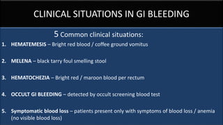
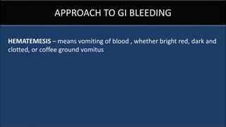
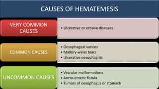
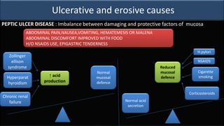
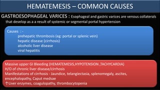
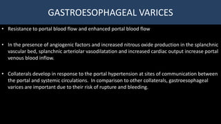
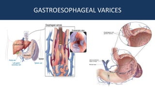
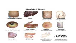
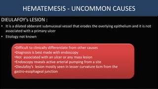
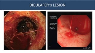
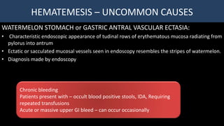
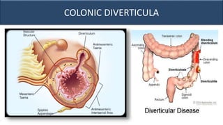
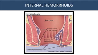
APPROACH TO GASTROINTESTINAL BLEEDING
SUJITHA TAMILSELVAM
III MBBS
PONDICHERRY INSTITUTE OF MEDICAL SCIENCES
CLINICAL SITUATIONS IN GI BLEEDING
5 Common clinical situations:
1. HEMATEMESIS – Bright red blood / coffee ground vomitus
2. MELENA – black tarry foul smelling stool
3. HEMATOCHEZIA – Bright red / maroon blood per rectum
4. OCCULT GI BLEEDING – detected by occult screening blood test
5. Symptomatic blood loss – patients present only with symptoms of blood loss / anemia
(no visible blood loss)
APPROACH TO GI BLEEDING
HEMATEMESIS – means vomiting of blood , whether bright red, dark and
clotted, or coffee ground vomitus
CAUSES OF HEMATEMESIS
• Ulcerative or erosive diseases
VERY COMMON
CAUSES
• Oesophageal varices
• Mallory-weiss tears
• Ulcerative oesophagitis
COMMON CAUSES
• Vascular malformations
• Aorto-enteric fistula
• Tumors of oesophagus or stomach
UNCOMMON CAUSES
Ulcerative and erosive causes
STRESS ULCERS :
• In critically ill patients, physiologic stress predisposes to ulcer
• Risk factors – Multiorgan failure, mechanical ventilation, hypotension (in sepsis), intracranial
injury(cushing ulcer), severe burns(curling ulcer)
Intracranial injury Direct vagal stimulation Acid hypersecretion
Systemic acidosis ↓ intracellular pH of
mucosal cells
Mucosal injury
Splanchnic
vasoconstriction
Hypoxia and ↓ blood
flow
Acute ulceration
•Hematemesis or bleed via nasogastric tube in ICU patient
• Falling hematocrit
• nasogastric aspirate of coffee ground material and melena
HEMATEMESIS – COMMON CAUSES
MALLORY - WEISS TEAR :
• Classic history of vomiting, retching ot coughing preceding hematemesis
• Seen in alcoholics
• Tears occur in the gastro-oesophageal junction
• Bleeding occurs in tears involving underlying esophageal or venous plexus
• Patients with portal hypertension – massive bleeding
Antecedent nausea, retching, vomiting followed by hematemesis
History of alcohol ingestion
Diagnosis made after ruling out other concominant signs and
symptoms
Ulcerative and erosive causes
PEPTIC ULCER DISEASE : Imbalance between damaging and protective factors of mucosa
Zollinger
ellison
syndrome
Hyperparat
hyroidism
Chronic renal
failure
↑ acid
production
Normal
mucosal
defence
Normal acid
secretion
H.pylori
NSAIDS
Cigarette
smoking
Corticosteroids
Reduced
mucosal
defence
ABDOMINAL PAIN,NAUSEA,VOMITING, HEMATEMESIS OR MALENA
ABDOMINAL DISCOMFORT IMPROVED WITH FOOD
H/O NSAIDS USE, EPIGASTRIC TENDERNESS
HEMATEMESIS – COMMON CAUSES
GASTROESOPHAGEAL VARICES : Esophageal and gastric varices are venous collaterals
that develop as a a result of systemic or segmental portal hypertension
Massive upper GI Bleeding (HEMATEMESIS,HYPOTENSION ,TACHYCARDIA)
H/O of chronic liver disease/cirrhosis
Manifestations of cirrhosis - Jaundice, telangiectasia, splenomegaly, ascites,
encephalopathy, Caput medsae
↑Liver enzymes, coagulopathy, thrombocytopenia
Causes : -
prehepatic thrombosis (eg: portal or splenic vein)
hepatic disease (cirrhosis)
alcoholic liver disease
viral hepatitis
GASTROESOPHAGEAL VARICES
• Resistance to portal blood flow and enhanced portal blood flow
• In the presence of angiogenic factors and increased nitrous oxide production in the splanchnic
vascular bed, splanchnic arteriolar vasodilatation and increased cardiac output increase portal
venous blood inflow.
• Collaterals develop in response to the portal hypertension at sites of communication between
the portal and systemic circulations. In comparison to other collaterals, gastroesophageal
varices are important due to their risk of rupture and bleeding.
GASTROESOPHAGEAL VARICES
HEMATEMESIS - UNCOMMON CAUSES
DIEULAFOY’s LESION :
• It is a dilated abberant submucosal vessel that erodes the overlying epithelium and it is not
associated with a primary ulcer
• Etiology not known
•Difficult to clinically differentiate from other causes
•Diagnosis is best made with endoscopy
•Not associated with an ulcer or any mass lesion
•Endoscopy reveals active arterial pumping from a site
•Dieulafoy’s lesion mostly seen in lesser curvature 6cm from the
gastro-esophageal junction
DIEULAFOY’s LESION
HEMATEMESIS – UNCOMMON CAUSES
WATERMELON STOMACH or GASTRIC ANTRAL VASCULAR ECTASIA:
• Characteristic endoscopic appearance of tudinal rows of erythematous mucosa radiating from
pylorus into antrum
• Ectatic or sacculated mucosal vessels seen in endoscopy resembles the stripes of watermelon.
• Diagnosis made by endoscopy
Chronic bleeding
Patients present with – occult blood positive stools, IDA, Requiring
repeated transfusions
Acute or massive upper GI bleed – can occur occasionally
WATERMELON STOMACH or GASTRIC ANTRAL VASCULAR
ECTASIA
HEMATEMESIS – UNCOMMON CAUSES
AORTO-ENTERIC FISTULA :
• Direct communication between the aorta and the gastro-intestinal tract
• Most common cause : Infected prosthetic aortic graft eroding into the intestine
• Other causes : penetrating ulcer, tumor invasion, foreign body perforation
• Though rare it is associated with high mortality rate if left undiagnosed and untreated
• 3rd or 4th portion of duodenum is the most common site.
•Hematemesis or hematochezia
•Followed by massive bleed resulting in hemorrhagic shock
•>50% - associated with back pain or abdominal pain
•<50% - Fever, association with sepsis
•H/O of prosthetic aortic graft or abdominal aortic aneurysms
AORTO-ENTERIC FISTULA
TUMORS OF OESOPHAGUS OR STOMACH
• Neoplasms account for less than 3% of acute upper GI bleed
• lipomas
• polyps
• Blue rubber bleb nevus syndrome
Benign lesions
• Adenocarcinoma
• Lymphoma
• Carcinoid tumor
• Colon cancer
• Lung cancer, breast cancer
Malignant tumors
(Primary or metastatic)
Luminal obstruction or ulceration causes DYSPHAGIA/ODYNOPHAGIA
Duodenal tumors – chronic nausea, vomiting, bezoar formation
HEMOCCULT – POSITIVE STOOLS, IDA, Endoscopy reveals an ulcerated mass,
anorexia, weight loss
Points to remember
• Amount of blood loss
• History of peptic ulcer disease
• Signs of chronic liver disease
• Recent ingestion of nsaids,aspirin,warfarin?
• History of retching before hematemesis(mallory-weiss)?
HEMATOCHEZIA
HEMATOCHEZIA
• Passage of bright red blood per rectum
• Also associated with passage of maroon stools
• Most common presentation of lower GI bleed
CAUSES:
•Colonic diverticula
•Internal hemorrhoids
•Colonic angiomas
•Colon cancer
•Ischemic colitis
•Inflammatory bowel disease
•Meckel’s diverticulum
COLONIC DIVERTICULA
• Diverticula are are herniations of colonic mucosa and submucosa through the muscular layers
of the colon
• Colonic diverticula are formed when colonic tissue is pushed by the intra-luminal pressure
• Common location – left colon
PAINLESS HEMATOCHEZIA
MILD LEFT LOWER QUADRANT DISCOMFORT
BLEEDING – MILD TO SEVERE
MOST COMMON CAUSE OF LOWER GI BLEED IN ADULTS
COLONIC DIVERTICULA
INTERNAL HAEMORRHOIDS
• PLEXUS OF VEINS JUST ABOVE THE SQUAMO COLUMNAR JUNCTION
• Characterised by bright red blood per rectum
• Blood often coats outside the stool
• Bleeding is usually painless
• Should be differentiated from external hemmorhoids, rectal varices, fissures.
• Particularly associated with constipation and straining stool
INTERNAL HEMORRHOIDS
COLON CANCER
• Most patients presents with occult gastrointestinal bleeding
• Hematochezia not very common
• In patients over 40 years
Recent change in bowel habits- constipation or diarrhea
Some cases palpable mass on abdominal or rectal examination
PAINLESS OCCULT BLEED –most common manifestation
IRON DEFICIENCY ANEMIA
INLAMMATORY BOWEL DISEASE
• Inflammatory bowel disease (IBD) is an immune-mediated chronic intestinal condition
• Ulcerative colitis (UC) and Crohn's disease (CD) are the two major types of IBD
• IBD is currently considered an inappropriate
• The major symptoms of UC are diarrhoea, rectal bleeding, tenesmus and crampy abdominal
pain
• Patients with proctitis usually pass fresh blood or blood-stained mucus, either mixed with
stool or streaked onto the surface of a normal or hard stool
• Look for Extraintestinal manifestations
Immune response to the endogenous commensal microbiota within the
intestines,intestinal epithelial dysfunction with or without some component of
autoimmunity,
INLAMMATORY BOWEL DISEASE
ISCHEMIC COLITIS
• Results from mucosal hypoxia
• Hypoperfusion of intramural vessels intestinal wall rather than large vessel occlusion
• Associated with atherosclerosis/vasculitis
• Invovlement is only segmentel because of collateral circulation
Sudden lower left quadrant pain with HAEMATOCHEZIA and DIARRHOEA
MECKEL’s DIVERTICULUM
• Commonest cause of gastrointestinal bleeding in children
• A congenital anomaly of gastrointestinal tract with incomplete obliteration of vitelline duct,
leaving an ileal diverticulum
• Classically 5cm long, 20cm from ileocecal valve
• Not all cases are associated with bleeding , 20% cases with heterotrophic mucosa
•Hemorrhage :in ectopic gastric mucosa present –peptic ulceration–painless
maroon rectal bleed or melaena
•Diverticulitis (presents like appendicitis)
•Intussusception
MECKEL’s DIVERTICULUM
POINTS TO REMEMBER IN BLOOD PER RECTUM
• Amount of bleed and colour of blood
• Blood on toilet paper(local anal pathology) ?
• Blood coated stool Pattern of bowel habits – hemorrhoids?
• Diarrhoea &mucus per rectum – IBD?
• Family history of colorectal ca
• Age – children(meckel diverticulum)
MELAENA
• All patients with active melaena assumed to be upper GI bleed
• Admission warranted – even haemodynamically stable because of suspicion of internal bleed
• Melaena - source may not be from upper GI – in such case polyps, cancer, diverticula
(by colonoscopy)
• If negative finding in colonoscopy and upper GI endoscopy – small bowel investigastion done
• Small bowel symptoms ? & evaluation
• IBD, Small bowel tumors, meckel’s diverticulum

Approach to Gastrointestinal bleeding

  • 1.
    APPROACH TO GASTROINTESTINALBLEEDING SUJITHA TAMILSELVAM III MBBS PONDICHERRY INSTITUTE OF MEDICAL SCIENCES
  • 2.
    CLINICAL SITUATIONS INGI BLEEDING 5 Common clinical situations: 1. HEMATEMESIS – Bright red blood / coffee ground vomitus 2. MELENA – black tarry foul smelling stool 3. HEMATOCHEZIA – Bright red / maroon blood per rectum 4. OCCULT GI BLEEDING – detected by occult screening blood test 5. Symptomatic blood loss – patients present only with symptoms of blood loss / anemia (no visible blood loss)
  • 3.
    APPROACH TO GIBLEEDING HEMATEMESIS – means vomiting of blood , whether bright red, dark and clotted, or coffee ground vomitus
  • 4.
    CAUSES OF HEMATEMESIS •Ulcerative or erosive diseases VERY COMMON CAUSES • Oesophageal varices • Mallory-weiss tears • Ulcerative oesophagitis COMMON CAUSES • Vascular malformations • Aorto-enteric fistula • Tumors of oesophagus or stomach UNCOMMON CAUSES
  • 5.
    Ulcerative and erosivecauses STRESS ULCERS : • In critically ill patients, physiologic stress predisposes to ulcer • Risk factors – Multiorgan failure, mechanical ventilation, hypotension (in sepsis), intracranial injury(cushing ulcer), severe burns(curling ulcer) Intracranial injury Direct vagal stimulation Acid hypersecretion Systemic acidosis ↓ intracellular pH of mucosal cells Mucosal injury Splanchnic vasoconstriction Hypoxia and ↓ blood flow Acute ulceration •Hematemesis or bleed via nasogastric tube in ICU patient • Falling hematocrit • nasogastric aspirate of coffee ground material and melena
  • 6.
    HEMATEMESIS – COMMONCAUSES MALLORY - WEISS TEAR : • Classic history of vomiting, retching ot coughing preceding hematemesis • Seen in alcoholics • Tears occur in the gastro-oesophageal junction • Bleeding occurs in tears involving underlying esophageal or venous plexus • Patients with portal hypertension – massive bleeding Antecedent nausea, retching, vomiting followed by hematemesis History of alcohol ingestion Diagnosis made after ruling out other concominant signs and symptoms
  • 8.
    Ulcerative and erosivecauses PEPTIC ULCER DISEASE : Imbalance between damaging and protective factors of mucosa Zollinger ellison syndrome Hyperparat hyroidism Chronic renal failure ↑ acid production Normal mucosal defence Normal acid secretion H.pylori NSAIDS Cigarette smoking Corticosteroids Reduced mucosal defence ABDOMINAL PAIN,NAUSEA,VOMITING, HEMATEMESIS OR MALENA ABDOMINAL DISCOMFORT IMPROVED WITH FOOD H/O NSAIDS USE, EPIGASTRIC TENDERNESS
  • 9.
    HEMATEMESIS – COMMONCAUSES GASTROESOPHAGEAL VARICES : Esophageal and gastric varices are venous collaterals that develop as a a result of systemic or segmental portal hypertension Massive upper GI Bleeding (HEMATEMESIS,HYPOTENSION ,TACHYCARDIA) H/O of chronic liver disease/cirrhosis Manifestations of cirrhosis - Jaundice, telangiectasia, splenomegaly, ascites, encephalopathy, Caput medsae ↑Liver enzymes, coagulopathy, thrombocytopenia Causes : - prehepatic thrombosis (eg: portal or splenic vein) hepatic disease (cirrhosis) alcoholic liver disease viral hepatitis
  • 10.
    GASTROESOPHAGEAL VARICES • Resistanceto portal blood flow and enhanced portal blood flow • In the presence of angiogenic factors and increased nitrous oxide production in the splanchnic vascular bed, splanchnic arteriolar vasodilatation and increased cardiac output increase portal venous blood inflow. • Collaterals develop in response to the portal hypertension at sites of communication between the portal and systemic circulations. In comparison to other collaterals, gastroesophageal varices are important due to their risk of rupture and bleeding.
  • 11.
  • 13.
    HEMATEMESIS - UNCOMMONCAUSES DIEULAFOY’s LESION : • It is a dilated abberant submucosal vessel that erodes the overlying epithelium and it is not associated with a primary ulcer • Etiology not known •Difficult to clinically differentiate from other causes •Diagnosis is best made with endoscopy •Not associated with an ulcer or any mass lesion •Endoscopy reveals active arterial pumping from a site •Dieulafoy’s lesion mostly seen in lesser curvature 6cm from the gastro-esophageal junction
  • 14.
  • 15.
    HEMATEMESIS – UNCOMMONCAUSES WATERMELON STOMACH or GASTRIC ANTRAL VASCULAR ECTASIA: • Characteristic endoscopic appearance of tudinal rows of erythematous mucosa radiating from pylorus into antrum • Ectatic or sacculated mucosal vessels seen in endoscopy resembles the stripes of watermelon. • Diagnosis made by endoscopy Chronic bleeding Patients present with – occult blood positive stools, IDA, Requiring repeated transfusions Acute or massive upper GI bleed – can occur occasionally
  • 16.
    WATERMELON STOMACH orGASTRIC ANTRAL VASCULAR ECTASIA
  • 17.
    HEMATEMESIS – UNCOMMONCAUSES AORTO-ENTERIC FISTULA : • Direct communication between the aorta and the gastro-intestinal tract • Most common cause : Infected prosthetic aortic graft eroding into the intestine • Other causes : penetrating ulcer, tumor invasion, foreign body perforation • Though rare it is associated with high mortality rate if left undiagnosed and untreated • 3rd or 4th portion of duodenum is the most common site. •Hematemesis or hematochezia •Followed by massive bleed resulting in hemorrhagic shock •>50% - associated with back pain or abdominal pain •<50% - Fever, association with sepsis •H/O of prosthetic aortic graft or abdominal aortic aneurysms
  • 18.
  • 19.
    TUMORS OF OESOPHAGUSOR STOMACH • Neoplasms account for less than 3% of acute upper GI bleed • lipomas • polyps • Blue rubber bleb nevus syndrome Benign lesions • Adenocarcinoma • Lymphoma • Carcinoid tumor • Colon cancer • Lung cancer, breast cancer Malignant tumors (Primary or metastatic) Luminal obstruction or ulceration causes DYSPHAGIA/ODYNOPHAGIA Duodenal tumors – chronic nausea, vomiting, bezoar formation HEMOCCULT – POSITIVE STOOLS, IDA, Endoscopy reveals an ulcerated mass, anorexia, weight loss
  • 20.
    Points to remember •Amount of blood loss • History of peptic ulcer disease • Signs of chronic liver disease • Recent ingestion of nsaids,aspirin,warfarin? • History of retching before hematemesis(mallory-weiss)?
  • 21.
  • 22.
    HEMATOCHEZIA • Passage ofbright red blood per rectum • Also associated with passage of maroon stools • Most common presentation of lower GI bleed CAUSES: •Colonic diverticula •Internal hemorrhoids •Colonic angiomas •Colon cancer •Ischemic colitis •Inflammatory bowel disease •Meckel’s diverticulum
  • 23.
    COLONIC DIVERTICULA • Diverticulaare are herniations of colonic mucosa and submucosa through the muscular layers of the colon • Colonic diverticula are formed when colonic tissue is pushed by the intra-luminal pressure • Common location – left colon PAINLESS HEMATOCHEZIA MILD LEFT LOWER QUADRANT DISCOMFORT BLEEDING – MILD TO SEVERE MOST COMMON CAUSE OF LOWER GI BLEED IN ADULTS
  • 24.
  • 25.
    INTERNAL HAEMORRHOIDS • PLEXUSOF VEINS JUST ABOVE THE SQUAMO COLUMNAR JUNCTION • Characterised by bright red blood per rectum • Blood often coats outside the stool • Bleeding is usually painless • Should be differentiated from external hemmorhoids, rectal varices, fissures. • Particularly associated with constipation and straining stool
  • 26.
  • 27.
    COLON CANCER • Mostpatients presents with occult gastrointestinal bleeding • Hematochezia not very common • In patients over 40 years Recent change in bowel habits- constipation or diarrhea Some cases palpable mass on abdominal or rectal examination PAINLESS OCCULT BLEED –most common manifestation IRON DEFICIENCY ANEMIA
  • 28.
    INLAMMATORY BOWEL DISEASE •Inflammatory bowel disease (IBD) is an immune-mediated chronic intestinal condition • Ulcerative colitis (UC) and Crohn's disease (CD) are the two major types of IBD • IBD is currently considered an inappropriate • The major symptoms of UC are diarrhoea, rectal bleeding, tenesmus and crampy abdominal pain • Patients with proctitis usually pass fresh blood or blood-stained mucus, either mixed with stool or streaked onto the surface of a normal or hard stool • Look for Extraintestinal manifestations Immune response to the endogenous commensal microbiota within the intestines,intestinal epithelial dysfunction with or without some component of autoimmunity,
  • 29.
  • 30.
    ISCHEMIC COLITIS • Resultsfrom mucosal hypoxia • Hypoperfusion of intramural vessels intestinal wall rather than large vessel occlusion • Associated with atherosclerosis/vasculitis • Invovlement is only segmentel because of collateral circulation Sudden lower left quadrant pain with HAEMATOCHEZIA and DIARRHOEA
  • 31.
    MECKEL’s DIVERTICULUM • Commonestcause of gastrointestinal bleeding in children • A congenital anomaly of gastrointestinal tract with incomplete obliteration of vitelline duct, leaving an ileal diverticulum • Classically 5cm long, 20cm from ileocecal valve • Not all cases are associated with bleeding , 20% cases with heterotrophic mucosa •Hemorrhage :in ectopic gastric mucosa present –peptic ulceration–painless maroon rectal bleed or melaena •Diverticulitis (presents like appendicitis) •Intussusception
  • 32.
  • 33.
    POINTS TO REMEMBERIN BLOOD PER RECTUM • Amount of bleed and colour of blood • Blood on toilet paper(local anal pathology) ? • Blood coated stool Pattern of bowel habits – hemorrhoids? • Diarrhoea &mucus per rectum – IBD? • Family history of colorectal ca • Age – children(meckel diverticulum)
  • 34.
    MELAENA • All patientswith active melaena assumed to be upper GI bleed • Admission warranted – even haemodynamically stable because of suspicion of internal bleed • Melaena - source may not be from upper GI – in such case polyps, cancer, diverticula (by colonoscopy) • If negative finding in colonoscopy and upper GI endoscopy – small bowel investigastion done • Small bowel symptoms ? & evaluation • IBD, Small bowel tumors, meckel’s diverticulum