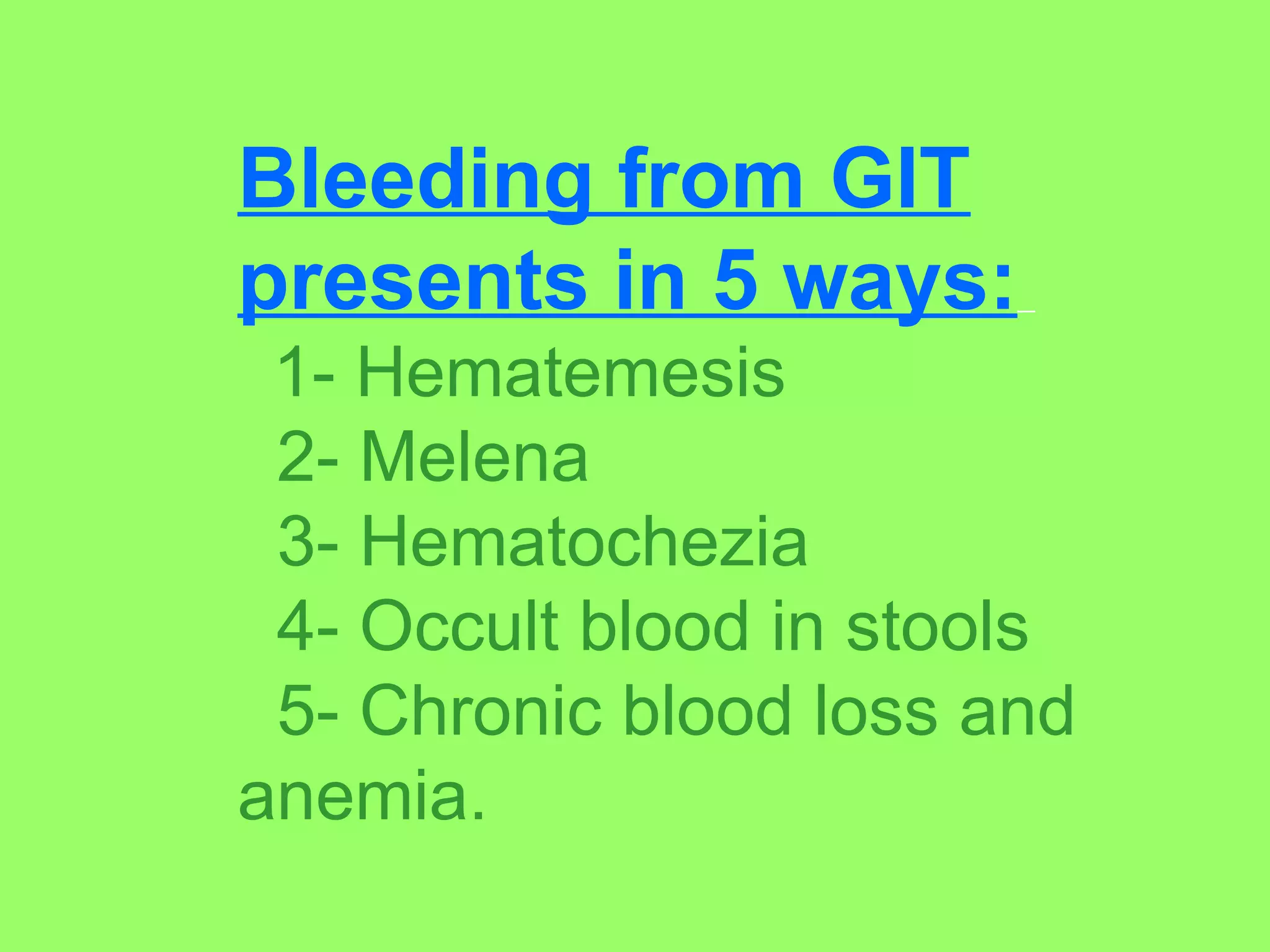
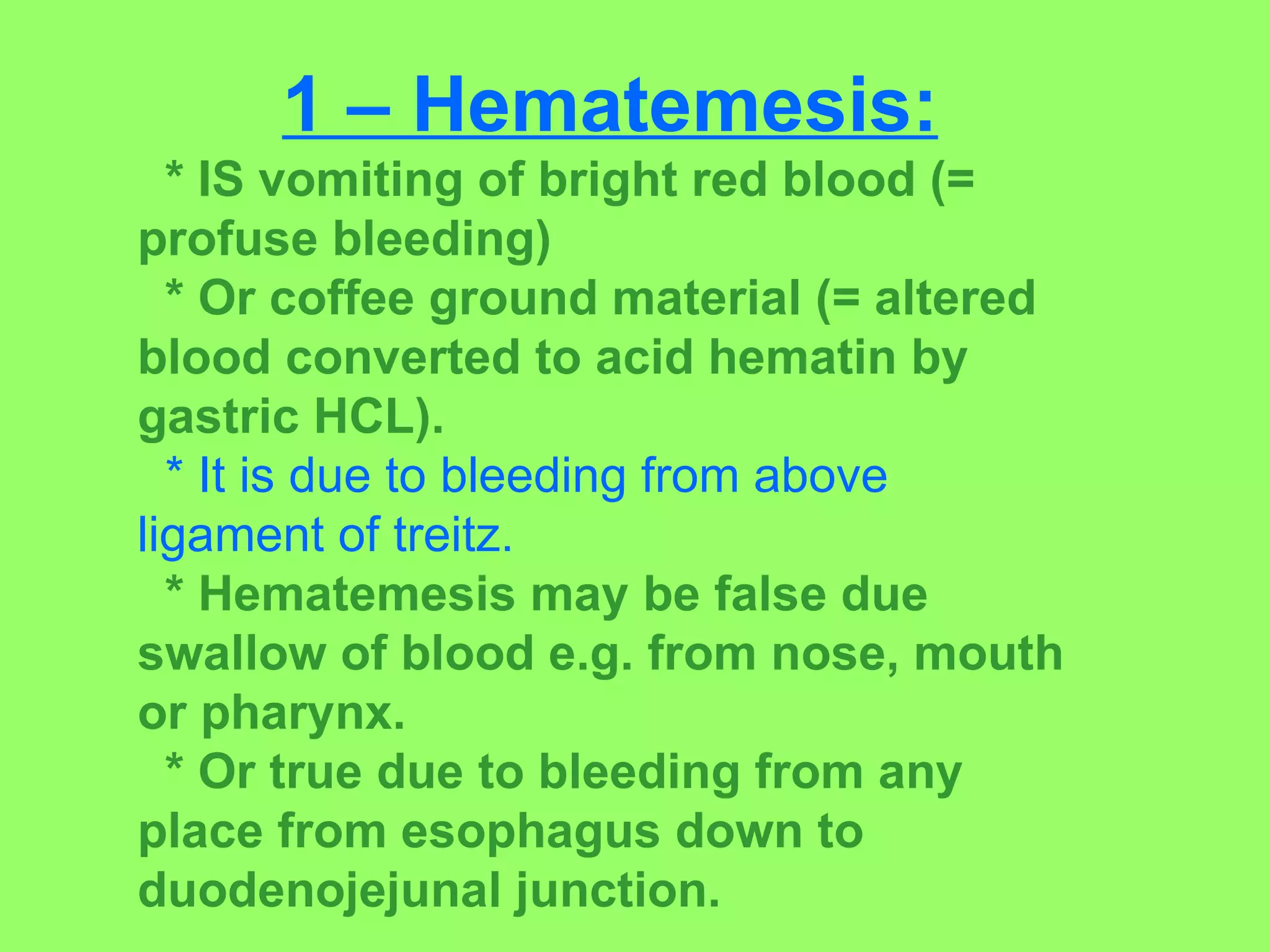
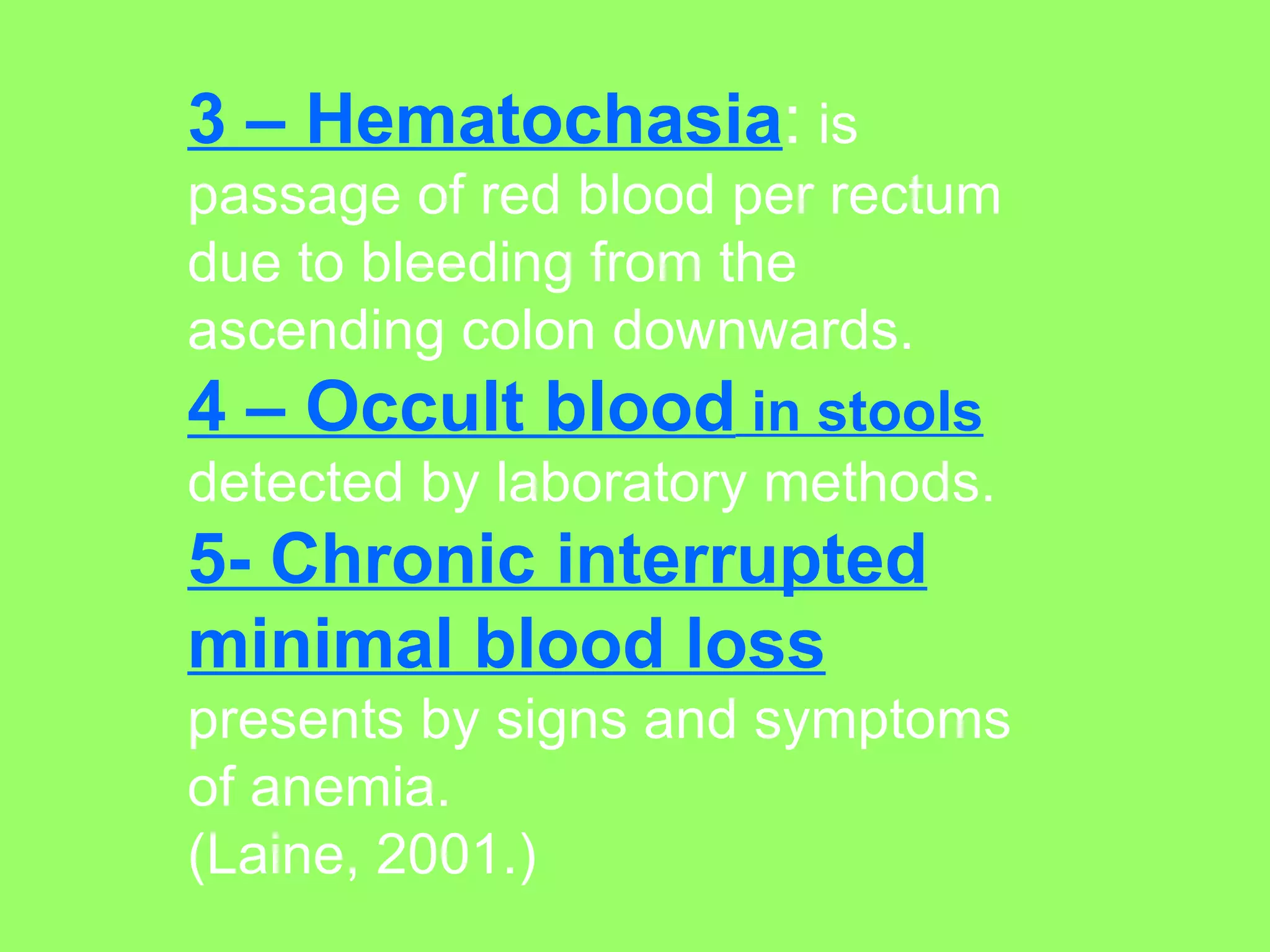
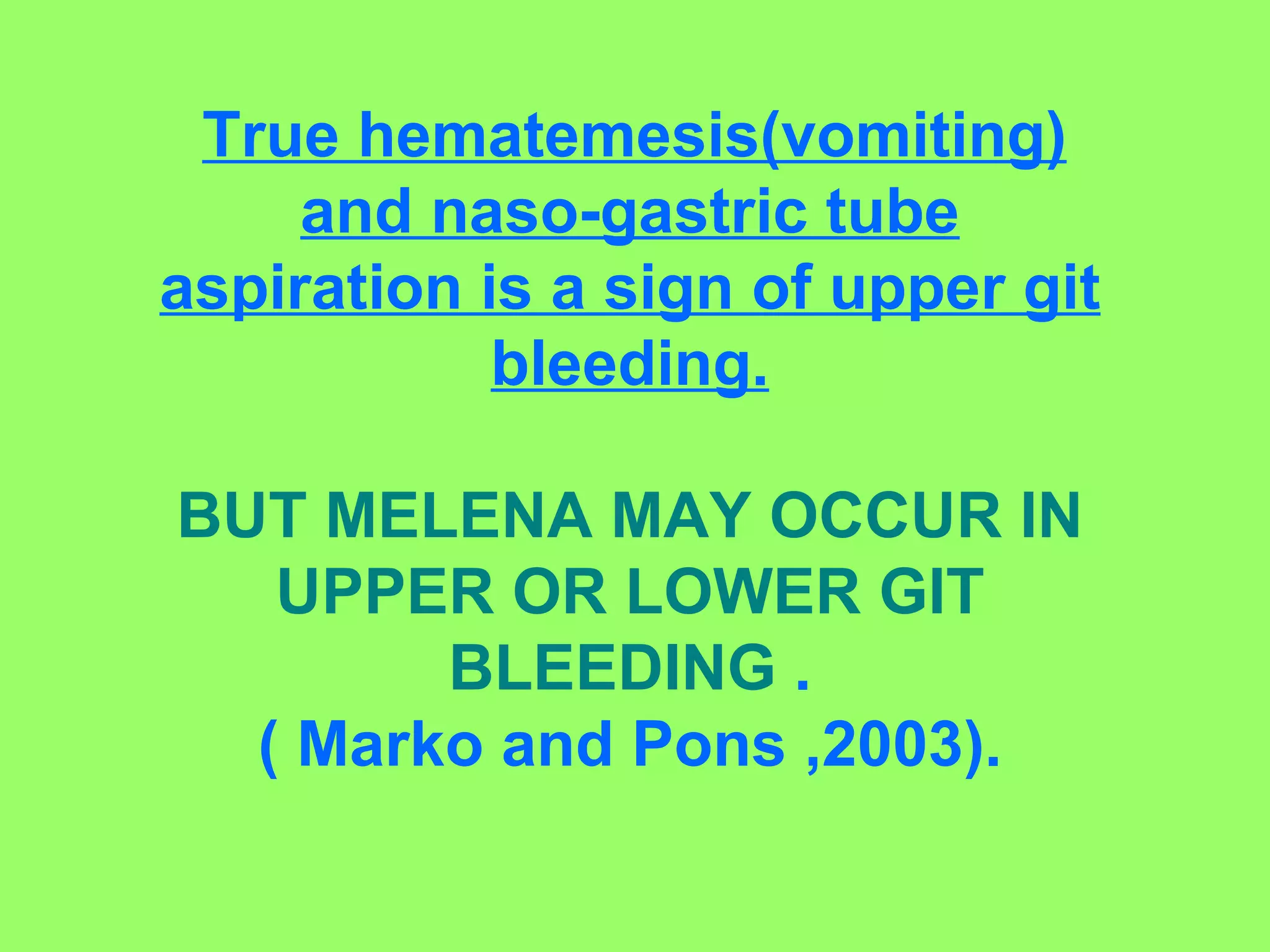
1. The document discusses gastrointestinal bleeding, describing its various presentations including hematemesis, melena, hematochezia, occult blood in stools, and chronic blood loss/anemia.
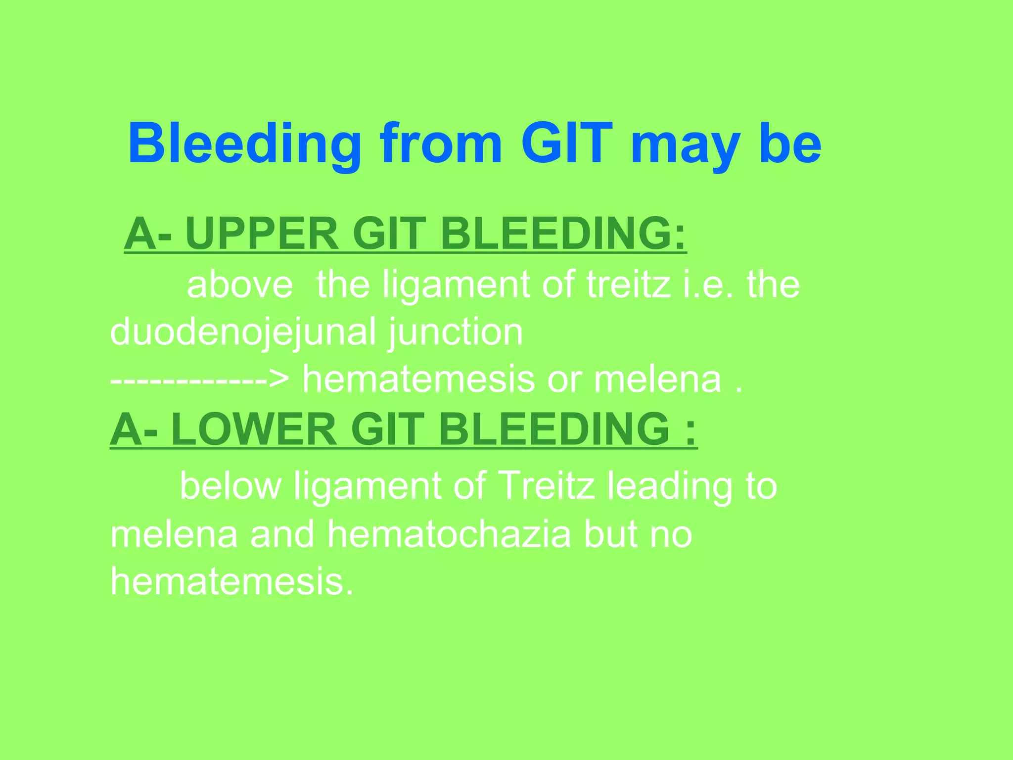
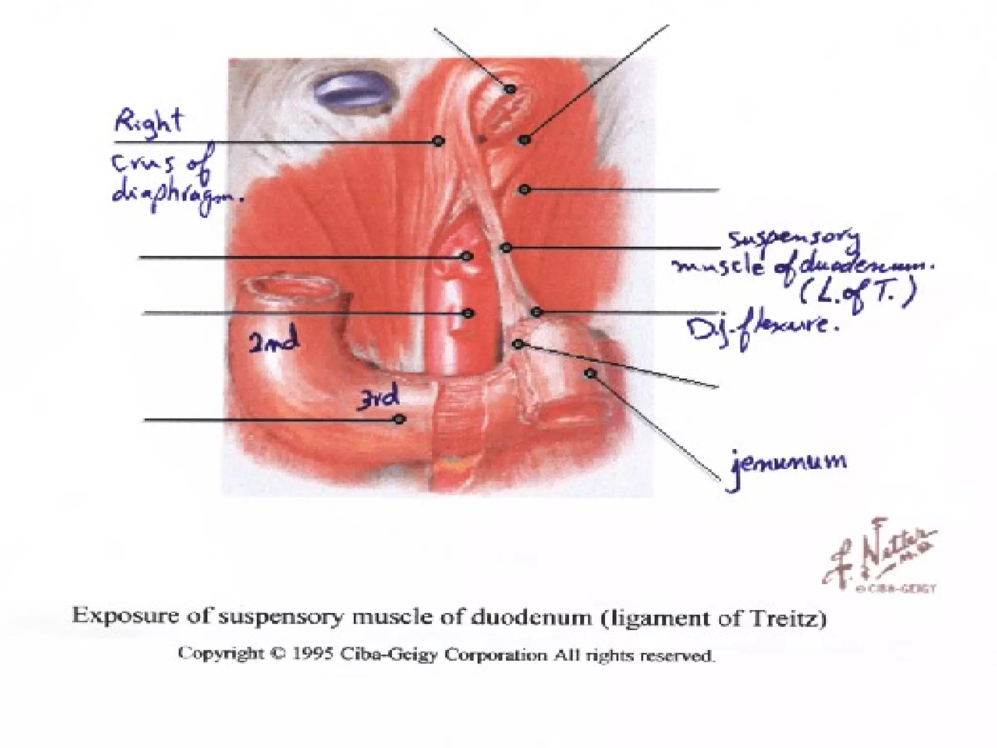
2. Upper GI bleeding occurs above the ligament of Treitz and can cause hematemesis or melena, while lower GI bleeding occurs below and causes melena and hematochezia but no hematemesis.
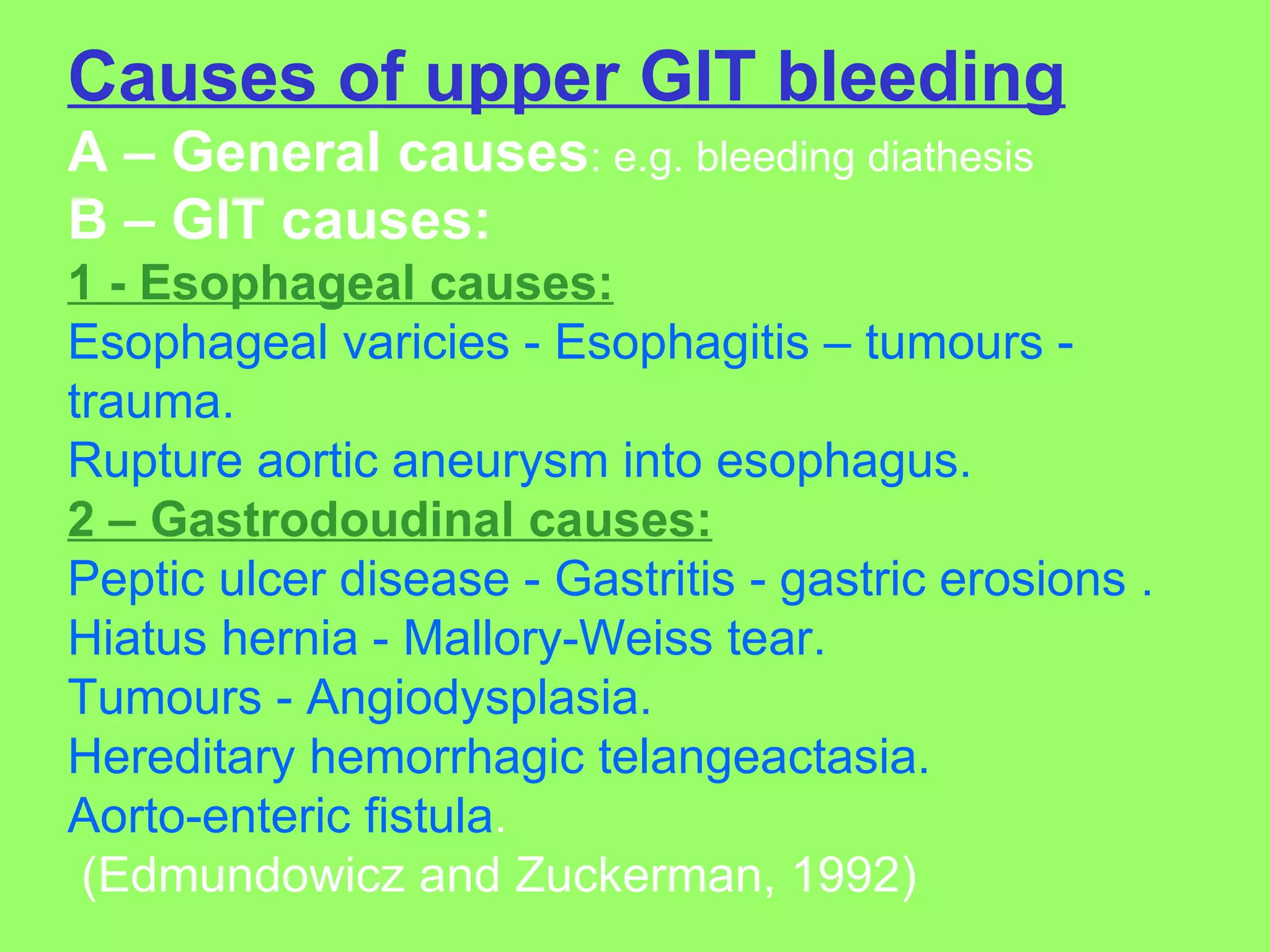
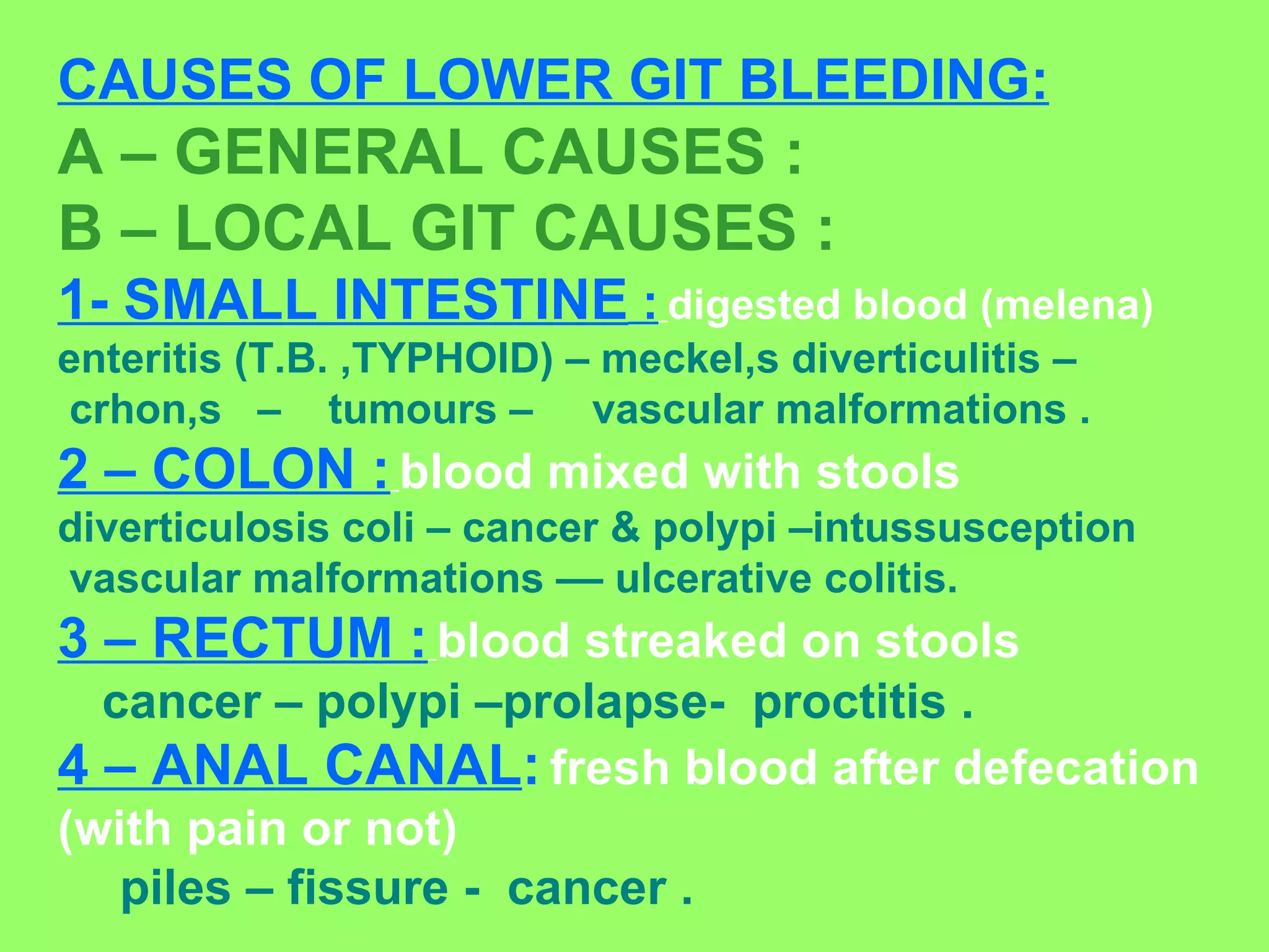
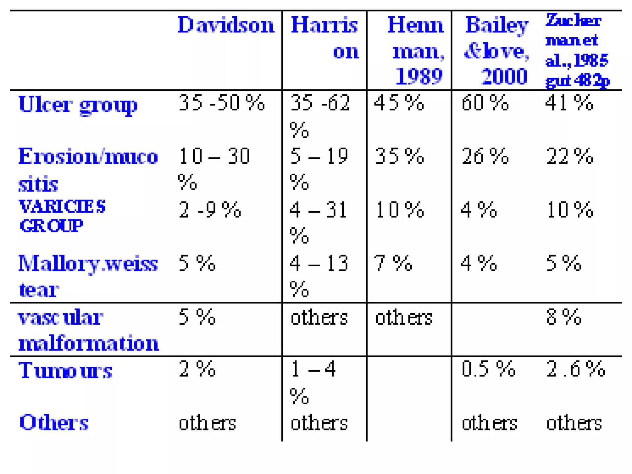
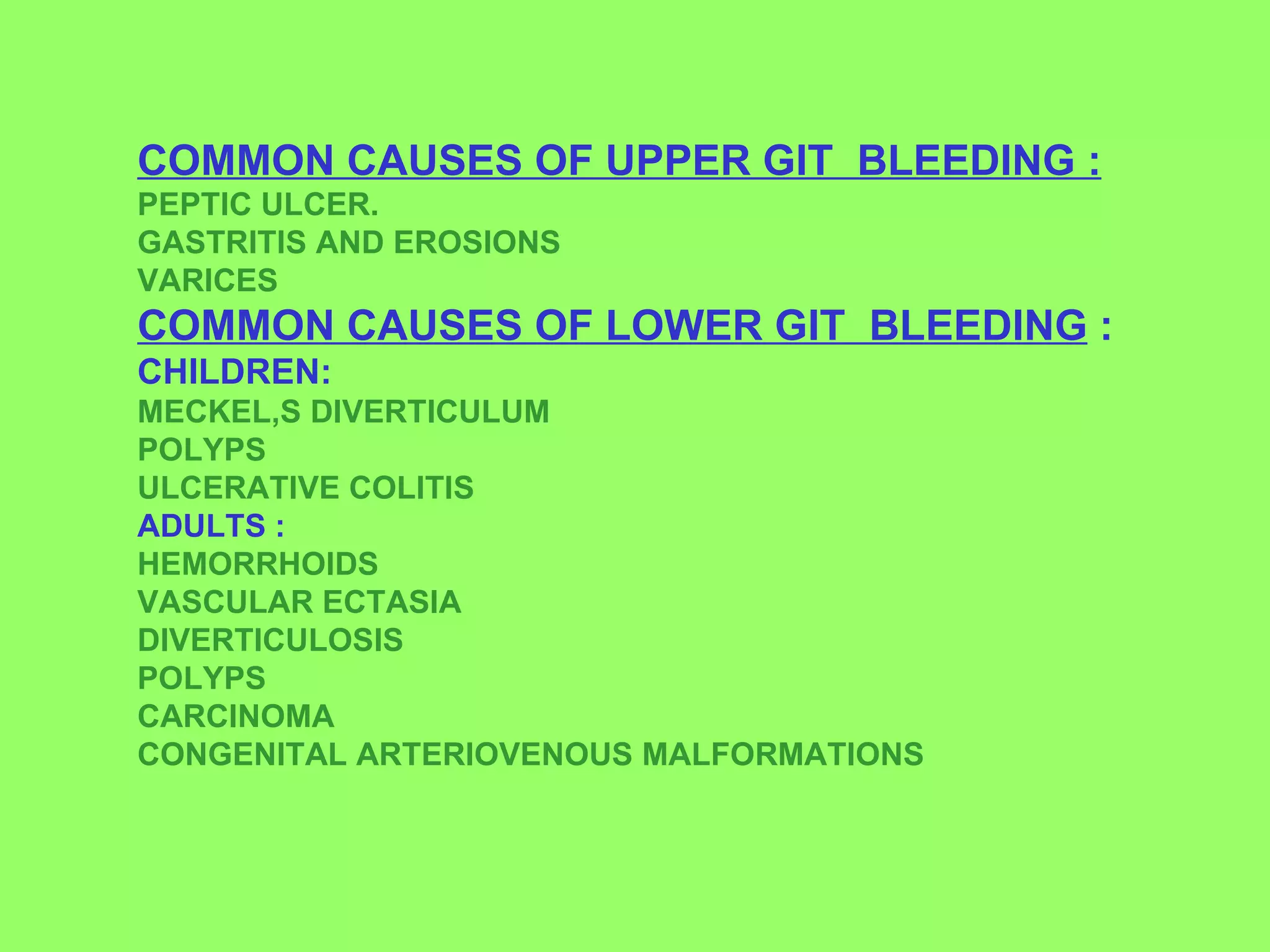
3. Common causes of upper GI bleeding include peptic ulcer disease, gastritis, and esophageal varices, while common causes of lower GI bleeding include hemorrhoids, diverticulosis, and polyps.
Seminar present the Upper Gastrointestinal Bleeding problems
Edited by : Dr. Inzar Yassen & Dr. Ammar L. Aldwaf
in Hawler Medical Uni. collage of medicine in 14/01/2014
Iraq - Kurdistan - Erbil
Approach to Management of Upper Gastrointestinal (GI) BleedingArun Vasireddy
Upper gastrointestinal bleeding is gastrointestinal bleeding in the upper gastrointestinal tract, commonly defined as bleeding arising from the esophagus, stomach, or duodenum. Blood may be observed in vomit (hematemesis) or in altered form in the stool (melena). Depending on the severity of the blood loss, there may be symptoms of insufficient circulating blood volume and shock. As a result, upper gastrointestinal bleeding is considered a medical emergency and typically requires hospital care for urgent diagnosis and treatment. Upper gastrointestinal bleeding can be caused by peptic ulcers, gastric erosions, esophageal varices, and some rarer causes such as gastric cancer.
The initial assessment includes measurement of the blood pressure and heart rate, as well as blood tests to determine hemoglobin concentration. In significant bleeding, fluid replacement is often required, as well as blood transfusion, before the source of bleeding can be determined by endoscopy of the upper digestive tract with an esophagogastroduodenoscopy. Depending on the source, endoscopic therapy can be applied to reduce rebleeding risk. Specific medical treatments (such as proton pump inhibitors for peptic ulcer disease) or procedures (such as TIPS for variceal hemorrhage) may be used. Recurrent or refractory bleeding may lead to need for surgery, although this has become uncommon as a result of improved endoscopic and medical treatment.
Explanation of what splenomegaly is in relation to its dimension deviation from normal spleen.Classification of splenomegaly according to it's size in adult and pediatric. The causes of splenomegaly along with the symptom that would manifest as a result of this anomaly. Lastly, diagnosis of splenomegaly
Hematemesis- vomiting of blood , a brief studymartinshaji
There can be many causes of hematemesis, such as: bleeding ulcers. prolonged and vigorous retching that causes tears in the esophageal mucosa (known as Mallory-Weiss Syndrome) gastric or intestinal varices.Haematemesis is simply defined as “vomiting blood”. It is caused by bleeding from part of the upper portion of the gastrointestinal tract. It has a wide range of possible causes, depending on the site of blood loss and the tissue that is actively bleeding. Hence it is necessary to analyse and treat the condition perfectly , this is brief study about all the aspects hematemesis ,vomiting of blood including etiology, definition,management ,treatment by drugs etc
please comment
thank u
Anti ulcer drugs and their Advance pharmacology ||
Anti-ulcer drugs are medications used to prevent and treat ulcers in the stomach and upper part of the small intestine (duodenal ulcers). These ulcers are often caused by an imbalance between stomach acid and the mucosal lining, which protects the stomach lining.
||Scope: Overview of various classes of anti-ulcer drugs, their mechanisms of action, indications, side effects, and clinical considerations.
Report Back from SGO 2024: What’s the Latest in Cervical Cancer?bkling
Are you curious about what’s new in cervical cancer research or unsure what the findings mean? Join Dr. Emily Ko, a gynecologic oncologist at Penn Medicine, to learn about the latest updates from the Society of Gynecologic Oncology (SGO) 2024 Annual Meeting on Women’s Cancer. Dr. Ko will discuss what the research presented at the conference means for you and answer your questions about the new developments.
Title: Sense of Taste
Presenter: Dr. Faiza, Assistant Professor of Physiology
Qualifications:
MBBS (Best Graduate, AIMC Lahore)
FCPS Physiology
ICMT, CHPE, DHPE (STMU)
MPH (GC University, Faisalabad)
MBA (Virtual University of Pakistan)
Learning Objectives:
Describe the structure and function of taste buds.
Describe the relationship between the taste threshold and taste index of common substances.
Explain the chemical basis and signal transduction of taste perception for each type of primary taste sensation.
Recognize different abnormalities of taste perception and their causes.
Key Topics:
Significance of Taste Sensation:
Differentiation between pleasant and harmful food
Influence on behavior
Selection of food based on metabolic needs
Receptors of Taste:
Taste buds on the tongue
Influence of sense of smell, texture of food, and pain stimulation (e.g., by pepper)
Primary and Secondary Taste Sensations:
Primary taste sensations: Sweet, Sour, Salty, Bitter, Umami
Chemical basis and signal transduction mechanisms for each taste
Taste Threshold and Index:
Taste threshold values for Sweet (sucrose), Salty (NaCl), Sour (HCl), and Bitter (Quinine)
Taste index relationship: Inversely proportional to taste threshold
Taste Blindness:
Inability to taste certain substances, particularly thiourea compounds
Example: Phenylthiocarbamide
Structure and Function of Taste Buds:
Composition: Epithelial cells, Sustentacular/Supporting cells, Taste cells, Basal cells
Features: Taste pores, Taste hairs/microvilli, and Taste nerve fibers
Location of Taste Buds:
Found in papillae of the tongue (Fungiform, Circumvallate, Foliate)
Also present on the palate, tonsillar pillars, epiglottis, and proximal esophagus
Mechanism of Taste Stimulation:
Interaction of taste substances with receptors on microvilli
Signal transduction pathways for Umami, Sweet, Bitter, Sour, and Salty tastes
Taste Sensitivity and Adaptation:
Decrease in sensitivity with age
Rapid adaptation of taste sensation
Role of Saliva in Taste:
Dissolution of tastants to reach receptors
Washing away the stimulus
Taste Preferences and Aversions:
Mechanisms behind taste preference and aversion
Influence of receptors and neural pathways
Impact of Sensory Nerve Damage:
Degeneration of taste buds if the sensory nerve fiber is cut
Abnormalities of Taste Detection:
Conditions: Ageusia, Hypogeusia, Dysgeusia (parageusia)
Causes: Nerve damage, neurological disorders, infections, poor oral hygiene, adverse drug effects, deficiencies, aging, tobacco use, altered neurotransmitter levels
Neurotransmitters and Taste Threshold:
Effects of serotonin (5-HT) and norepinephrine (NE) on taste sensitivity
Supertasters:
25% of the population with heightened sensitivity to taste, especially bitterness
Increased number of fungiform papillae
HOT NEW PRODUCT! BIG SALES FAST SHIPPING NOW FROM CHINA!! EU KU DB BK substit...GL Anaacs
Contact us if you are interested:
Email / Skype : kefaya1771@gmail.com
Threema: PXHY5PDH
New BATCH Ku !!! MUCH IN DEMAND FAST SALE EVERY BATCH HAPPY GOOD EFFECT BIG BATCH !
Contact me on Threema or skype to start big business!!
Hot-sale products:
NEW HOT EUTYLONE WHITE CRYSTAL!!
5cl-adba precursor (semi finished )
5cl-adba raw materials
ADBB precursor (semi finished )
ADBB raw materials
APVP powder
5fadb/4f-adb
Jwh018 / Jwh210
Eutylone crystal
Protonitazene (hydrochloride) CAS: 119276-01-6
Flubrotizolam CAS: 57801-95-3
Metonitazene CAS: 14680-51-4
Payment terms: Western Union,MoneyGram,Bitcoin or USDT.
Deliver Time: Usually 7-15days
Shipping method: FedEx, TNT, DHL,UPS etc.Our deliveries are 100% safe, fast, reliable and discreet.
Samples will be sent for your evaluation!If you are interested in, please contact me, let's talk details.
We specializes in exporting high quality Research chemical, medical intermediate, Pharmaceutical chemicals and so on. Products are exported to USA, Canada, France, Korea, Japan,Russia, Southeast Asia and other countries.
Explore natural remedies for syphilis treatment in Singapore. Discover alternative therapies, herbal remedies, and lifestyle changes that may complement conventional treatments. Learn about holistic approaches to managing syphilis symptoms and supporting overall health.
TEST BANK for Operations Management, 14th Edition by William J. Stevenson, Ve...kevinkariuki227
TEST BANK for Operations Management, 14th Edition by William J. Stevenson, Verified Chapters 1 - 19, Complete Newest Version.pdf
Lung Cancer: Artificial Intelligence, Synergetics, Complex System Analysis, S...Oleg Kshivets
RESULTS: Overall life span (LS) was 2252.1±1742.5 days and cumulative 5-year survival (5YS) reached 73.2%, 10 years – 64.8%, 20 years – 42.5%. 513 LCP lived more than 5 years (LS=3124.6±1525.6 days), 148 LCP – more than 10 years (LS=5054.4±1504.1 days).199 LCP died because of LC (LS=562.7±374.5 days). 5YS of LCP after bi/lobectomies was significantly superior in comparison with LCP after pneumonectomies (78.1% vs.63.7%, P=0.00001 by log-rank test). AT significantly improved 5YS (66.3% vs. 34.8%) (P=0.00000 by log-rank test) only for LCP with N1-2. Cox modeling displayed that 5YS of LCP significantly depended on: phase transition (PT) early-invasive LC in terms of synergetics, PT N0—N12, cell ratio factors (ratio between cancer cells- CC and blood cells subpopulations), G1-3, histology, glucose, AT, blood cell circuit, prothrombin index, heparin tolerance, recalcification time (P=0.000-0.038). Neural networks, genetic algorithm selection and bootstrap simulation revealed relationships between 5YS and PT early-invasive LC (rank=1), PT N0—N12 (rank=2), thrombocytes/CC (3), erythrocytes/CC (4), eosinophils/CC (5), healthy cells/CC (6), lymphocytes/CC (7), segmented neutrophils/CC (8), stick neutrophils/CC (9), monocytes/CC (10); leucocytes/CC (11). Correct prediction of 5YS was 100% by neural networks computing (area under ROC curve=1.0; error=0.0).
CONCLUSIONS: 5YS of LCP after radical procedures significantly depended on: 1) PT early-invasive cancer; 2) PT N0--N12; 3) cell ratio factors; 4) blood cell circuit; 5) biochemical factors; 6) hemostasis system; 7) AT; 8) LC characteristics; 9) LC cell dynamics; 10) surgery type: lobectomy/pneumonectomy; 11) anthropometric data. Optimal diagnosis and treatment strategies for LC are: 1) screening and early detection of LC; 2) availability of experienced thoracic surgeons because of complexity of radical procedures; 3) aggressive en block surgery and adequate lymph node dissection for completeness; 4) precise prediction; 5) adjuvant chemoimmunoradiotherapy for LCP with unfavorable prognosis.
Ethanol (CH3CH2OH), or beverage alcohol, is a two-carbon alcohol
that is rapidly distributed in the body and brain. Ethanol alters many
neurochemical systems and has rewarding and addictive properties. It
is the oldest recreational drug and likely contributes to more morbidity,
mortality, and public health costs than all illicit drugs combined. The
5th edition of the Diagnostic and Statistical Manual of Mental Disorders
(DSM-5) integrates alcohol abuse and alcohol dependence into a single
disorder called alcohol use disorder (AUD), with mild, moderate,
and severe subclassifications (American Psychiatric Association, 2013).
In the DSM-5, all types of substance abuse and dependence have been
combined into a single substance use disorder (SUD) on a continuum
from mild to severe. A diagnosis of AUD requires that at least two of
the 11 DSM-5 behaviors be present within a 12-month period (mild
AUD: 2–3 criteria; moderate AUD: 4–5 criteria; severe AUD: 6–11 criteria).
The four main behavioral effects of AUD are impaired control over
drinking, negative social consequences, risky use, and altered physiological
effects (tolerance, withdrawal). This chapter presents an overview
of the prevalence and harmful consequences of AUD in the U.S.,
the systemic nature of the disease, neurocircuitry and stages of AUD,
comorbidities, fetal alcohol spectrum disorders, genetic risk factors, and
pharmacotherapies for AUD.
3. Bleeding from GIT presents in 5 ways: 1- Hematemesis 2- Melena 3- Hematochezia 4- Occult blood in stools 5- Chronic blood loss and anemia.
4. 1 – Hematemesis: * IS vomiting of bright red blood (= profuse bleeding) * Or coffee ground material (= altered blood converted to acid hematin by gastric HCL). * It is due to bleeding from above ligament of treitz. * Hematemesis may be false due swallow of blood e.g. from nose, mouth or pharynx. * Or true due to bleeding from any place from esophagus down to duodenojejunal junction.
5. 2 - Melena: * the passage of black tarry loose stools containing digested blood by the action of digestive enzymes and bacteria. *It is due to bleeding from any place above and including caecum . *If bleeding is sever, red blood clots may pass in stools.
6. 3 – Hematochasia : is passage of red blood per rectum due to bleeding from the ascending colon downwards. 4 – Occult blood in stools detected by laboratory methods. 5- Chronic interrupted minimal blood loss presents by signs and symptoms of anemia. (Laine, 2001.)
7. Bleeding from GIT may be A- UPPER GIT BLEEDING: above the ligament of treitz i.e. the duodenojejunal junction ------------> hematemesis or melena . A- LOWER GIT BLEEDING : below ligament of Treitz leading to melena and hematochazia but no hematemesis.
8.
9. True hematemesis(vomiting) and naso-gastric tube aspiration is a sign of upper git bleeding. BUT MELENA MAY OCCUR IN UPPER OR LOWER GIT BLEEDING . ( Marko and Pons ,2003).
10. Causes of upper GIT bleeding A – General causes : e.g. bleeding diathesis B – GIT causes: 1 - Esophageal causes: Esophageal varicies - Esophagitis – tumours - trauma. Rupture aortic aneurysm into esophagus. 2 – Gastrodoudinal causes: Peptic ulcer disease - Gastritis - gastric erosions . Hiatus hernia - Mallory-Weiss tear. Tumours - Angiodysplasia. Hereditary hemorrhagic telangeactasia. Aorto-enteric fistula . (Edmundowicz and Zuckerman, 1992)
11. CAUSES OF LOWER GIT BLEEDING: A – GENERAL CAUSES : B – LOCAL GIT CAUSES : 1- SMALL INTESTINE : digested blood (melena) enteritis (T.B. ,TYPHOID) – meckel,s diverticulitis – crhon,s – tumours – vascular malformations . 2 – COLON : blood mixed with stools diverticulosis coli – cancer & polypi –intussusception vascular malformations –– ulcerative colitis. 3 – RECTUM : blood streaked on stools cancer – polypi –prolapse- proctitis . 4 – ANAL CANAL : fresh blood after defecation (with pain or not) piles – fissure - cancer .
12.
13.
14. COMMON CAUSES OF UPPER GIT BLEEDING : PEPTIC ULCER. GASTRITIS AND EROSIONS VARICES COMMON CAUSES OF LOWER GIT BLEEDING : CHILDREN: MECKEL,S DIVERTICULUM POLYPS ULCERATIVE COLITIS ADULTS : HEMORRHOIDS VASCULAR ECTASIA DIVERTICULOSIS POLYPS CARCINOMA CONGENITAL ARTERIOVENOUS MALFORMATIONS
16. EVALUATION OF THE CASE : 1 – IS THERE HEMODYNAMIC CMPROMISE ? 2 – IS THERE ACTIVE BLEEDING? 3 – IS THIS A HIGH RISK PATIENT ? 4 – IS THIS UPPER OR LOWER GIT BLEEDING?
17.
18. CALCULATION OF AMOUNT OF BLOOD LOSS AND RESUSCETAION FLUIDS MARINO ( 1998) : STEP 1 1 – CALCULATION OF BLOOD VOLUME AND BODY FLIUDS :
23. USE OF OXYGEN EXTRACTION % TO EVALUATE HYPOVOLAEMIA : *MEASURE ( SaO2) BY PULSE OXIMETRY . *Measure O2 SATURATION IN VENOUS BLOOD GASES
24.
25.
26. Clinical picture of hypovolaemic shock Rapid weak pulse : - 1 *catecholamine release , *mary,s law =tachycardia with hypotension , *stimulated cardiac accelerating center directly by hypoxia and reflexly by carotid and aortic body chemoreceptor . 2- Hypotension and low pulse pressure : Decrease in blood volume= decrease in venous return = decrease in cardiac output = decrease in ABP. 3 -Subnormal temperature : vasoconstriction and decreased tissue metabolism . 4 - Increased rate and depth of respiration : Due to tissue hypoxia and hypotension .
27. Continue,hypovol.shock: 5 -Pale (vasoconstriction of capillaries), cold (vasoconstriction of arterioles) , clammy skin (sweat secretion ) = sympathetic over activity . 6 -Collapsed viens and decreased CVP . 7 -Oliguria : decreased renal blood flow and ADH release . 8 -Thirst sensation : 9 - Restlessness early with mild to moderate hypovoleamia and lethargy with moderate to sever hypovoleamia . 10 – CLINICAL PICTURE OF THE CAUSE :
28.
29. LABORATORY INVESTIGATIONS: 1- BLOOD GROUP AND CROSS MATCHING: FOR 4 – 8 UNITS ACCOIRDING TO SUSPECTING REBLEEDING STORE PLASMA FOR ONGOING CROSS MATCHING TAKE SAMPLE BEFORE COLLOID USE 2-CBC: HB%, PCV: CHANGED ONLY IN MASSIVE GIT BLEEDING, GIVES IDEA ABOUT PREVIOUS FITTNESS OF PATIENS. WBCS: IF MORE THAN 15000 CONFIRM ABOUT ANY SEPSIS. PLATELATS COUNT: if less than 50000 consider platelet support. 3-Urea and electrolytes: may be elevated inspite of normal creatinine due to increased protein absorption AND RETURNS AFTER VOLUME RESTORATION.. 4-Blood glucose : may decrease in liver disease. 5-PT, PTT AND LFTS : CHANGED IN LIVER DISEASE AND IN PATIENTS TAKING WARFARIN . 6-Monitor Arterial Blood . gases in morbid conditions. OCCULT BLOOD IN STOOL in minimal bleeding
30. DeterminATION OF SITE OF BLEEDING : 1 – History: DETERMINE DEGREE OF BLOOD LOSS BUT NOT SO ACCURATE ,LEVEL OF BLEEDING ,ETIOLOGY OF BLEEDING,PRECIPITATING FACTOR,PREVIOUS BLEEDING. 2 – Ryle tube and PR: 3 – Upper endoscopy, anorectosegmoidoscopy and colonoscopy : 4 – RADIOISOTOPIC Scanning by technetium labelled Rbcs: FOR SCREENING BEFORE ARTERIOGRAPHY ,IT CAN DETECT BLEEDING LESS THAN 0.5ML /MIN,A POSITIVE SCAN POINT TO CANDIDATE OF ARTERIOGRAPHY,NEGATIVE SCAN INDICATES SHORT TERM GOOD PROGNOSIS . 5 – Selective arteriography : DETERMINES THE SITE OF BLEEDING NOT THE CAUSE. USED FOR THERAPEUTIC INTRA-ARTERIAL INJECTION OF VASOPRESSIN OR ARTERIAL EMBOLISATION BY GELFOAM
31. PRIMARY EVALUATION AND RESSUSCITATION: IF IMPENDING HYPOVOLEMIC SHOCK: A airway protection and consider endotracheal tube if aspiration is suspected . B BREATHING SUPPORT C circulatory support : 1- wide pore venous access . 2 – appropriate fluid transfusion according to patient condition and facilities . 3 – contact with surgeons and emergency endoscopic team early . insert retained urinary cath.and calculate urine hourly. 4- insert ryle tube to detect hematemesis and or do gastric wash according to cause . 5 – in compromised patients cvp and intensive care measurements is considered according to every case .
32. Vasopressin : constrict splanchnic arterioles 0.4 u/min. for one day then 0.2 u /min . for another day. Better given with nitroglycerin. Glypressin:long duration ,less side effects 2mg iv every hour till bleeding stops then 1 mg every 6 hours octreotide : selective splanchnic arteriolar vasoconstriction 50 microgr iv bolus then 50 microgram every 6 hours for 48 hours
33. CERTAIN PRECAUTIONS * HB% OF 7-8 gm.WILL GIVE ADEQUATE OXYGENATION FOR NORMOVOLEMIC BUT IN HYPOVOLEMIC OR COMPROMISED PATIENT 9-10 gm. IS BETTER ACHIEVED. * GIVE PACKED RBS IN CARDIAC RISKY PATIENTS PLATELETS FOR MASSIVE BLOOD TRANSFUSION * FFP FOR COAGULATION DISORDERS * PLATELET CONCENTRATE FOR THROMBOCYTOPENIA less than 50,000. * BLOOD GROUP O NEGATIVE EVEN WITHOUT CROSS MATCHING FOTR LIFE THREATENING CONDITIONS . * CALCIUM ONE AMPULE FOR EVERY FOUR UNITS. * CHECK FOR BLOOD HAEMOLYSIS IN UNCONSCIOUS PATIENTS.
34. Hypovolaemia and shock: * 500 ml. of blood loss leads to minimal clinical finding. * 1000 ml. of blood loss causes positive tilt test. * 2000 ml. of blood loss presents with features of shock. * Rapid loss of 50% of blood volume is usually fatal. * Elders cannot accommodate for hypovolaemia properly. * Mild hypovolaemia = compensatory vasoconstriction to maintain blood pressure. * More hypovolaemia = hypotension, increase in peripheral vascular resistance, capillary and venous bed collapse, and all of these leads to more tissue hypoxia.
35.
36. Low risk criteria : Henneman,2003 . 1 – No co morbid diseases. 2 – Normal vital signs. 3 – Normal or trace positive stool guaiac. 4 - Negative gastric aspirate. 5 – Normal or near normal HB%&hematocrit. 6 – No problem to ask for medical help on need. 7 – Proper understanding of S. &S. of bleeding. 8 – No high risk factors and easy medical follow up.
37. HIGH RISK PATIENTS : VELAYO,2003. 1 – AGE > 60 YEARS . 2 – COMORBID CONDITIONS : D.M. , RENAL, CARDIAC, HEPATIC FAILURE, IHD,CANCER. 3 – PERSISTENT HYPOTENSION . MORE THAN 4 UNITS OF TRANSFUSION. - 4 5 – BLEEDING OR REBLEEDING DURING HOSPITALISATION. 6 – BLOODY NASOGASTRIC ASPIRATE . 7 – NEED FOR EMERGENCY SURGERY . 8 – HIGH RISK LESIONS : ESPGHAGIAL VARECES , A-E FISTULA ,BIGACTIVELY BLEEDING ULCERS IN POSTERIOR PULP OF DUODINUM.