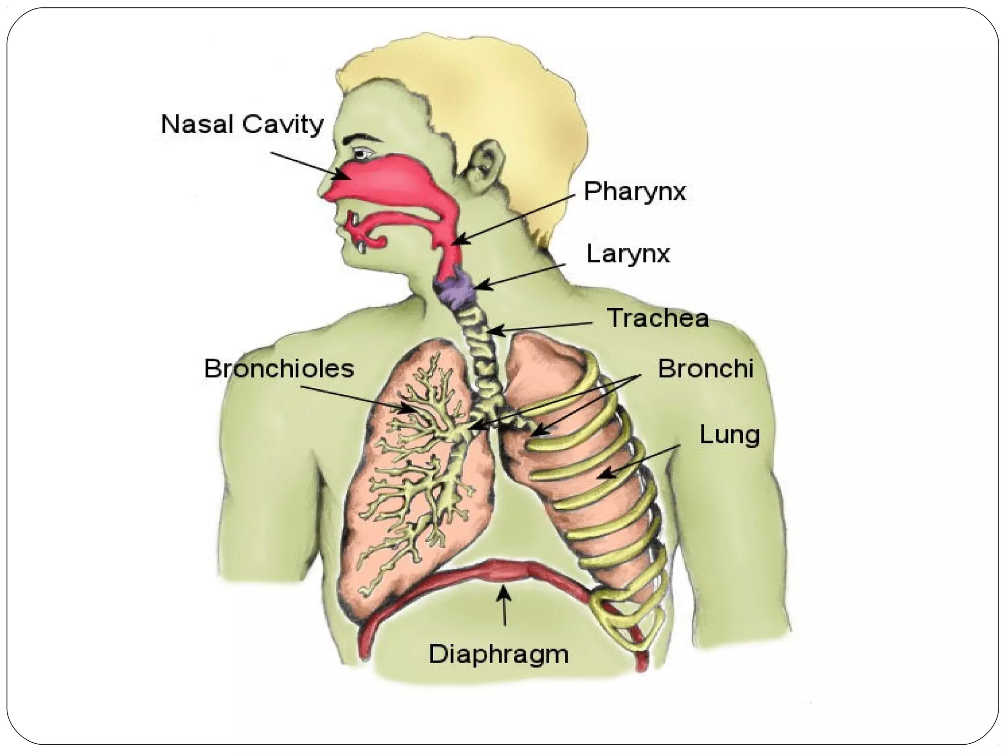
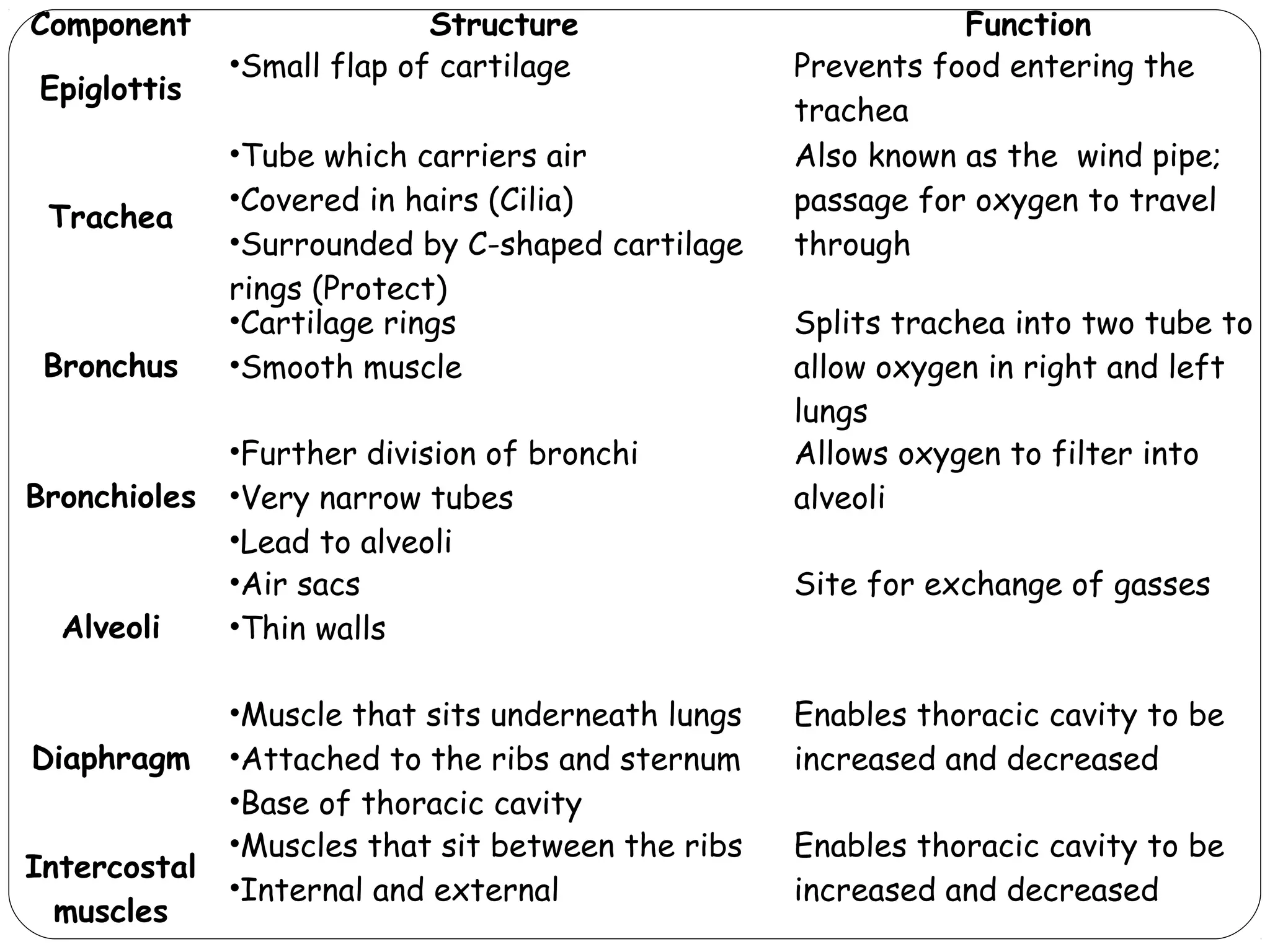
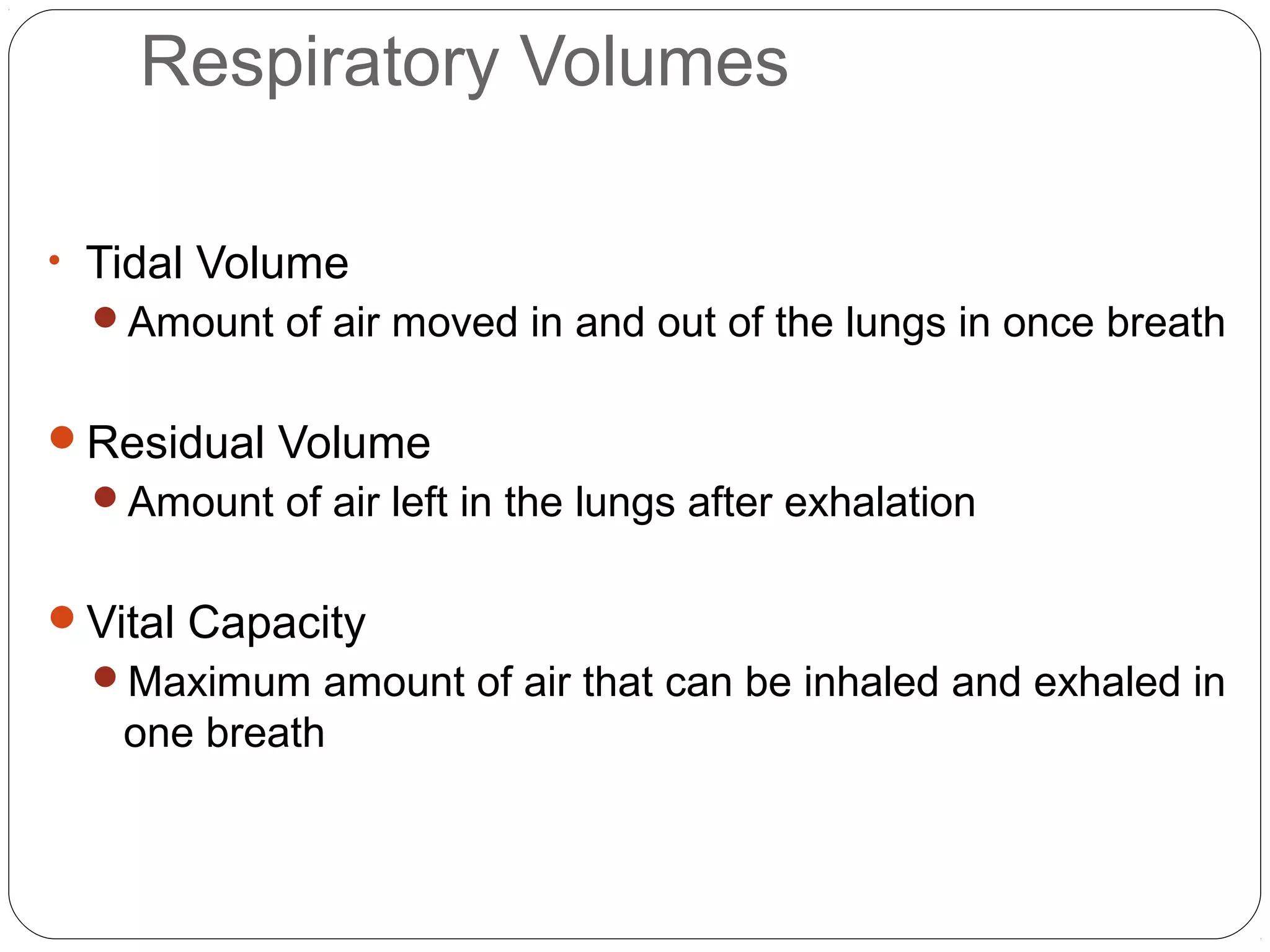
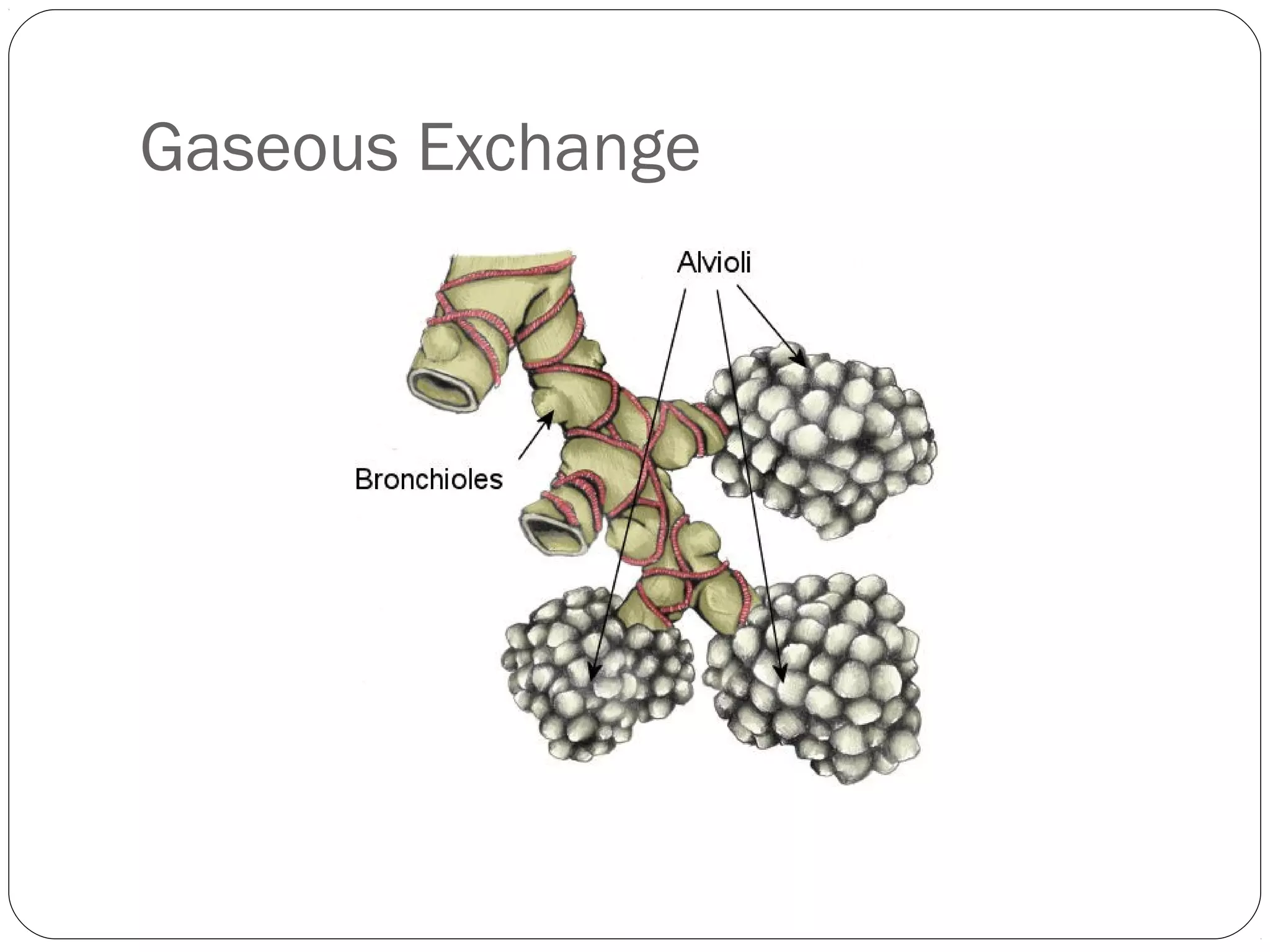
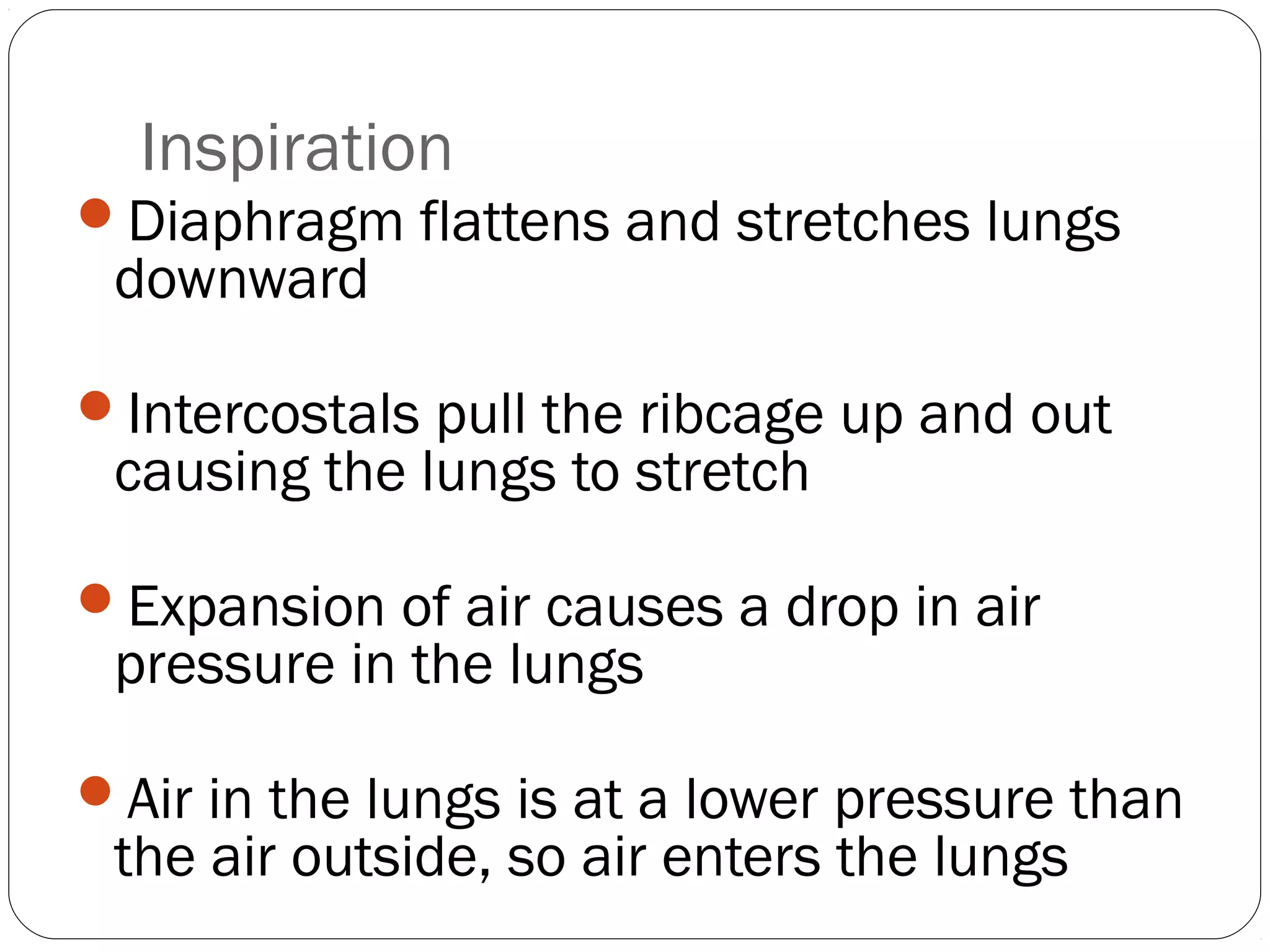
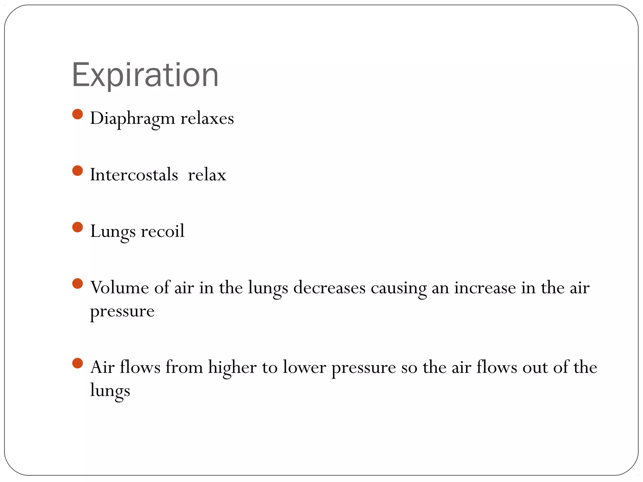
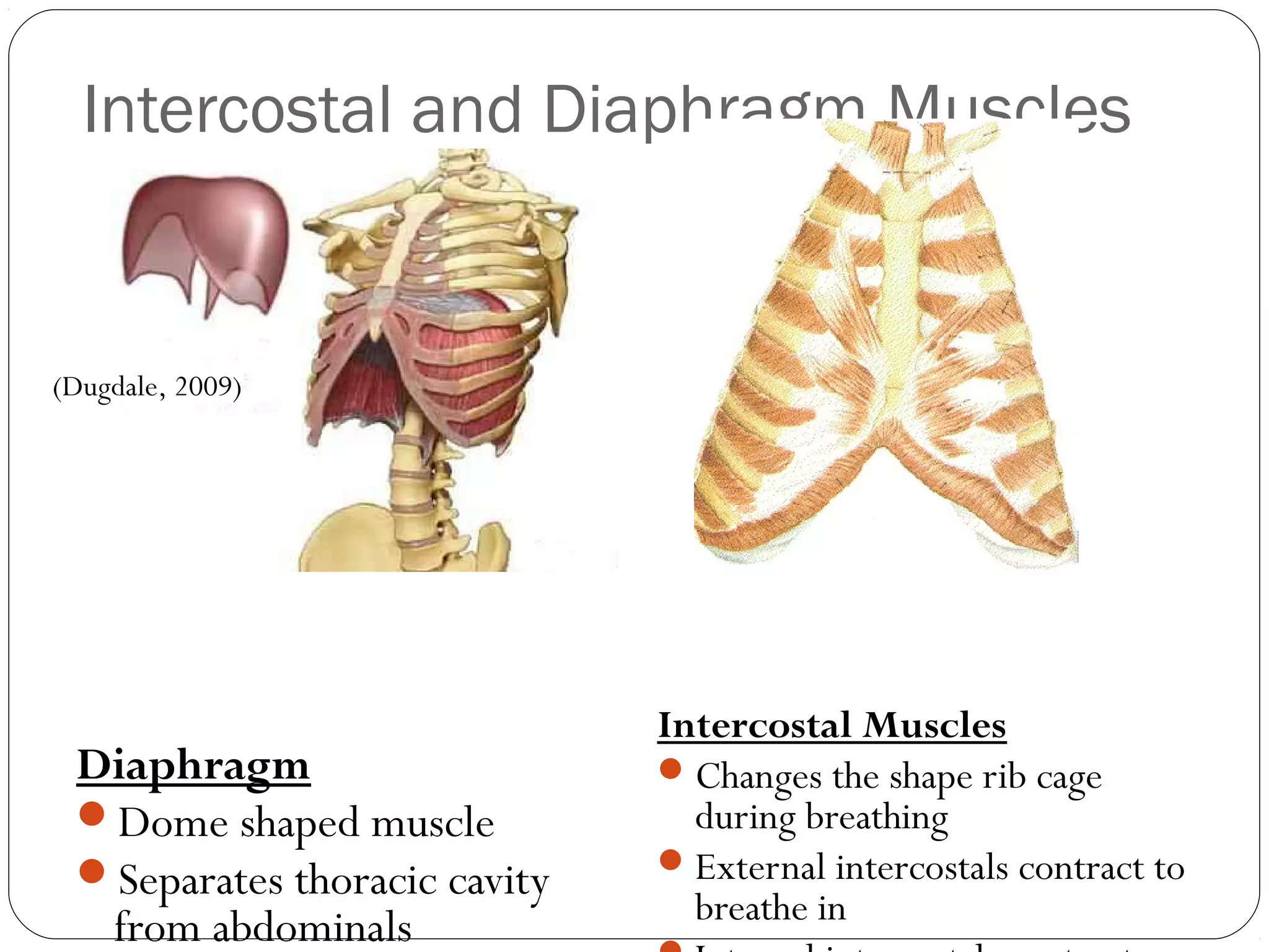
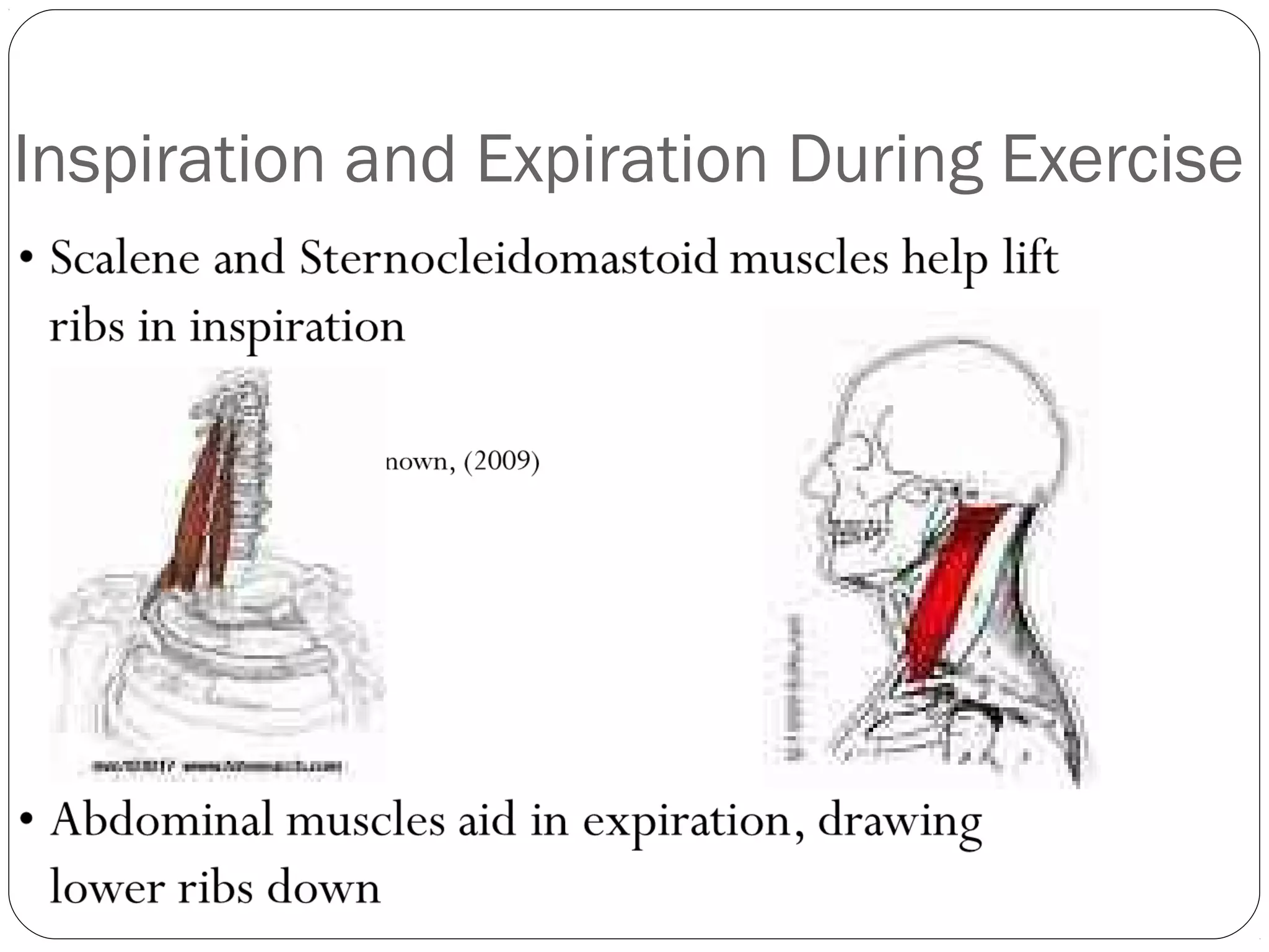
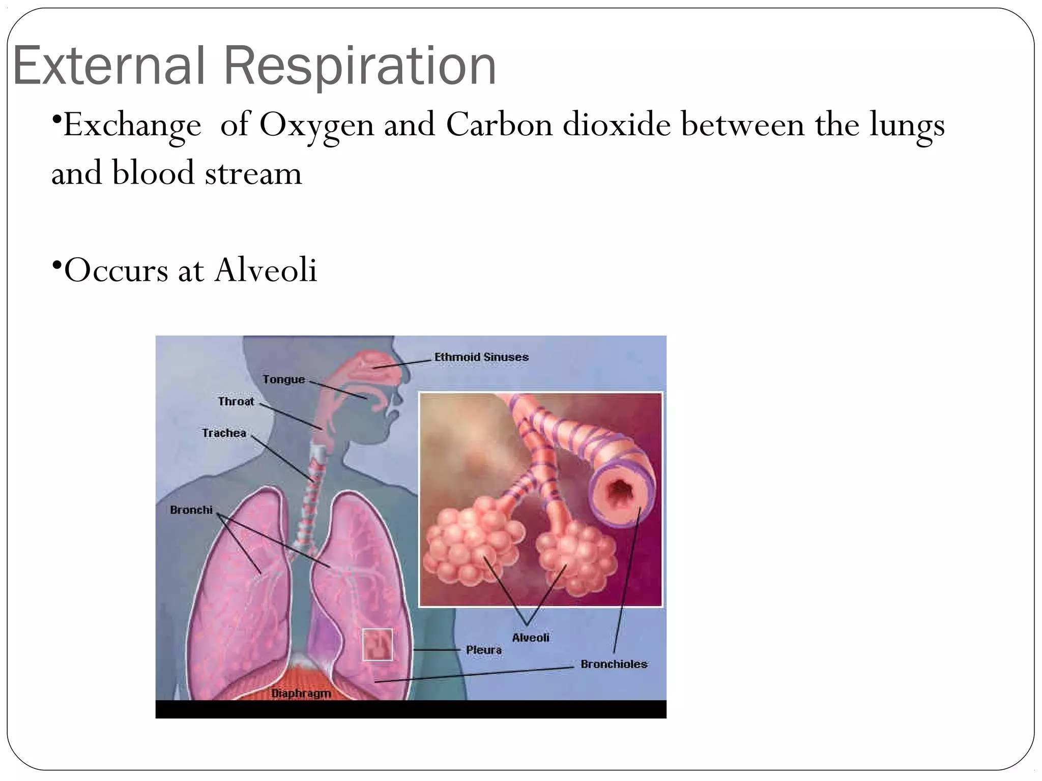
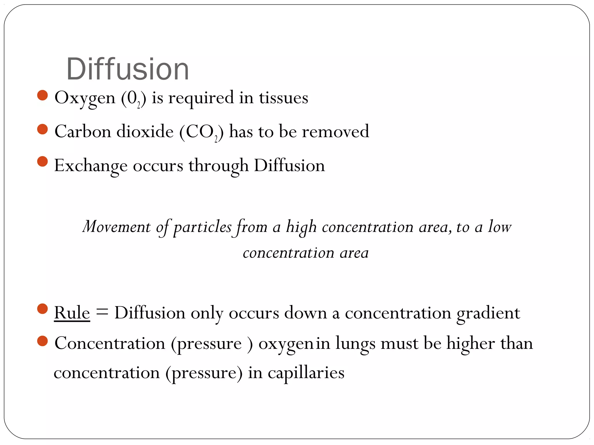
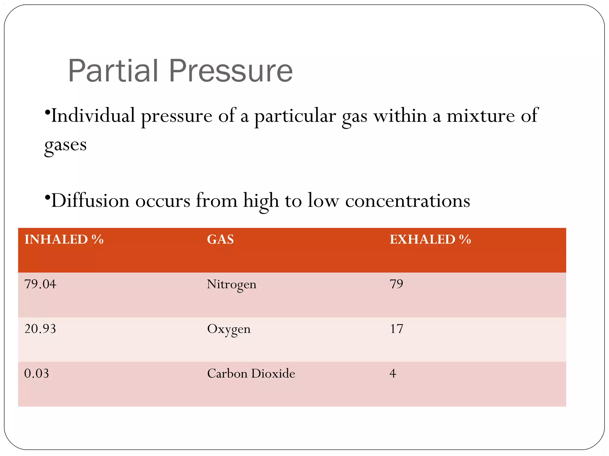
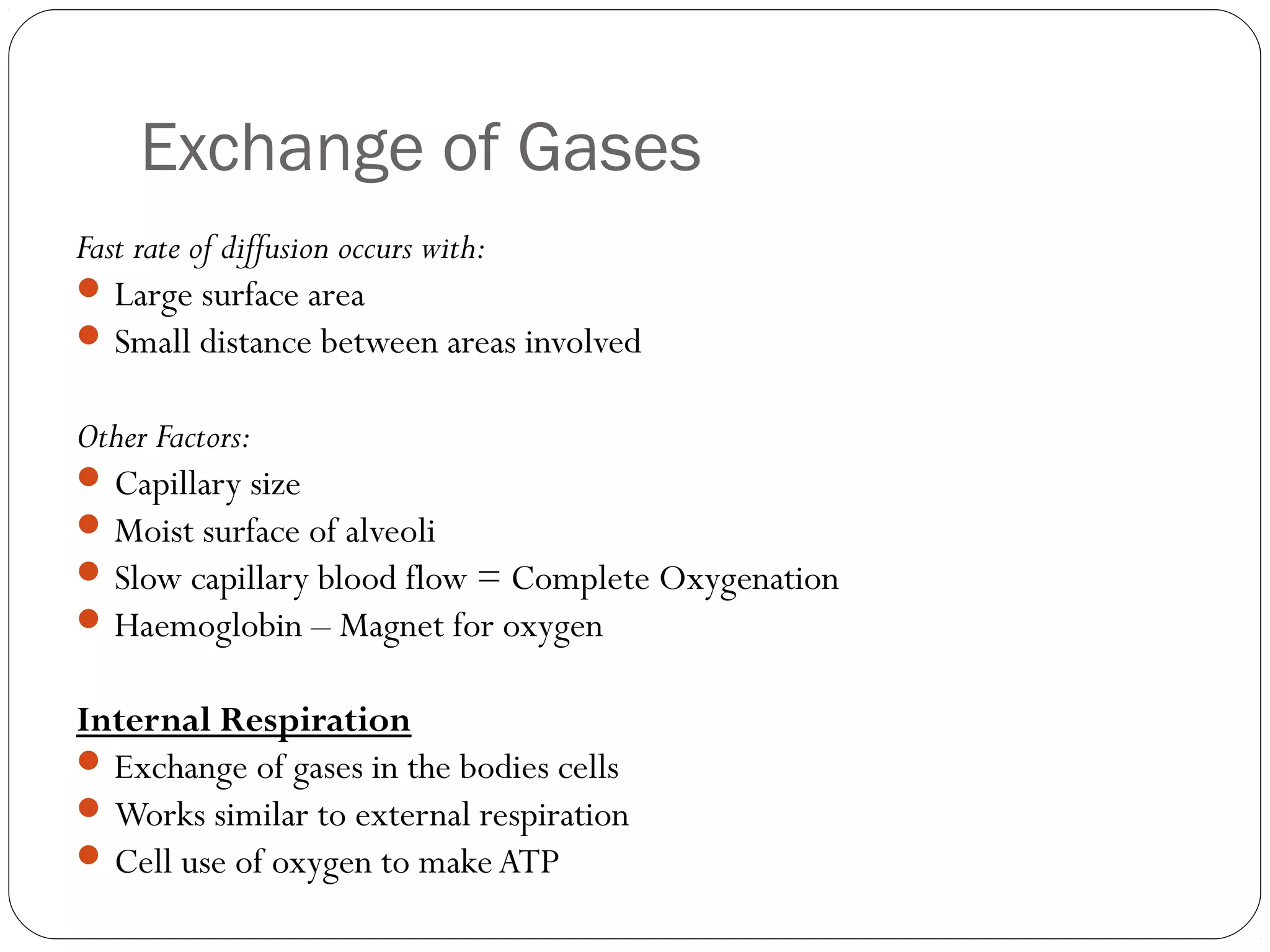
Cardio - respiratory System
Structure and Function of the Lungs
Lloyd Dean
Aims
 Describe the structure of the lungs
 Identify the function of the lungs
 Recall how lungs perform their function
Recap
Concave Discs

Venule

Artery

Platelets

Arterioles

Capillary

Plasma

CO2

Lumen

Tunica Media (Smooth

Muscle)
TIME TO DO SOME COLOURING IN! (1 Pack per study
buddy)
Rule 1. Have fun
Rule 2. If you find a term that you do not know then research
it on your mobile device
Rule 3. Make as many notes on the sheets as possible to help
with your video!
Component
Epiglottis

Trachea

Bronchus

Bronchioles

Alveoli

Diaphragm
Intercostal
muscles

Structure
•Small flap of cartilage
•Tube which carriers air
•Covered in hairs (Cilia)
•Surrounded by C-shaped cartilage
rings (Protect)
•Cartilage rings
•Smooth muscle
•Further division of bronchi
•Very narrow tubes
•Lead to alveoli
•Air sacs
•Thin walls
•Muscle that sits underneath lungs
•Attached to the ribs and sternum
•Base of thoracic cavity
•Muscles that sit between the ribs
•Internal and external

Function
Prevents food entering the
trachea
Also known as the wind pipe;
passage for oxygen to travel
through
Splits trachea into two tube to
allow oxygen in right and left
lungs
Allows oxygen to filter into
alveoli
Site for exchange of gasses

Enables thoracic cavity to be
increased and decreased
Enables thoracic cavity to be
increased and decreased
Respiratory Volumes
• Tidal Volume
Amount of air moved in and out of the lungs in once breath

Residual Volume
Amount of air left in the lungs after exhalation

Vital Capacity
Maximum amount of air that can be inhaled and exhaled in

one breath
Gaseous Exchange
Inspiration
Diaphragm flattens and stretches lungs

downward

Intercostals pull the ribcage up and out

causing the lungs to stretch

Expansion of air causes a drop in air

pressure in the lungs

Air in the lungs is at a lower pressure than

the air outside, so air enters the lungs
Expiration
 Diaphragm relaxes
 Intercostals relax
 Lungs recoil
 Volume of air in the lungs decreases causing an increase in the air

pressure

 Air flows from higher to lower pressure so the air flows out of the

lungs
Intercostal and Diaphragm Muscles

(Dugdale, 2009)

Diaphragm
Dome shaped muscle
Separates thoracic cavity
from abdominals

Intercostal Muscles
 Changes the shape rib cage
during breathing
 External intercostals contract to
breathe in
Inspiration and Expiration During Exercise
Inhalation and Exhalation
External Respiration
•Exchange of Oxygen and Carbon dioxide between the lungs

and blood stream
•Occurs at Alveoli

(WebMD, 2009)
Diffusion

Oxygen (02) is required in tissues
Carbon dioxide (CO2) has to be removed
Exchange occurs through Diffusion

Movement of particles from a high concentration area, to a low
concentration area
Rule = Diffusion only occurs down a concentration gradient
Concentration (pressure ) oxygen in lungs must be higher than

concentration (pressure) in capillaries
Partial Pressure
•Individual pressure of a particular gas within a mixture of

gases
•Diffusion occurs from high to low concentrations
INHALED %

GAS

EXHALED %

79.04

Nitrogen

79

20.93

Oxygen

17

0.03

Carbon Dioxide

4
Exchange of Gases
Fast rate of diffusion occurs with:
 Large surface area
 Small distance between areas involved
Other Factors:
 Capillary size
 Moist surface of alveoli
 Slow capillary blood flow = Complete Oxygenation
 Haemoglobin – Magnet for oxygen
Internal Respiration
 Exchange of gases in the bodies cells
 Works similar to external respiration
 Cell use of oxygen to make ATP
Gas Exchange
Neural and Chemical Control of
Breathing
Medulla Oblongata (Respiratory centre)
Impulses
Stretch receptors
Chemoreceptors
Lactic acid

Structure and function of the lungs

  • 1.
    Cardio - respiratorySystem Structure and Function of the Lungs Lloyd Dean
  • 2.
    Aims  Describe thestructure of the lungs  Identify the function of the lungs  Recall how lungs perform their function
  • 3.
  • 4.
    TIME TO DOSOME COLOURING IN! (1 Pack per study buddy) Rule 1. Have fun Rule 2. If you find a term that you do not know then research it on your mobile device Rule 3. Make as many notes on the sheets as possible to help with your video!
  • 6.
    Component Epiglottis Trachea Bronchus Bronchioles Alveoli Diaphragm Intercostal muscles Structure •Small flap ofcartilage •Tube which carriers air •Covered in hairs (Cilia) •Surrounded by C-shaped cartilage rings (Protect) •Cartilage rings •Smooth muscle •Further division of bronchi •Very narrow tubes •Lead to alveoli •Air sacs •Thin walls •Muscle that sits underneath lungs •Attached to the ribs and sternum •Base of thoracic cavity •Muscles that sit between the ribs •Internal and external Function Prevents food entering the trachea Also known as the wind pipe; passage for oxygen to travel through Splits trachea into two tube to allow oxygen in right and left lungs Allows oxygen to filter into alveoli Site for exchange of gasses Enables thoracic cavity to be increased and decreased Enables thoracic cavity to be increased and decreased
  • 7.
    Respiratory Volumes • TidalVolume Amount of air moved in and out of the lungs in once breath Residual Volume Amount of air left in the lungs after exhalation Vital Capacity Maximum amount of air that can be inhaled and exhaled in one breath
  • 8.
  • 9.
    Inspiration Diaphragm flattens andstretches lungs downward Intercostals pull the ribcage up and out causing the lungs to stretch Expansion of air causes a drop in air pressure in the lungs Air in the lungs is at a lower pressure than the air outside, so air enters the lungs
  • 10.
    Expiration  Diaphragm relaxes Intercostals relax  Lungs recoil  Volume of air in the lungs decreases causing an increase in the air pressure  Air flows from higher to lower pressure so the air flows out of the lungs
  • 11.
    Intercostal and DiaphragmMuscles (Dugdale, 2009) Diaphragm Dome shaped muscle Separates thoracic cavity from abdominals Intercostal Muscles  Changes the shape rib cage during breathing  External intercostals contract to breathe in
  • 12.
  • 13.
  • 14.
    External Respiration •Exchange ofOxygen and Carbon dioxide between the lungs and blood stream •Occurs at Alveoli (WebMD, 2009)
  • 15.
    Diffusion Oxygen (02) isrequired in tissues Carbon dioxide (CO2) has to be removed Exchange occurs through Diffusion Movement of particles from a high concentration area, to a low concentration area Rule = Diffusion only occurs down a concentration gradient Concentration (pressure ) oxygen in lungs must be higher than concentration (pressure) in capillaries
  • 16.
    Partial Pressure •Individual pressureof a particular gas within a mixture of gases •Diffusion occurs from high to low concentrations INHALED % GAS EXHALED % 79.04 Nitrogen 79 20.93 Oxygen 17 0.03 Carbon Dioxide 4
  • 17.
    Exchange of Gases Fastrate of diffusion occurs with:  Large surface area  Small distance between areas involved Other Factors:  Capillary size  Moist surface of alveoli  Slow capillary blood flow = Complete Oxygenation  Haemoglobin – Magnet for oxygen Internal Respiration  Exchange of gases in the bodies cells  Works similar to external respiration  Cell use of oxygen to make ATP
  • 18.
  • 19.
    Neural and ChemicalControl of Breathing Medulla Oblongata (Respiratory centre) Impulses Stretch receptors Chemoreceptors Lactic acid

Editor's Notes

  • #10 Page 6 in Books
  • #11 Page 6 in Books
  • #12 Page 6 in Books
  • #15 Link Breathing to Gaseous exchange. Where does it occur? How Does it occur?
  • #16 Page 7 in Books
  • #17 Page 7 in Books ACTIVITY - Turn Two Tables Upside Down Table 1 (ALVEOLI) 3 Students being “Oxygen” 1 Student being “Carbon Dioxide” Table 2 (CAPILLARIES) 3 Students being “Carbon Dioxide” 1 Student being “Oxygen” Which partial pressure of Carbon Dioxide and Oxygen has highest concentration? Let them diffuse Link to oxygenated and deoxygenated blood
  • #18 Page 7 in Books