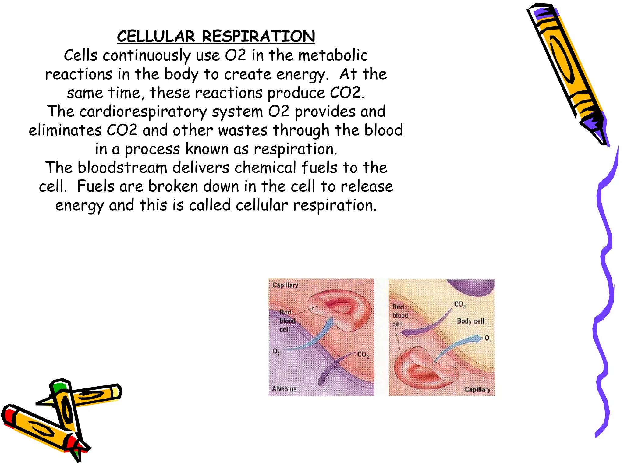
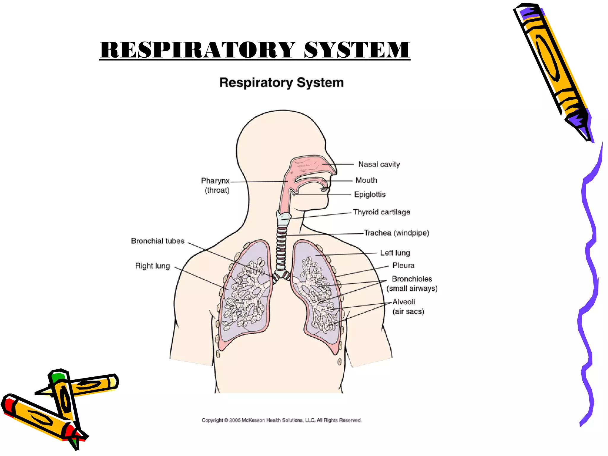
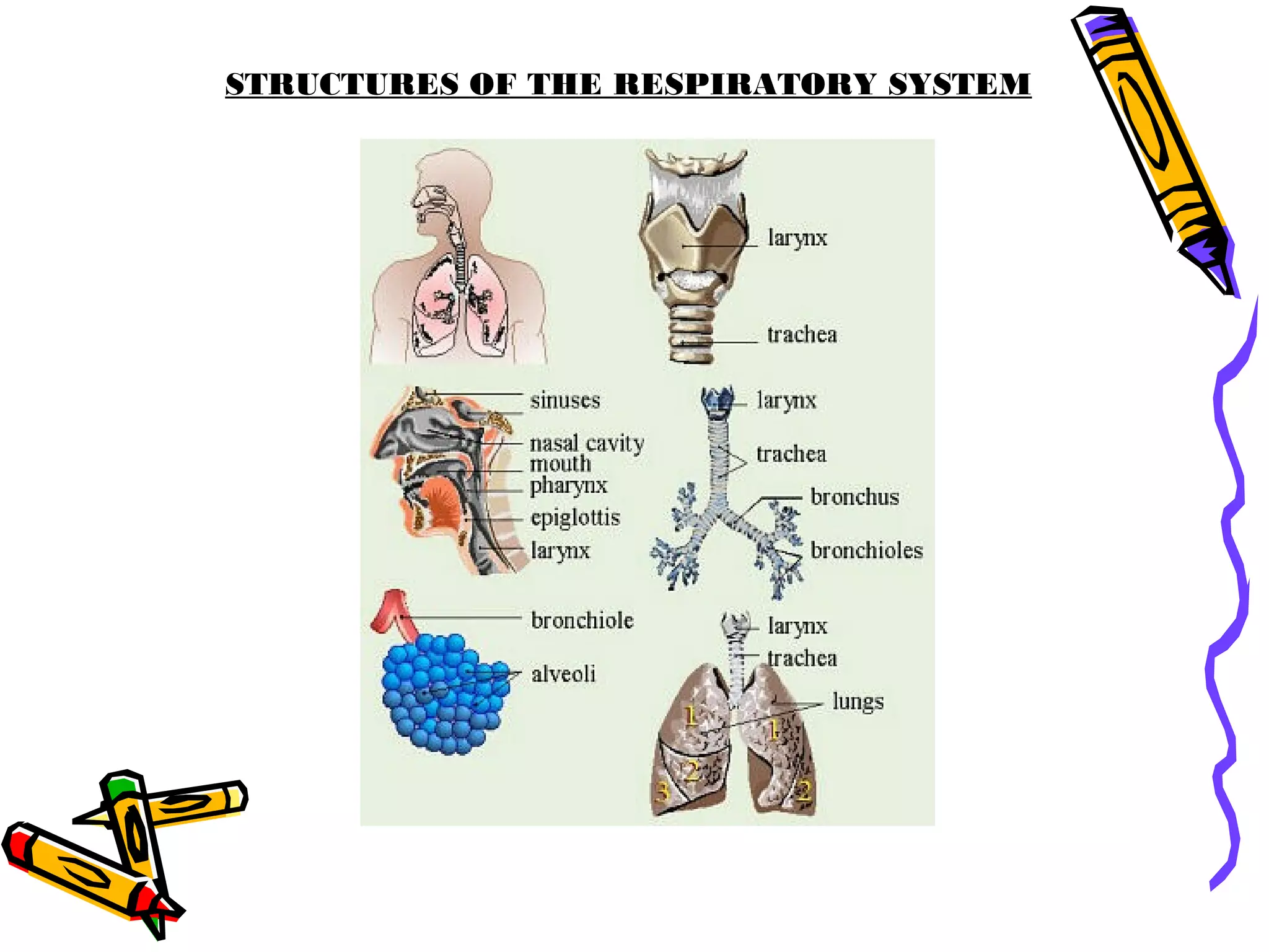
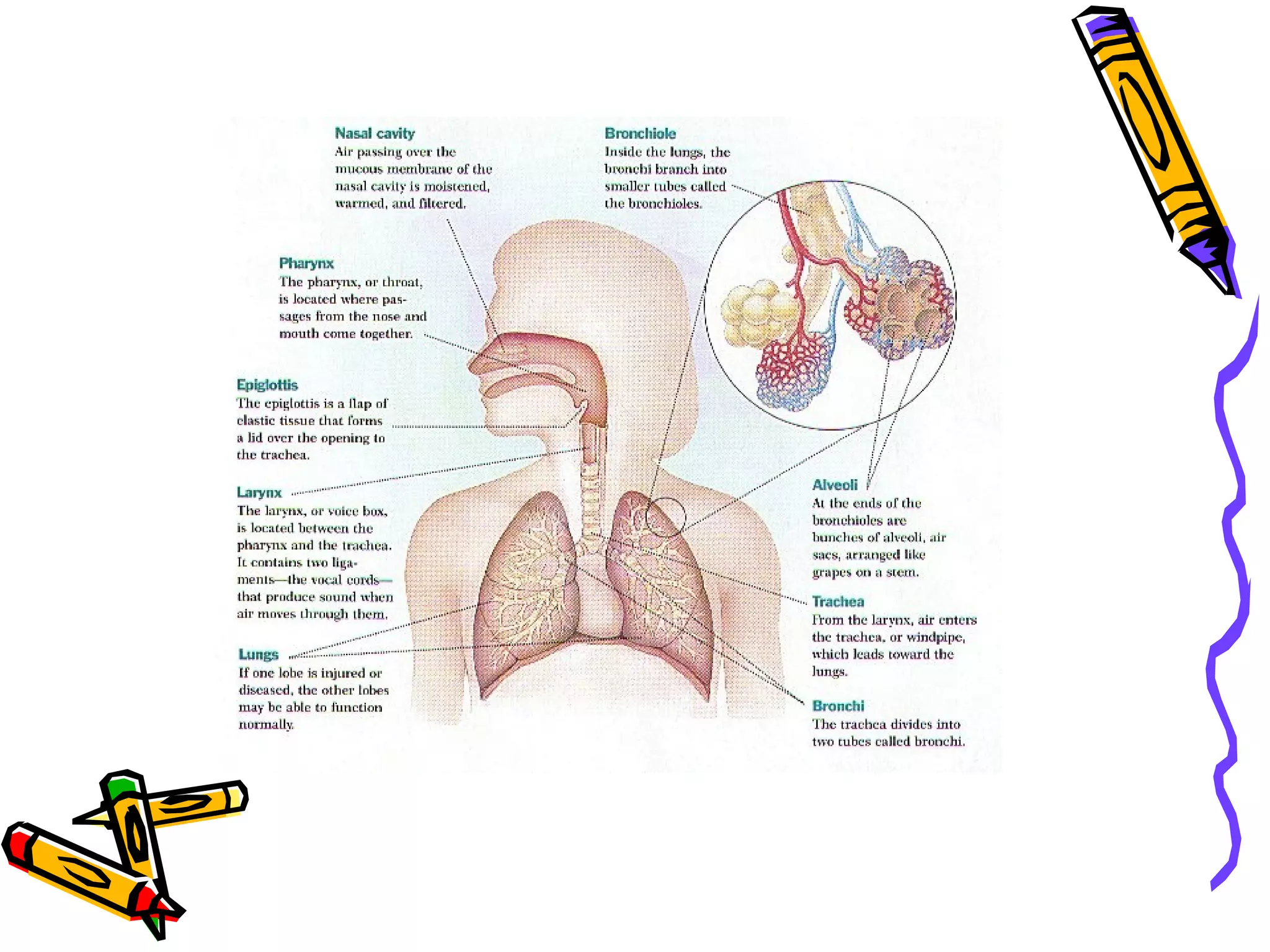
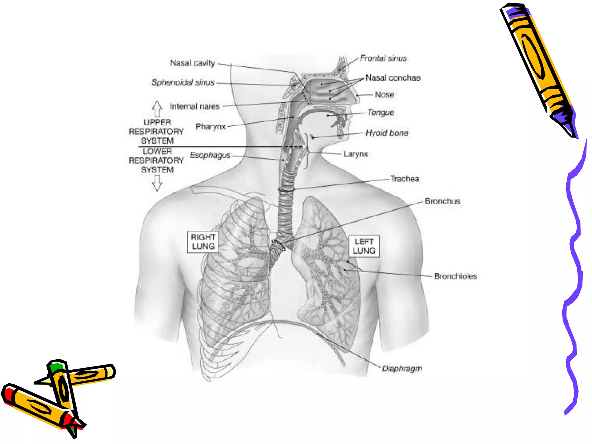
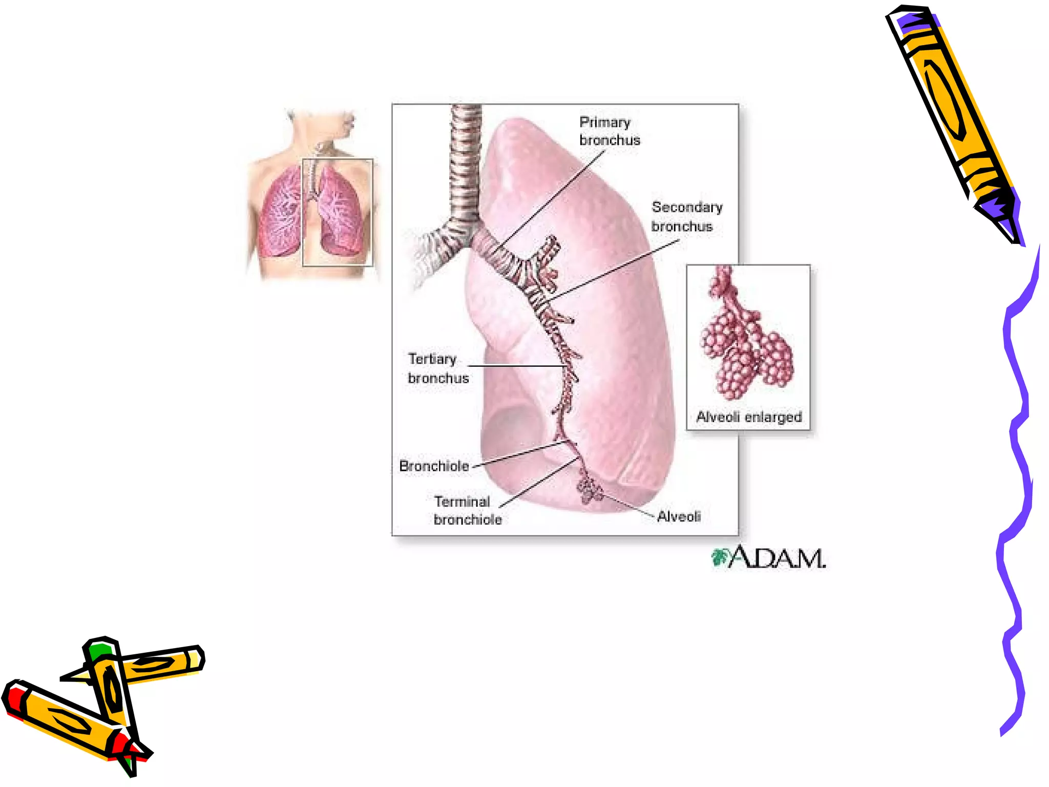
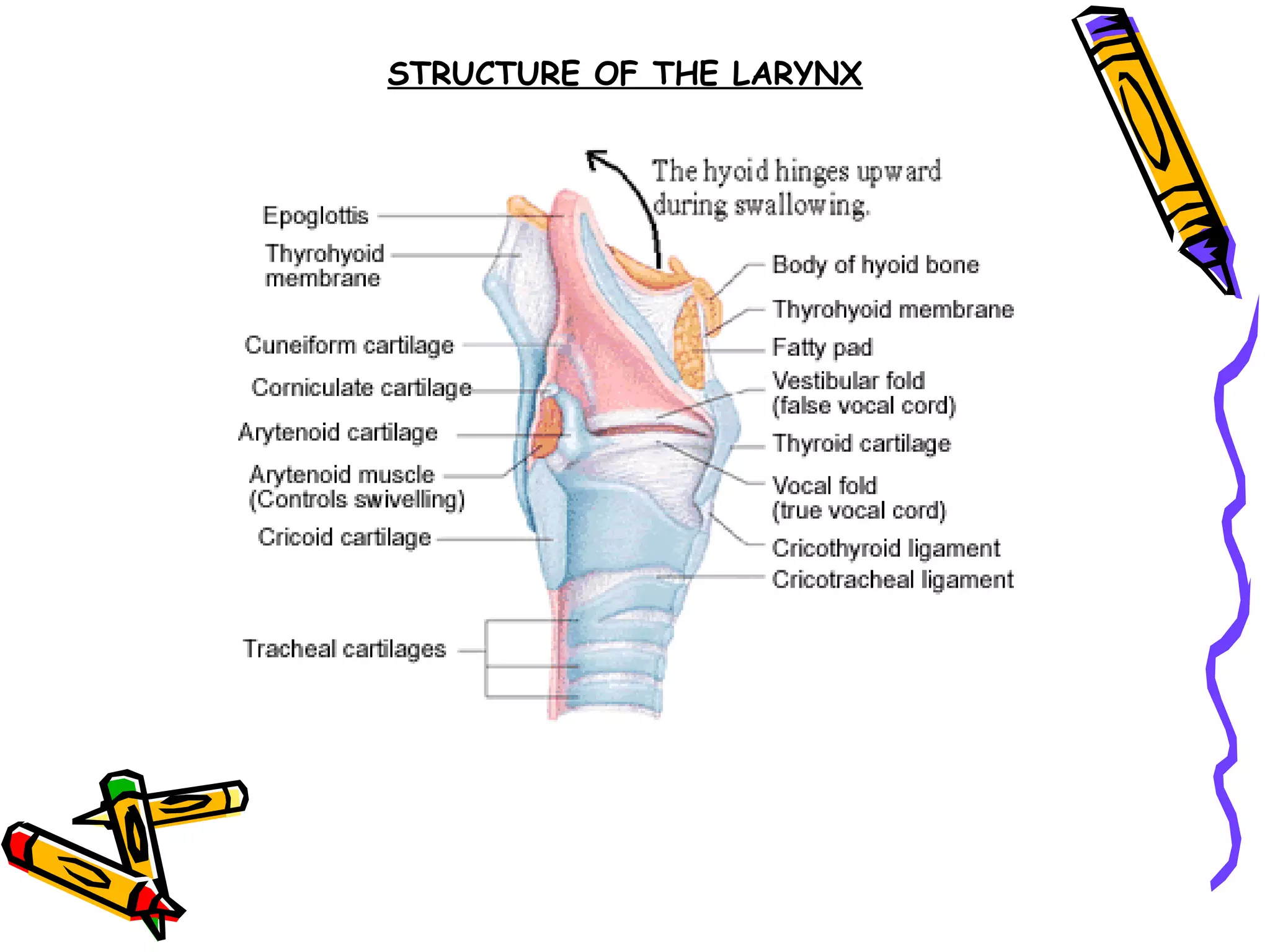
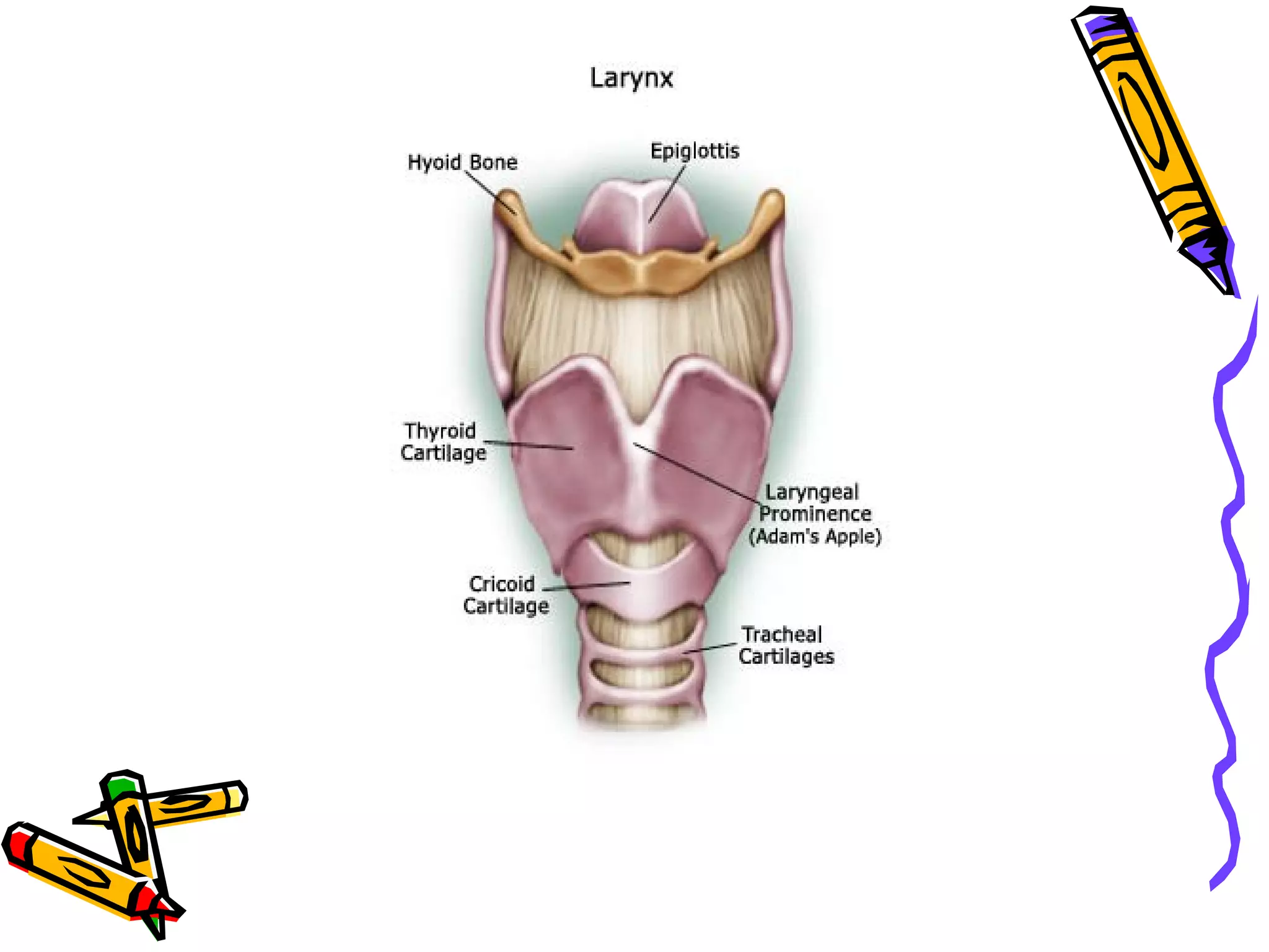
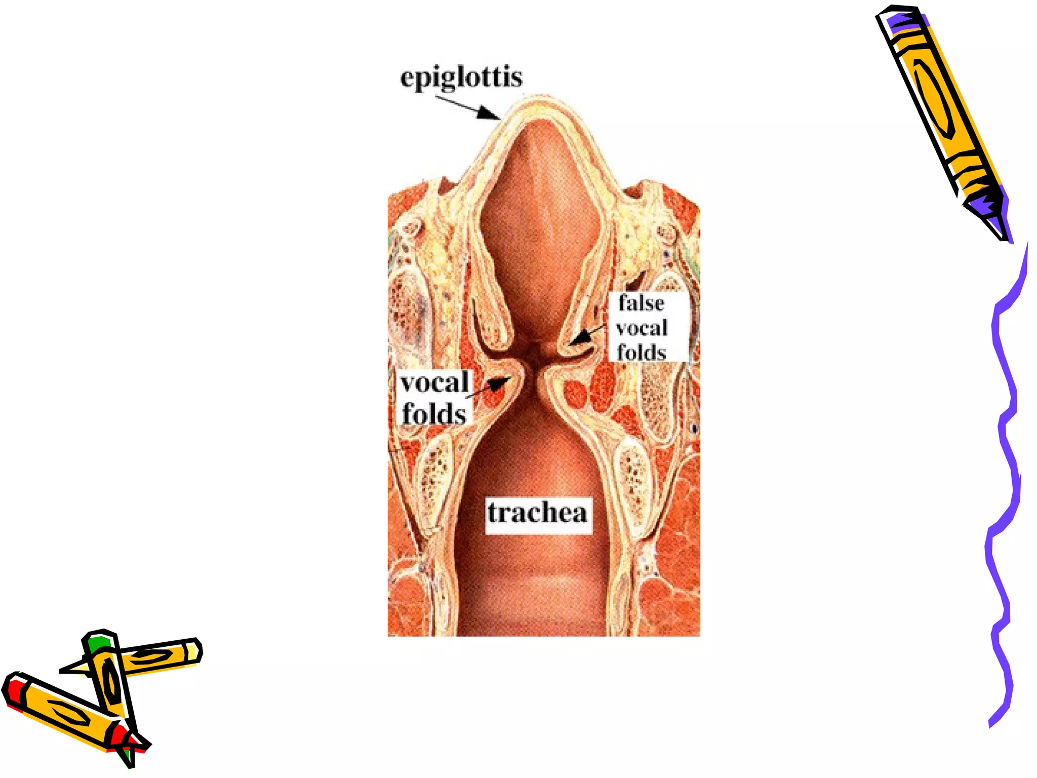
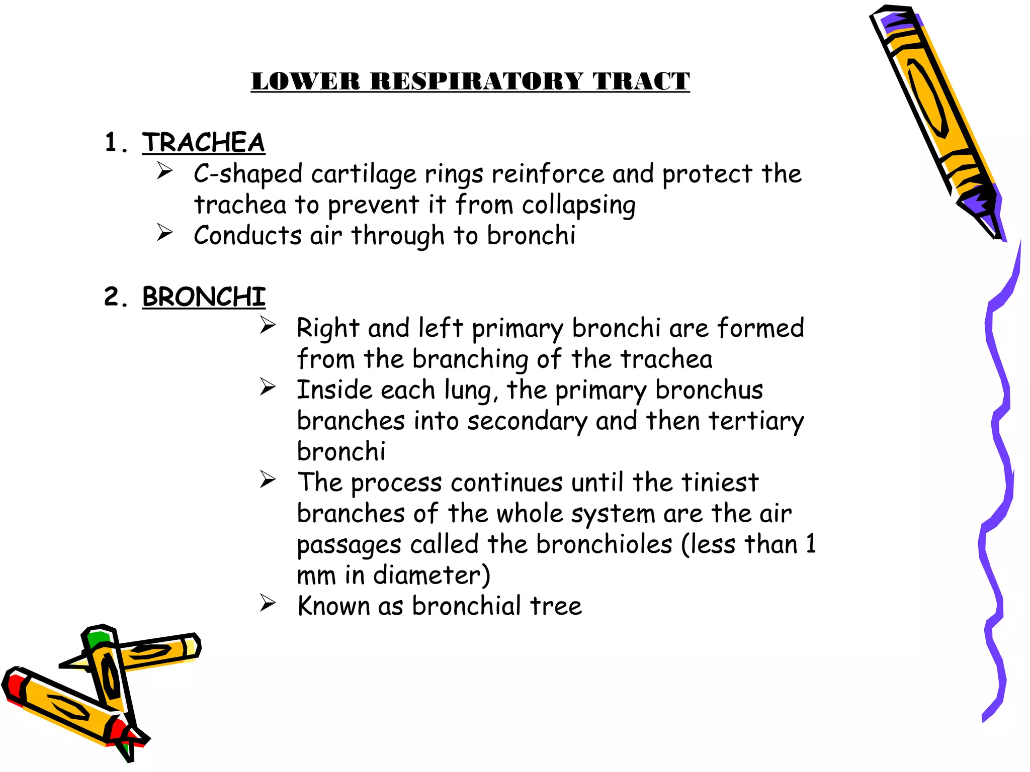
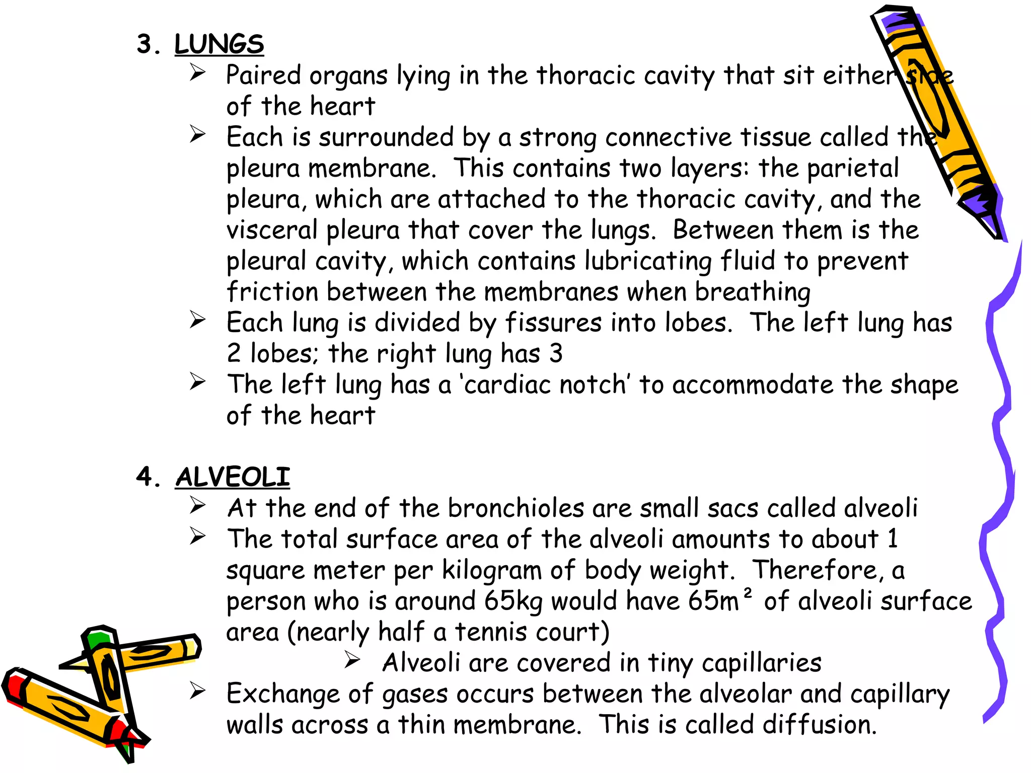
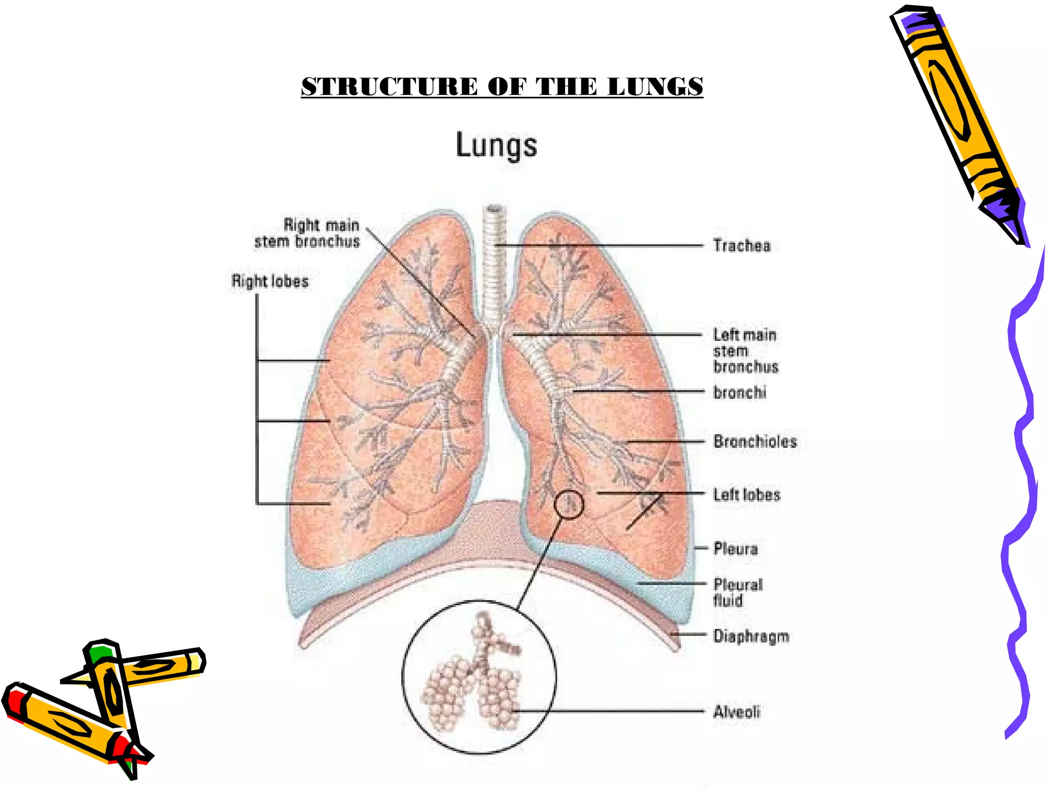
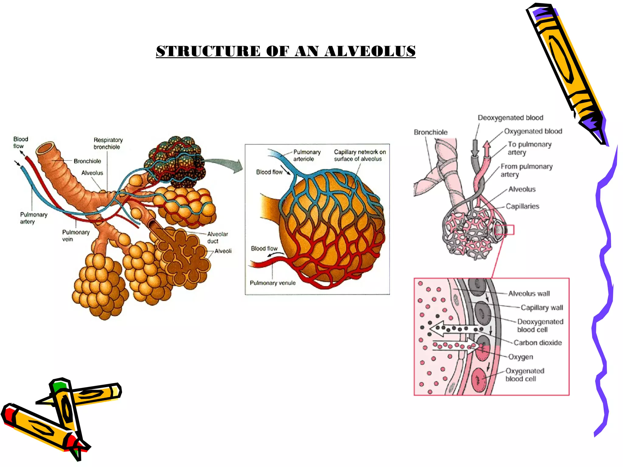
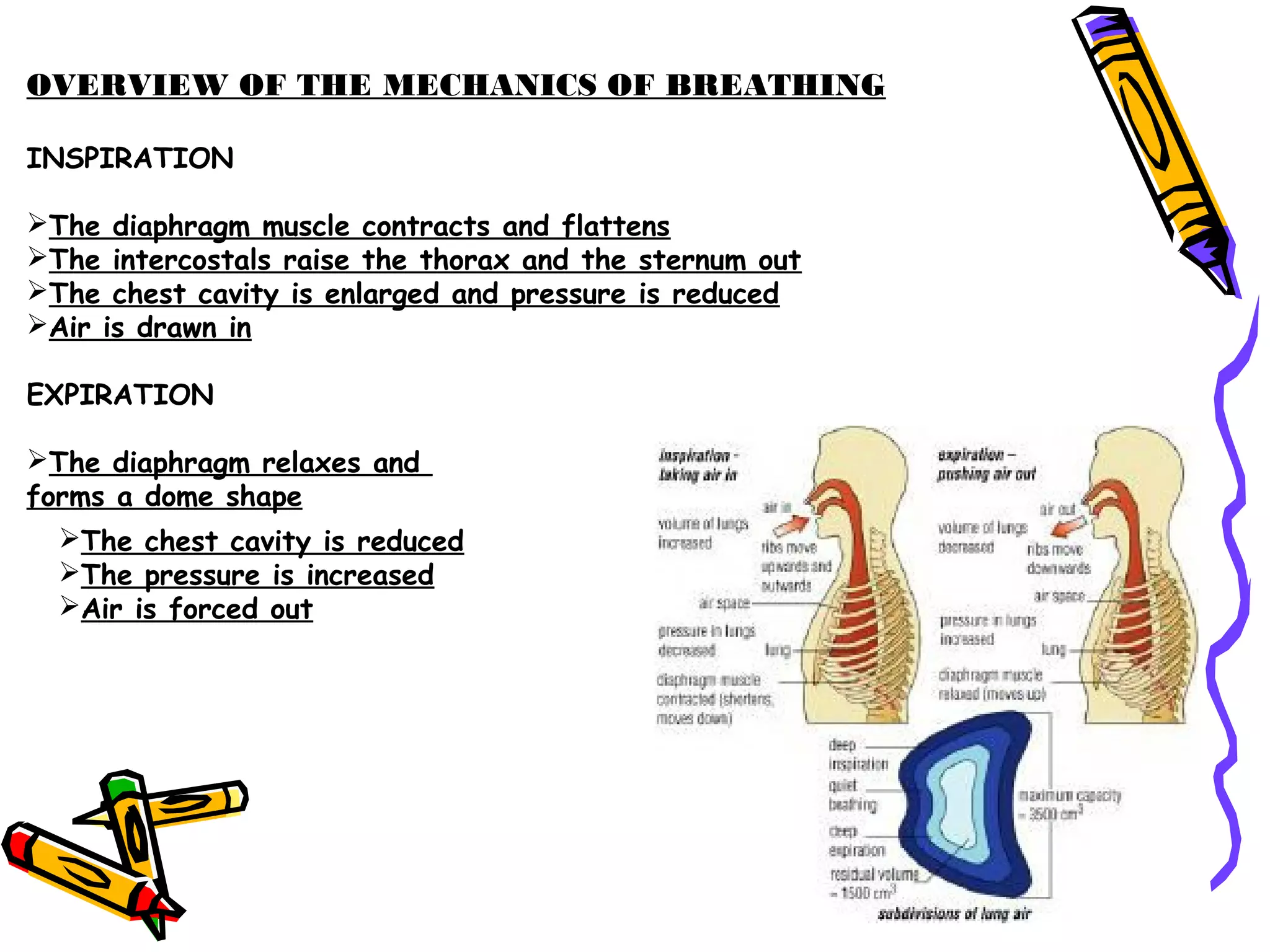
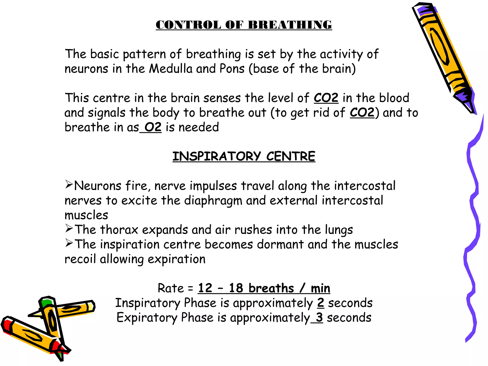
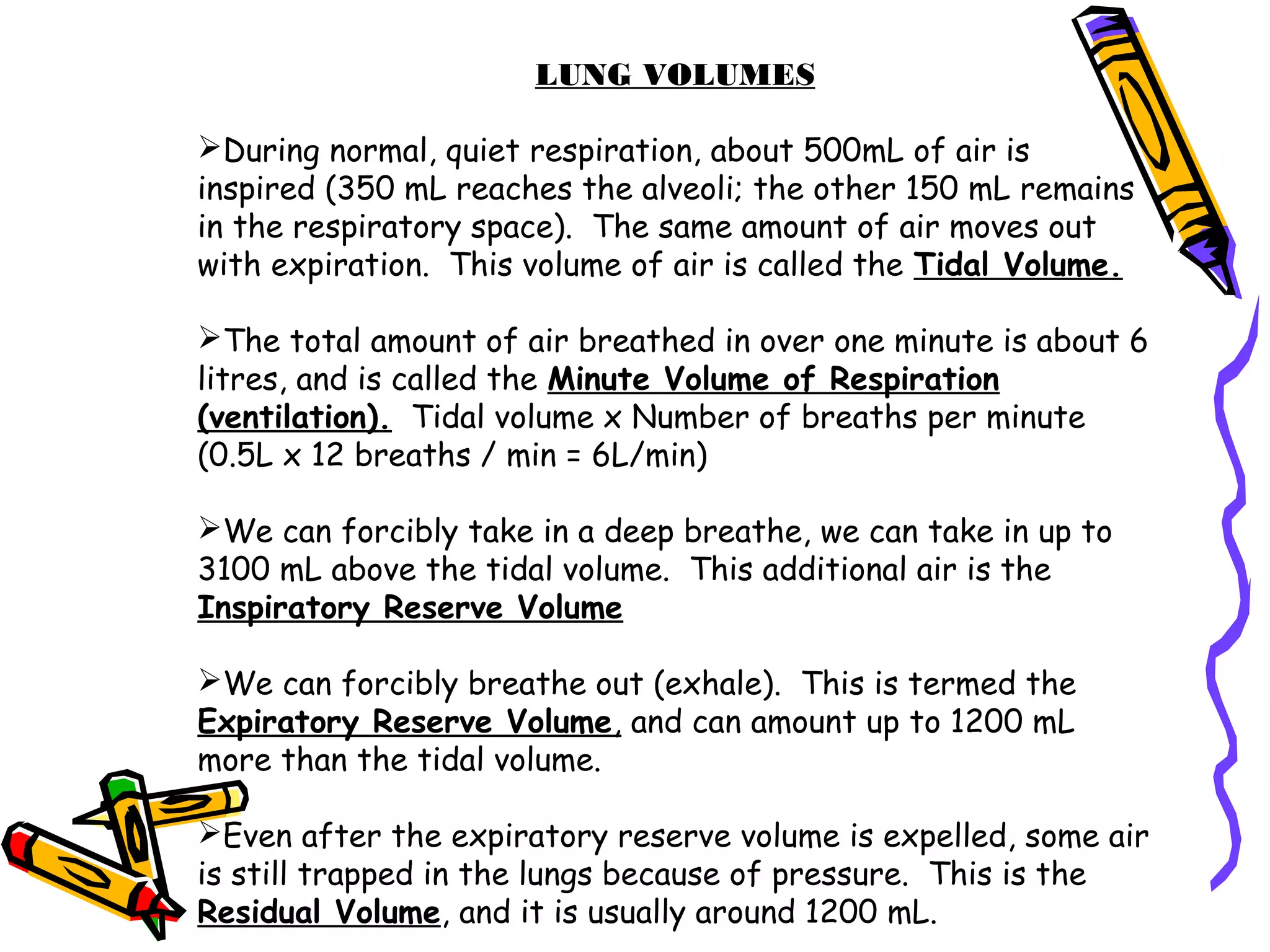
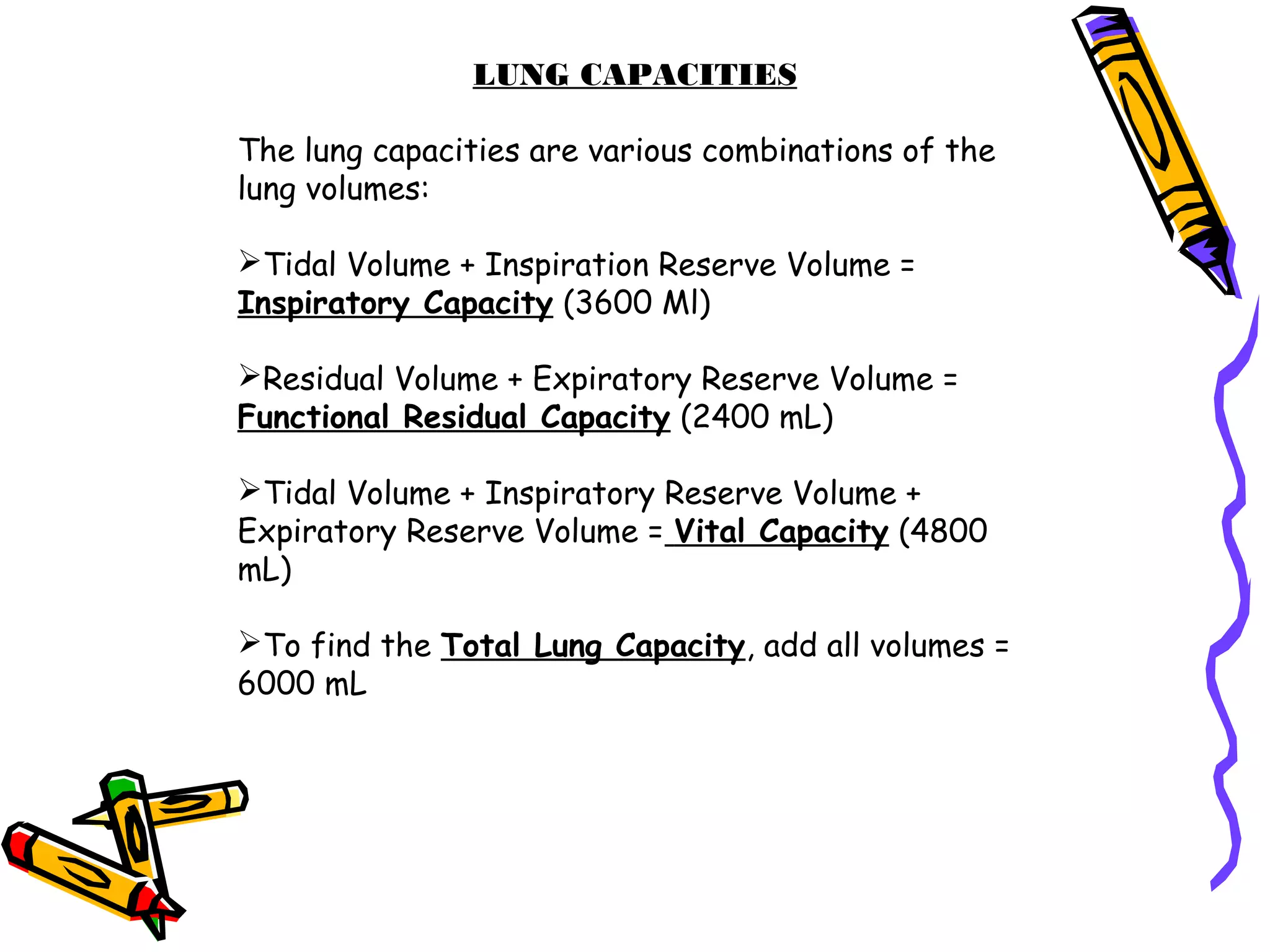
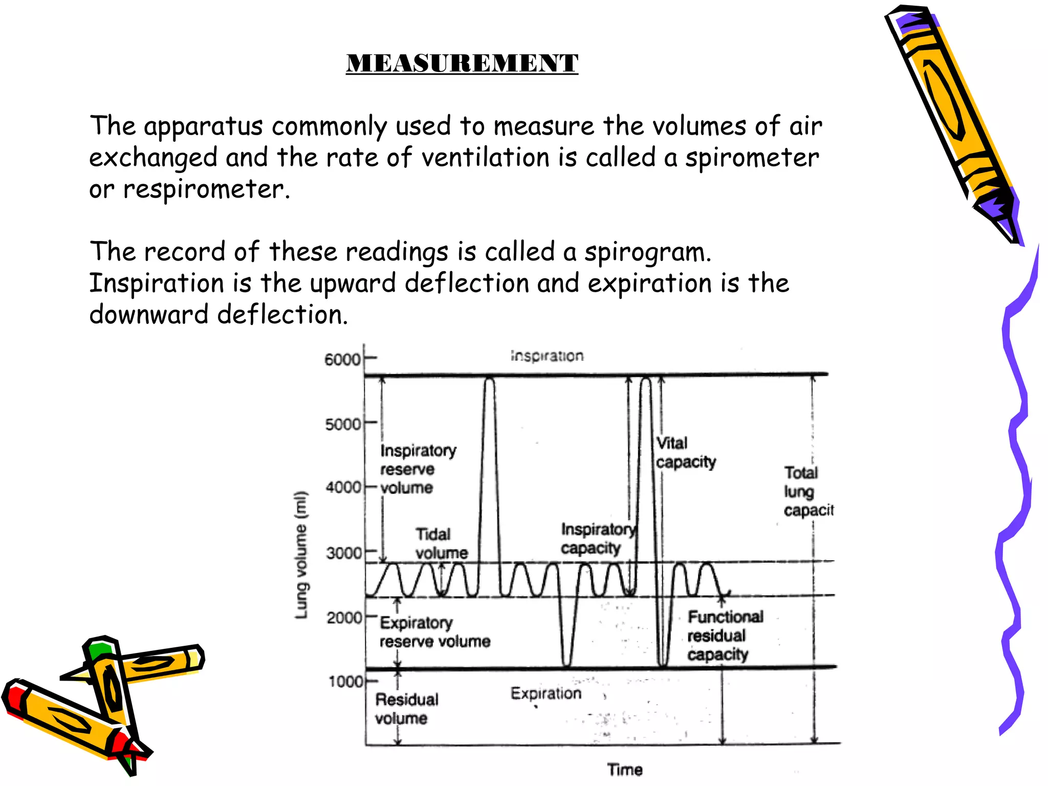
Basic Anatomy andBasic Anatomy and
PhysiologyPhysiology
Respiratory SystemRespiratory System
CELLULAR RESPIRATION
Cells continuously use O2 in the metabolic
reactions in the body to create energy. At the
same time, these reactions produce CO2.
The cardiorespiratory system O2 provides and
eliminates CO2 and other wastes through the blood
in a process known as respiration.
The bloodstream delivers chemical fuels to the
cell. Fuels are broken down in the cell to release
energy and this is called cellular respiration.
As CO2 in the cell accumulates, the concentration is
higher than that in the bloodstream. The
CO2 diffuses to the bloodstream.
As O2 concentration is higher in the bloodstream, O2
diffuses to the cells.
How does the circulatory system release CO2 from
the body into the atmosphere?
How does O2 get into the blood?
This occurs in the lungs.
Glucose + O2 → CO2 + H2O + Energy
RESPIRATORY SYSTEM
STRUCTURES OF THE RESPIRATORY SYSTEM
UPPER RESPIRATORY TRACT
1. NASAL PASSAGES
 Air enters through nostrils
 Small hairs filter out dust and large particles
 Air passes through to the nasal passageways
2. NASOPHARYNX
 Mucus layer traps finer particles, which are carried by cilia to the
pharynx (throat) to be swallowed
3. PHARYNX
 Passageway for air and food and to provide a resonating chamber
for speech sounds
4. OROPHARYNX and LARYNGOPHARYNX
 Oropharynx connects nasopharynx to laryngopharynx
 Laryngopharynx extends to the oesophagus and larynx
5. LARYNX
 Contains the vocal cords and connects the pharynx with the
trachea
 Thyroid cartilage (Adam’s Apple) gives its triangular shape
 Epiglottis is a large leaf shaped piece of cartilage lying on
the top of the larynx. It forms a lid over the trachea to
prevent foreign bodies and liquid going down.
STRUCTURE OF THE LARYNX
LOWER RESPIRATORY TRACT
1. TRACHEA
 C-shaped cartilage rings reinforce and protect the
trachea to prevent it from collapsing
 Conducts air through to bronchi
2. BRONCHI
 Right and left primary bronchi are formed
from the branching of the trachea
 Inside each lung, the primary bronchus
branches into secondary and then tertiary
bronchi
 The process continues until the tiniest
branches of the whole system are the air
passages called the bronchioles (less than 1
mm in diameter)
 Known as bronchial tree
3. LUNGS
 Paired organs lying in the thoracic cavity that sit either side
of the heart
 Each is surrounded by a strong connective tissue called the
pleura membrane. This contains two layers: the parietal
pleura, which are attached to the thoracic cavity, and the
visceral pleura that cover the lungs. Between them is the
pleural cavity, which contains lubricating fluid to prevent
friction between the membranes when breathing
 Each lung is divided by fissures into lobes. The left lung has
2 lobes; the right lung has 3
 The left lung has a ‘cardiac notch’ to accommodate the shape
of the heart
4. ALVEOLI
 At the end of the bronchioles are small sacs called alveoli
 The total surface area of the alveoli amounts to about 1
square meter per kilogram of body weight. Therefore, a
person who is around 65kg would have 65m² of alveoli surface
area (nearly half a tennis court)
 Alveoli are covered in tiny capillaries
 Exchange of gases occurs between the alveolar and capillary
walls across a thin membrane. This is called diffusion.
STRUCTURE OF THE LUNGS
STRUCTURE OF AN ALVEOLUS
LUNG FUNCTION
Respiration is the exchange of gases between the cells, blood
and atmosphere
It involves four processes
1. Pulmonary Ventilation (breathing) – the movement of air
from the atmosphere into the alveoli
2. Pulmonary Diffusion – exchange of O2 and CO2 between the
lungs and the blood
3. Transport of Respiration Gases – transportation of O2 and
CO2 between the lungs and the tissue cells of the body via
the blood
4. Internal Respiration – gas exchange between the blood
capillaries and the tissue cells.
INSPIRATION AND EXPIRATION
Pulmonary ventilation (breathing) allows a continuous flow
of air from the outside into and out of the lung alveoli
Air flows in to the body and out of it for the same reason
that blood flows through the body – a pressure gradient
exists.
Gases will generally move from areas of high pressure
into areas of low pressure
Breathing in (inspiration) occurs because the air outside
has a higher pressure than the air in the lungs as your
muscles have increased the size and the volume inside the
lungs
When the diaphragm and external intercostals relax, the
pressure inside the lungs is greater than that outside as
the lung size and volume has decreased. Hence we breathe
out in the process of expiration.
OVERVIEW OF THE MECHANICS OF BREATHING
INSPIRATION
The diaphragm muscle contracts and flattens
The intercostals raise the thorax and the sternum out
The chest cavity is enlarged and pressure is reduced
Air is drawn in
EXPIRATION
The diaphragm relaxes and
forms a dome shape
The chest cavity is reduced
The pressure is increased
Air is forced out
CONTROL OF BREATHING
The basic pattern of breathing is set by the activity of
neurons in the Medulla and Pons (base of the brain)
This centre in the brain senses the level of CO2 in the blood
and signals the body to breathe out (to get rid of CO2) and to
breathe in as O2 is needed
INSPIRATORY CENTRE
Neurons fire, nerve impulses travel along the intercostal
nerves to excite the diaphragm and external intercostal
muscles
The thorax expands and air rushes into the lungs
The inspiration centre becomes dormant and the muscles
recoil allowing expiration
Rate = 12 – 18 breaths / min
Inspiratory Phase is approximately 2 seconds
Expiratory Phase is approximately 3 seconds
LUNG VOLUMES
During normal, quiet respiration, about 500mL of air is
inspired (350 mL reaches the alveoli; the other 150 mL remains
in the respiratory space). The same amount of air moves out
with expiration. This volume of air is called the Tidal Volume.
The total amount of air breathed in over one minute is about 6
litres, and is called the Minute Volume of Respiration
(ventilation). Tidal volume x Number of breaths per minute
(0.5L x 12 breaths / min = 6L/min)
We can forcibly take in a deep breathe, we can take in up to
3100 mL above the tidal volume. This additional air is the
Inspiratory Reserve Volume
We can forcibly breathe out (exhale). This is termed the
Expiratory Reserve Volume, and can amount up to 1200 mL
more than the tidal volume.
Even after the expiratory reserve volume is expelled, some air
is still trapped in the lungs because of pressure. This is the
Residual Volume, and it is usually around 1200 mL.
LUNG CAPACITIES
The lung capacities are various combinations of the
lung volumes:
Tidal Volume + Inspiration Reserve Volume =
Inspiratory Capacity (3600 Ml)
Residual Volume + Expiratory Reserve Volume =
Functional Residual Capacity (2400 mL)
Tidal Volume + Inspiratory Reserve Volume +
Expiratory Reserve Volume = Vital Capacity (4800
mL)
To find the Total Lung Capacity, add all volumes =
6000 mL
MEASUREMENT
The apparatus commonly used to measure the volumes of air
exchanged and the rate of ventilation is called a spirometer
or respirometer.
The record of these readings is called a spirogram.
Inspiration is the upward deflection and expiration is the
downward deflection.
During exercise:
Tidal volume (TV) increases
Inspiratory Reserve volume (IRV) decrease
Expiratory Reserve volume (ERV) slightly decreases
Residual volume (RV) slightly increases
Total lung capacity (TLC) slightly decreases
Vital capacity (VC) slightly decrease
When at rest, a total of 6 litres of blood passes through your lungs
every minute. This figure increases enormously during exercise.
(Davis et al, p.67, 1986)
IMMEDIATE RESPONSES TO EXERCISE
Before exercise, rate and depth of breathing increases as
nervous activity is increased.
Once exercising, rate and depth increase greatly because
increased amounts of CO2 in the blood trigger greater
respiratory activity.
The increase in frequency (rate) and depth (TV) provides
greater ventilation and occurs in proportion to the exercise
effort
Lung volumes also change:
Increased TV – up to 5 or 6 times
EVR and IRV utilised more, thus decrease in volume
Blood flow is greater, therefore there are more open
capillaries at the alveolar-capillary membrane, increasing the
surface area in the lungs for gaseous exchange
RECOVERY PERIOD
When exercise ceases, the body returns to pre-exercise
condition or recovery period.
Here:
Ventilation decreases
CO2 is reduced
Stimulation from muscles and joints is reduced
RESPIRATORY ADAPTATIONS TO EXERCISE
1. EFFECTS ON VENTILATION
 Ventilation increases from 6L/min at rest to more than 100L/min
during exercise. This is achieved by increases in:
Respiration rate – from 15 to 40 or more breaths/min
Tidal Volume – from 10% of vital capacity to more than 50%
2. EFFECTS ON LUNG DIFFUSION
 During strenuous exercise there is a threefold increase in oxygen
diffusion from the alveoli to the blood
3. EFFECTS ON OXYGEN UPTAKE (VO2)
 Oxygen uptake is the amount of oxygen taken up and used by the
body. It reflects the total amount of work being done by the
body.
 During strenuous exercise there can be a 20-fold increase
in VO2, which increases linearly with the increase of
exercise intensities. As a person approaches exhaustion,
their VO2 will reach a maximum that will not go any
higher. This is a person’s VO2 max, the largest amount of
oxygen that a person can utilise within a given time.
LONG TERM RESPONSES TO EXERCISE
Regular exercise causes adjustments to the respiratory system
ie adaptation and benefit = improved lung function
The muscles involved in breathing are conditioned and
strengthened and the chest therefore has a greater ability to
expand
Trained athletes have improved lung volume and lung function
and can therefore take in more air per breath, require less
breathing work to maintain same levels of O2 in the blood and can
utilise air more efficiently.
Increased lung volume results in improved diffusion of O2 from
lungs to the blood.
Lung volume at rest also becomes greater
These adjustments increase efficiency of gaseous
exchange and thus improve the amount of O2 to be
transported to body cells.
OXYGEN DEBT AND DEFICIT
These terms refer to a lack of oxygen while training/racing and after such
activity is over. To go into these areas of exercise are normal, but the
goal is to not go too far into either category. Below is a brief description
of each and a chart which will detail the process more clearly than we can
explain with words alone.
Oxygen Deficit – while exercising intensely the body is sometimes unable
to fulfil all of its energy needs. Specifically, it is unable to intake and
absorbs enough oxygen to adequately ‘fee’ the muscles and amounts of
energy needed to perform the tasks the athlete is requesting from the
body. In order to make up the difference without sacrificing the output,
the body must tap into its anaerobic metabolism. This is where the body
goes into a mix of aerobic and anaerobic energy production. While not
hugely detrimental, oxygen deficit can grow to a level that the anaerobic
energy system cannot cover. This can cause performance to deteriorate.
Oxygen Debt – this term describes how the body pays back its debt
incurred above after the exercise is over. You will notice that even after
you have finished racing, you will continue to breathe hard. At this point
your body is still trying to repay the oxygen debt that was created when
you were working hard. Technically, it is excessive
post-exercise oxygen consumption.
Check out the illustration below for a graphical
description of these terms.
RESPIRATORY PROBLEMS
SMOKING
The vital capacity of the lungs can be reduced by 10 –
15% after once cigarette.
Narrowing of the airways increases the mucus
secretion.
Prolonged smoking results in tar and irritants coating
the lungs and reducing the elasticity of the alveoli. This
increases the resistance to airflow and decreases the
oxygen transporting capacity of the blood.
More oxygen is used for breathing when you are a
smoker. As a result there is less available to the working
muscles, which leads to fatigue at a faster rate.
ASTHMA
Asthma is the result of an allergic reaction to pollen,
dust mite, and animals and/or cold, exercise, fumes,
smog, viral infection and anxiety.
Small muscles surrounding the bronchi
constrict and there is an over secretion of
mucus. The walls of the bronchi can swell
and narrow the airways causing coughing,
wheezing and shortness of breath.
BRONCHITIS
Bronchitis is caused by bacterial infections, pollution and smoking.
The symptoms include increased mucus, coughing, wheezing and
expiration difficulties.
Treatment is by antibiotics (usually penicillin)
EMPHYSEMA
Emphysema is caused by smoking and prolonged exposure to pollution
(eg coal miners)
The lining of the airways is damaged (cilia) and cannot move dirt and
mucus. This then accumulates and the alveoli become less elastic.
Oxygen uptake is then decreased and breathing (expiration) becomes
difficult.
PULMONARY FIBROSIS
Pulmonary fibrosis is an abnormal formation of fibre-like scar
tissue in the alveoli (air sacs which take oxygen to the lungs
and expel carbon dioxide) and interstitial tissue (the tissues
between and surrounding the alveoli) of the lungs. It is a
chronic lung disease associated with inflammation.
Pulmonary Fibrosis Cont.
Symptoms
• Breathlessness, especially during exercise
• Chronic dry cough
• Shortness of breath
• Chest discomfort
• Reduced VC and IRV
• Causes
• Exposure to cigarette smoke
• Inhaled environmental pollutants
• Occupational pollutants, including asbestos, ground stone, metal
dust or mouldy hay
Exercise at Altitude
Exercise at altitude can have a major effect on performance
Endurance performance is usually diminished and anaerobic
performance unaffected. This is because ascent to altitude results in
lower oxygen in inspired air, and therefore less oxygen delivery to
active muscles.
The percentage of oxygen in the air is at the same at sea level, but
the total amount of air is less.
A moderate altitude around 15oom can start to affect the athlete,
and over 5000m can be extreme.
An increase of altitude results in a decrease of 2°C per 300m, so
the cold also has to be dealt with.
Physiological responses to exercise:
Decreased arterial oxygen content
Increased ventilation
Increased CO2 output
Increased stroke volume
Decreased sub-max HR
Decreased VO2 max
Progressive dehydration – increased breathing of colder / dryer air
and increased urine.
Overcoming the Effects of Altitude
Live high / train low – maximise the resting adaptations to altitude
and minimise the disruption to training caused by altitude
Daily transit – between high and low altitude
Nitrogen houses – decreases O2 in the air by increasing nitrogen.
Mimic high altitude conditions and you can set the height you require
Altitude tents – portable and have a decreased O2.
Acclimatisation at Altitude
Adaptations occur over 2 – 3 weeks:
Increased red blood cell concentration – over days due to dehydration
and weeks / months due to increased cell production
Partial restoration of plasma volume
Increased muscle capillarisation
Increased muscle enzyme activity
Altitude Sickness
This generally occurs above 3000m and is most likely to happen in:
Unacclimatised individuals
After rapid ascent
In combination with exercise
Symptoms:
Headache
Dizziness
Nausea
Sleep disturbances due to cardiovascular responses to
hyperventilation
* Pulmonary / cerebral edema may occur in extreme
circumstances and requires immediate return to lower
altitudes.
BA&P Respiratory

BA&P Respiratory

  • 1.
    Basic Anatomy andBasicAnatomy and PhysiologyPhysiology Respiratory SystemRespiratory System
  • 2.
    CELLULAR RESPIRATION Cells continuouslyuse O2 in the metabolic reactions in the body to create energy. At the same time, these reactions produce CO2. The cardiorespiratory system O2 provides and eliminates CO2 and other wastes through the blood in a process known as respiration. The bloodstream delivers chemical fuels to the cell. Fuels are broken down in the cell to release energy and this is called cellular respiration.
  • 3.
    As CO2 inthe cell accumulates, the concentration is higher than that in the bloodstream. The CO2 diffuses to the bloodstream. As O2 concentration is higher in the bloodstream, O2 diffuses to the cells. How does the circulatory system release CO2 from the body into the atmosphere? How does O2 get into the blood? This occurs in the lungs. Glucose + O2 → CO2 + H2O + Energy
  • 4.
  • 5.
    STRUCTURES OF THERESPIRATORY SYSTEM
  • 9.
    STRUCTURES OF THERESPIRATORY SYSTEM UPPER RESPIRATORY TRACT 1. NASAL PASSAGES  Air enters through nostrils  Small hairs filter out dust and large particles  Air passes through to the nasal passageways 2. NASOPHARYNX  Mucus layer traps finer particles, which are carried by cilia to the pharynx (throat) to be swallowed 3. PHARYNX  Passageway for air and food and to provide a resonating chamber for speech sounds 4. OROPHARYNX and LARYNGOPHARYNX  Oropharynx connects nasopharynx to laryngopharynx  Laryngopharynx extends to the oesophagus and larynx 5. LARYNX  Contains the vocal cords and connects the pharynx with the trachea  Thyroid cartilage (Adam’s Apple) gives its triangular shape  Epiglottis is a large leaf shaped piece of cartilage lying on the top of the larynx. It forms a lid over the trachea to prevent foreign bodies and liquid going down.
  • 10.
  • 13.
    LOWER RESPIRATORY TRACT 1.TRACHEA  C-shaped cartilage rings reinforce and protect the trachea to prevent it from collapsing  Conducts air through to bronchi 2. BRONCHI  Right and left primary bronchi are formed from the branching of the trachea  Inside each lung, the primary bronchus branches into secondary and then tertiary bronchi  The process continues until the tiniest branches of the whole system are the air passages called the bronchioles (less than 1 mm in diameter)  Known as bronchial tree
  • 14.
    3. LUNGS  Pairedorgans lying in the thoracic cavity that sit either side of the heart  Each is surrounded by a strong connective tissue called the pleura membrane. This contains two layers: the parietal pleura, which are attached to the thoracic cavity, and the visceral pleura that cover the lungs. Between them is the pleural cavity, which contains lubricating fluid to prevent friction between the membranes when breathing  Each lung is divided by fissures into lobes. The left lung has 2 lobes; the right lung has 3  The left lung has a ‘cardiac notch’ to accommodate the shape of the heart 4. ALVEOLI  At the end of the bronchioles are small sacs called alveoli  The total surface area of the alveoli amounts to about 1 square meter per kilogram of body weight. Therefore, a person who is around 65kg would have 65m² of alveoli surface area (nearly half a tennis court)  Alveoli are covered in tiny capillaries  Exchange of gases occurs between the alveolar and capillary walls across a thin membrane. This is called diffusion.
  • 15.
  • 16.
  • 17.
    LUNG FUNCTION Respiration isthe exchange of gases between the cells, blood and atmosphere It involves four processes 1. Pulmonary Ventilation (breathing) – the movement of air from the atmosphere into the alveoli 2. Pulmonary Diffusion – exchange of O2 and CO2 between the lungs and the blood 3. Transport of Respiration Gases – transportation of O2 and CO2 between the lungs and the tissue cells of the body via the blood 4. Internal Respiration – gas exchange between the blood capillaries and the tissue cells.
  • 18.
    INSPIRATION AND EXPIRATION Pulmonaryventilation (breathing) allows a continuous flow of air from the outside into and out of the lung alveoli Air flows in to the body and out of it for the same reason that blood flows through the body – a pressure gradient exists. Gases will generally move from areas of high pressure into areas of low pressure Breathing in (inspiration) occurs because the air outside has a higher pressure than the air in the lungs as your muscles have increased the size and the volume inside the lungs When the diaphragm and external intercostals relax, the pressure inside the lungs is greater than that outside as the lung size and volume has decreased. Hence we breathe out in the process of expiration.
  • 19.
    OVERVIEW OF THEMECHANICS OF BREATHING INSPIRATION The diaphragm muscle contracts and flattens The intercostals raise the thorax and the sternum out The chest cavity is enlarged and pressure is reduced Air is drawn in EXPIRATION The diaphragm relaxes and forms a dome shape The chest cavity is reduced The pressure is increased Air is forced out
  • 20.
    CONTROL OF BREATHING Thebasic pattern of breathing is set by the activity of neurons in the Medulla and Pons (base of the brain) This centre in the brain senses the level of CO2 in the blood and signals the body to breathe out (to get rid of CO2) and to breathe in as O2 is needed INSPIRATORY CENTRE Neurons fire, nerve impulses travel along the intercostal nerves to excite the diaphragm and external intercostal muscles The thorax expands and air rushes into the lungs The inspiration centre becomes dormant and the muscles recoil allowing expiration Rate = 12 – 18 breaths / min Inspiratory Phase is approximately 2 seconds Expiratory Phase is approximately 3 seconds
  • 21.
    LUNG VOLUMES During normal,quiet respiration, about 500mL of air is inspired (350 mL reaches the alveoli; the other 150 mL remains in the respiratory space). The same amount of air moves out with expiration. This volume of air is called the Tidal Volume. The total amount of air breathed in over one minute is about 6 litres, and is called the Minute Volume of Respiration (ventilation). Tidal volume x Number of breaths per minute (0.5L x 12 breaths / min = 6L/min) We can forcibly take in a deep breathe, we can take in up to 3100 mL above the tidal volume. This additional air is the Inspiratory Reserve Volume We can forcibly breathe out (exhale). This is termed the Expiratory Reserve Volume, and can amount up to 1200 mL more than the tidal volume. Even after the expiratory reserve volume is expelled, some air is still trapped in the lungs because of pressure. This is the Residual Volume, and it is usually around 1200 mL.
  • 22.
    LUNG CAPACITIES The lungcapacities are various combinations of the lung volumes: Tidal Volume + Inspiration Reserve Volume = Inspiratory Capacity (3600 Ml) Residual Volume + Expiratory Reserve Volume = Functional Residual Capacity (2400 mL) Tidal Volume + Inspiratory Reserve Volume + Expiratory Reserve Volume = Vital Capacity (4800 mL) To find the Total Lung Capacity, add all volumes = 6000 mL
  • 23.
    MEASUREMENT The apparatus commonlyused to measure the volumes of air exchanged and the rate of ventilation is called a spirometer or respirometer. The record of these readings is called a spirogram. Inspiration is the upward deflection and expiration is the downward deflection.
  • 24.
    During exercise: Tidal volume(TV) increases Inspiratory Reserve volume (IRV) decrease Expiratory Reserve volume (ERV) slightly decreases Residual volume (RV) slightly increases Total lung capacity (TLC) slightly decreases Vital capacity (VC) slightly decrease When at rest, a total of 6 litres of blood passes through your lungs every minute. This figure increases enormously during exercise. (Davis et al, p.67, 1986)
  • 25.
    IMMEDIATE RESPONSES TOEXERCISE Before exercise, rate and depth of breathing increases as nervous activity is increased. Once exercising, rate and depth increase greatly because increased amounts of CO2 in the blood trigger greater respiratory activity. The increase in frequency (rate) and depth (TV) provides greater ventilation and occurs in proportion to the exercise effort Lung volumes also change: Increased TV – up to 5 or 6 times EVR and IRV utilised more, thus decrease in volume Blood flow is greater, therefore there are more open capillaries at the alveolar-capillary membrane, increasing the surface area in the lungs for gaseous exchange RECOVERY PERIOD When exercise ceases, the body returns to pre-exercise condition or recovery period. Here: Ventilation decreases CO2 is reduced Stimulation from muscles and joints is reduced
  • 26.
    RESPIRATORY ADAPTATIONS TOEXERCISE 1. EFFECTS ON VENTILATION  Ventilation increases from 6L/min at rest to more than 100L/min during exercise. This is achieved by increases in: Respiration rate – from 15 to 40 or more breaths/min Tidal Volume – from 10% of vital capacity to more than 50% 2. EFFECTS ON LUNG DIFFUSION  During strenuous exercise there is a threefold increase in oxygen diffusion from the alveoli to the blood 3. EFFECTS ON OXYGEN UPTAKE (VO2)  Oxygen uptake is the amount of oxygen taken up and used by the body. It reflects the total amount of work being done by the body.  During strenuous exercise there can be a 20-fold increase in VO2, which increases linearly with the increase of exercise intensities. As a person approaches exhaustion, their VO2 will reach a maximum that will not go any higher. This is a person’s VO2 max, the largest amount of oxygen that a person can utilise within a given time.
  • 27.
    LONG TERM RESPONSESTO EXERCISE Regular exercise causes adjustments to the respiratory system ie adaptation and benefit = improved lung function The muscles involved in breathing are conditioned and strengthened and the chest therefore has a greater ability to expand Trained athletes have improved lung volume and lung function and can therefore take in more air per breath, require less breathing work to maintain same levels of O2 in the blood and can utilise air more efficiently. Increased lung volume results in improved diffusion of O2 from lungs to the blood. Lung volume at rest also becomes greater These adjustments increase efficiency of gaseous exchange and thus improve the amount of O2 to be transported to body cells.
  • 28.
    OXYGEN DEBT ANDDEFICIT These terms refer to a lack of oxygen while training/racing and after such activity is over. To go into these areas of exercise are normal, but the goal is to not go too far into either category. Below is a brief description of each and a chart which will detail the process more clearly than we can explain with words alone. Oxygen Deficit – while exercising intensely the body is sometimes unable to fulfil all of its energy needs. Specifically, it is unable to intake and absorbs enough oxygen to adequately ‘fee’ the muscles and amounts of energy needed to perform the tasks the athlete is requesting from the body. In order to make up the difference without sacrificing the output, the body must tap into its anaerobic metabolism. This is where the body goes into a mix of aerobic and anaerobic energy production. While not hugely detrimental, oxygen deficit can grow to a level that the anaerobic energy system cannot cover. This can cause performance to deteriorate. Oxygen Debt – this term describes how the body pays back its debt incurred above after the exercise is over. You will notice that even after you have finished racing, you will continue to breathe hard. At this point your body is still trying to repay the oxygen debt that was created when you were working hard. Technically, it is excessive post-exercise oxygen consumption. Check out the illustration below for a graphical description of these terms.
  • 30.
    RESPIRATORY PROBLEMS SMOKING The vitalcapacity of the lungs can be reduced by 10 – 15% after once cigarette. Narrowing of the airways increases the mucus secretion. Prolonged smoking results in tar and irritants coating the lungs and reducing the elasticity of the alveoli. This increases the resistance to airflow and decreases the oxygen transporting capacity of the blood. More oxygen is used for breathing when you are a smoker. As a result there is less available to the working muscles, which leads to fatigue at a faster rate. ASTHMA Asthma is the result of an allergic reaction to pollen, dust mite, and animals and/or cold, exercise, fumes, smog, viral infection and anxiety. Small muscles surrounding the bronchi constrict and there is an over secretion of mucus. The walls of the bronchi can swell and narrow the airways causing coughing, wheezing and shortness of breath.
  • 31.
    BRONCHITIS Bronchitis is causedby bacterial infections, pollution and smoking. The symptoms include increased mucus, coughing, wheezing and expiration difficulties. Treatment is by antibiotics (usually penicillin) EMPHYSEMA Emphysema is caused by smoking and prolonged exposure to pollution (eg coal miners) The lining of the airways is damaged (cilia) and cannot move dirt and mucus. This then accumulates and the alveoli become less elastic. Oxygen uptake is then decreased and breathing (expiration) becomes difficult. PULMONARY FIBROSIS Pulmonary fibrosis is an abnormal formation of fibre-like scar tissue in the alveoli (air sacs which take oxygen to the lungs and expel carbon dioxide) and interstitial tissue (the tissues between and surrounding the alveoli) of the lungs. It is a chronic lung disease associated with inflammation.
  • 32.
    Pulmonary Fibrosis Cont. Symptoms •Breathlessness, especially during exercise • Chronic dry cough • Shortness of breath • Chest discomfort • Reduced VC and IRV • Causes • Exposure to cigarette smoke • Inhaled environmental pollutants • Occupational pollutants, including asbestos, ground stone, metal dust or mouldy hay
  • 33.
    Exercise at Altitude Exerciseat altitude can have a major effect on performance Endurance performance is usually diminished and anaerobic performance unaffected. This is because ascent to altitude results in lower oxygen in inspired air, and therefore less oxygen delivery to active muscles. The percentage of oxygen in the air is at the same at sea level, but the total amount of air is less. A moderate altitude around 15oom can start to affect the athlete, and over 5000m can be extreme. An increase of altitude results in a decrease of 2°C per 300m, so the cold also has to be dealt with.
  • 34.
    Physiological responses toexercise: Decreased arterial oxygen content Increased ventilation Increased CO2 output Increased stroke volume Decreased sub-max HR Decreased VO2 max Progressive dehydration – increased breathing of colder / dryer air and increased urine. Overcoming the Effects of Altitude Live high / train low – maximise the resting adaptations to altitude and minimise the disruption to training caused by altitude Daily transit – between high and low altitude Nitrogen houses – decreases O2 in the air by increasing nitrogen. Mimic high altitude conditions and you can set the height you require Altitude tents – portable and have a decreased O2.
  • 35.
    Acclimatisation at Altitude Adaptationsoccur over 2 – 3 weeks: Increased red blood cell concentration – over days due to dehydration and weeks / months due to increased cell production Partial restoration of plasma volume Increased muscle capillarisation Increased muscle enzyme activity Altitude Sickness This generally occurs above 3000m and is most likely to happen in: Unacclimatised individuals After rapid ascent In combination with exercise Symptoms: Headache Dizziness Nausea Sleep disturbances due to cardiovascular responses to hyperventilation * Pulmonary / cerebral edema may occur in extreme circumstances and requires immediate return to lower altitudes.