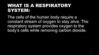
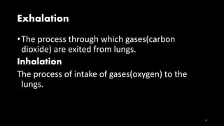
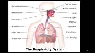
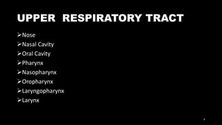
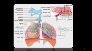
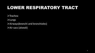
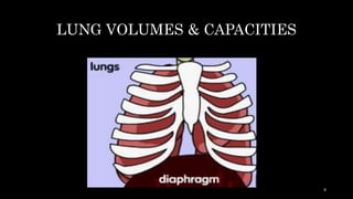
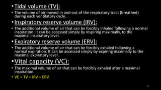
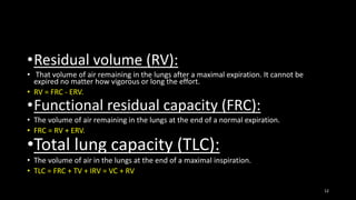
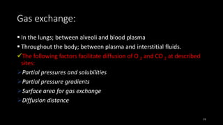
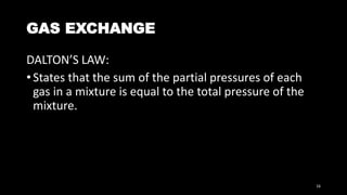
The respiratory system provides oxygen to the body's cells while removing carbon dioxide through the processes of inhalation and exhalation. It consists of an upper respiratory tract including the nose, mouth and larynx, and a lower respiratory tract including the trachea, bronchi, bronchioles and alveoli in the lungs. The lungs contain volumes of air that can be measured including tidal volume, inspiratory reserve volume, expiratory reserve volume and residual volume. Gas exchange occurs through diffusion between the alveoli and blood plasma, and between plasma and tissues throughout the body, facilitated by factors like partial pressures and surface area.
The respiratory system is the network of organs and tissues that help you breathe. It includes your airways, lungs, and blood vessels. The muscles that power your lungs are also part of the respiratory system. These parts work together to move oxygen throughout the body and clean out waste gases like carbon dioxide.
Anatomically, respiratory tract is divided into upper (organ outside thorax - nose, pharynx and larynx) and lower respiratory tract (organ within thorax - trachea, bronchi, bronchioles, alveolar duct and alveoli).
This PPT covers Anatomy and Physiology of respiratory system. Anatomy of respiratory organs, Mechanism of respiration, Internal Respiration, external respiration, Transport of oxygen in blood, Transport of carbon dioxide in blood, Regulation of respiration, lung volume and lung capacities are explained.
The respiratory system consists of all the organs involved in breathing. These include the nose, pharynx, larynx, trachea, bronchi and lungs. The respiratory system does two very important things: it brings oxygen into our bodies, which we need for our cells to live and function properly; and it helps us get rid of carbon dioxide, which is a waste product of cellular function. The nose, pharynx, larynx, trachea and bronchi all work like a system of pipes through which the air is funnelled down into our lungs. There, in very small air sacs called alveoli, oxygen is brought into the bloodstream and carbon dioxide is pushed from the blood out into the air. When something goes wrong with part of the respiratory system, such as an infection like pneumonia, it makes it harder for us to get the oxygen we need and to get rid of the waste product carbon dioxide. Common respiratory symptoms include breathlessness, cough, and chest pain.
"Discover the latest and most comprehensive PowerPoint presentation on the Respiratory System! Our presentation is perfect for students, educators, and healthcare professionals.
Features:
Detailed diagrams and animations to help you visualize the inner workings of the respiratory system
Accurate and up-to-date information to keep you informed on the latest research and discoveries
User-friendly layout that makes it easy to navigate and understand
Additional materials, such as quizzes and worksheets to supplement your learning
Don't miss out on this essential resource for your education or practice. Get your hands on the Respiratory System PowerPoint presentation today!"
Please note the above is an example, some characteristics of PPT or resource you are selling might be different and you need to adjust the content accordingly.
Breathing and Exchange of Gases Class 11thNehaRohtagi1
Created By: NehaRohtagi1
Class 11th CBSE [NCERT]
Biology Chapter 17
Notes on the topic: Breathing and Exchange of Gases
For Class - 11th
I hope that you will found this presentation useful and it will help you out for your concept understanding.
Thank You!
Please give feedbacks and suggestions to get presentations on more interesting topics.
Breathing and exchange of gases are important for Class 11 students. Breathing or Respiration, is vital for all living organisms as it facilitates the exchange of gases between the body and the environment. In humans, the respiratory system consists of various organs and structures that work together to ensure the intake of oxygen and the removal of carbon dioxide. Understanding the mechanisms of breathing and gas exchange is crucial for comprehending how organisms obtain oxygen for cellular respiration and expel waste carbon dioxide. Let's delve into the intricacies of this fundamental physiological process.
For more information, visit-www.vavaclasses.com
respiratory system, respiratory mechanism and volumes.pptxGarimaMittal46
respiratory system, respiratory mechanism and volumes and capacities, Human Respiratory system, gaseous exchange, pulmonary system,
submitted by Garima Mittal
the beautiful thing about learning is that no one can take it away from you...so study and hard .....i hope it is helpful to you and its useful for study...best of luck
ANATOMY AND PHYSIOLOGY OF RESPIRATORY SYSTEM.pptxAyurgyan2077
Anatomy and physiology of respiratory system basics. The structural and functional unit of life are called cells. The group of cells with similar structure and function constitute a tissue and similar group of tissues constitute an organ. Likewise, the similar functioning organs constutute the body system.
The current Presentation is about Hypoxia, Asphyxia, Lung function tests, and Regulation of Respiration under the Physiology of the Respiratory System.
Anatomy of respiratory system with special reference to anatomy of lungs,
mechanism of respiration, regulation of respiration
Lung Volumes and capacities transport of respiratory gases, artificial respiration,
and resuscitation methods.
Travis Hills' Endeavors in Minnesota: Fostering Environmental and Economic Pr...Travis Hills MN
Travis Hills of Minnesota developed a method to convert waste into high-value dry fertilizer, significantly enriching soil quality. By providing farmers with a valuable resource derived from waste, Travis Hills helps enhance farm profitability while promoting environmental stewardship. Travis Hills' sustainable practices lead to cost savings and increased revenue for farmers by improving resource efficiency and reducing waste.
DERIVATION OF MODIFIED BERNOULLI EQUATION WITH VISCOUS EFFECTS AND TERMINAL V...Wasswaderrick3
In this book, we use conservation of energy techniques on a fluid element to derive the Modified Bernoulli equation of flow with viscous or friction effects. We derive the general equation of flow/ velocity and then from this we derive the Pouiselle flow equation, the transition flow equation and the turbulent flow equation. In the situations where there are no viscous effects , the equation reduces to the Bernoulli equation. From experimental results, we are able to include other terms in the Bernoulli equation. We also look at cases where pressure gradients exist. We use the Modified Bernoulli equation to derive equations of flow rate for pipes of different cross sectional areas connected together. We also extend our techniques of energy conservation to a sphere falling in a viscous medium under the effect of gravity. We demonstrate Stokes equation of terminal velocity and turbulent flow equation. We look at a way of calculating the time taken for a body to fall in a viscous medium. We also look at the general equation of terminal velocity.
Nucleophilic Addition of carbonyl compounds.pptxSSR02
Nucleophilic addition is the most important reaction of carbonyls. Not just aldehydes and ketones, but also carboxylic acid derivatives in general.
Carbonyls undergo addition reactions with a large range of nucleophiles.
Comparing the relative basicity of the nucleophile and the product is extremely helpful in determining how reversible the addition reaction is. Reactions with Grignards and hydrides are irreversible. Reactions with weak bases like halides and carboxylates generally don’t happen.
Electronic effects (inductive effects, electron donation) have a large impact on reactivity.
Large groups adjacent to the carbonyl will slow the rate of reaction.
Neutral nucleophiles can also add to carbonyls, although their additions are generally slower and more reversible. Acid catalysis is sometimes employed to increase the rate of addition.
Professional air quality monitoring systems provide immediate, on-site data for analysis, compliance, and decision-making.
Monitor common gases, weather parameters, particulates.
What is greenhouse gasses and how many gasses are there to affect the Earth.moosaasad1975
What are greenhouse gasses how they affect the earth and its environment what is the future of the environment and earth how the weather and the climate effects.
Comparing Evolved Extractive Text Summary Scores of Bidirectional Encoder Rep...University of Maribor
Slides from:
11th International Conference on Electrical, Electronics and Computer Engineering (IcETRAN), Niš, 3-6 June 2024
Track: Artificial Intelligence
https://www.etran.rs/2024/en/home-english/
Deep Behavioral Phenotyping in Systems Neuroscience for Functional Atlasing a...Ana Luísa Pinho
Functional Magnetic Resonance Imaging (fMRI) provides means to characterize brain activations in response to behavior. However, cognitive neuroscience has been limited to group-level effects referring to the performance of specific tasks. To obtain the functional profile of elementary cognitive mechanisms, the combination of brain responses to many tasks is required. Yet, to date, both structural atlases and parcellation-based activations do not fully account for cognitive function and still present several limitations. Further, they do not adapt overall to individual characteristics. In this talk, I will give an account of deep-behavioral phenotyping strategies, namely data-driven methods in large task-fMRI datasets, to optimize functional brain-data collection and improve inference of effects-of-interest related to mental processes. Key to this approach is the employment of fast multi-functional paradigms rich on features that can be well parametrized and, consequently, facilitate the creation of psycho-physiological constructs to be modelled with imaging data. Particular emphasis will be given to music stimuli when studying high-order cognitive mechanisms, due to their ecological nature and quality to enable complex behavior compounded by discrete entities. I will also discuss how deep-behavioral phenotyping and individualized models applied to neuroimaging data can better account for the subject-specific organization of domain-general cognitive systems in the human brain. Finally, the accumulation of functional brain signatures brings the possibility to clarify relationships among tasks and create a univocal link between brain systems and mental functions through: (1) the development of ontologies proposing an organization of cognitive processes; and (2) brain-network taxonomies describing functional specialization. To this end, tools to improve commensurability in cognitive science are necessary, such as public repositories, ontology-based platforms and automated meta-analysis tools. I will thus discuss some brain-atlasing resources currently under development, and their applicability in cognitive as well as clinical neuroscience.
ESR spectroscopy in liquid food and beverages.pptxPRIYANKA PATEL
With increasing population, people need to rely on packaged food stuffs. Packaging of food materials requires the preservation of food. There are various methods for the treatment of food to preserve them and irradiation treatment of food is one of them. It is the most common and the most harmless method for the food preservation as it does not alter the necessary micronutrients of food materials. Although irradiated food doesn’t cause any harm to the human health but still the quality assessment of food is required to provide consumers with necessary information about the food. ESR spectroscopy is the most sophisticated way to investigate the quality of the food and the free radicals induced during the processing of the food. ESR spin trapping technique is useful for the detection of highly unstable radicals in the food. The antioxidant capability of liquid food and beverages in mainly performed by spin trapping technique.
Observation of Io’s Resurfacing via Plume Deposition Using Ground-based Adapt...Sérgio Sacani
Since volcanic activity was first discovered on Io from Voyager images in 1979, changes
on Io’s surface have been monitored from both spacecraft and ground-based telescopes.
Here, we present the highest spatial resolution images of Io ever obtained from a groundbased telescope. These images, acquired by the SHARK-VIS instrument on the Large
Binocular Telescope, show evidence of a major resurfacing event on Io’s trailing hemisphere. When compared to the most recent spacecraft images, the SHARK-VIS images
show that a plume deposit from a powerful eruption at Pillan Patera has covered part
of the long-lived Pele plume deposit. Although this type of resurfacing event may be common on Io, few have been detected due to the rarity of spacecraft visits and the previously low spatial resolution available from Earth-based telescopes. The SHARK-VIS instrument ushers in a new era of high resolution imaging of Io’s surface using adaptive
optics at visible wavelengths.
BREEDING METHODS FOR DISEASE RESISTANCE.pptxRASHMI M G
Plant breeding for disease resistance is a strategy to reduce crop losses caused by disease. Plants have an innate immune system that allows them to recognize pathogens and provide resistance. However, breeding for long-lasting resistance often involves combining multiple resistance genes
ANAMOLOUS SECONDARY GROWTH IN DICOT ROOTS.pptxRASHMI M G
Abnormal or anomalous secondary growth in plants. It defines secondary growth as an increase in plant girth due to vascular cambium or cork cambium. Anomalous secondary growth does not follow the normal pattern of a single vascular cambium producing xylem internally and phloem externally.
The ability to recreate computational results with minimal effort and actionable metrics provides a solid foundation for scientific research and software development. When people can replicate an analysis at the touch of a button using open-source software, open data, and methods to assess and compare proposals, it significantly eases verification of results, engagement with a diverse range of contributors, and progress. However, we have yet to fully achieve this; there are still many sociotechnical frictions.
Inspired by David Donoho's vision, this talk aims to revisit the three crucial pillars of frictionless reproducibility (data sharing, code sharing, and competitive challenges) with the perspective of deep software variability.
Our observation is that multiple layers — hardware, operating systems, third-party libraries, software versions, input data, compile-time options, and parameters — are subject to variability that exacerbates frictions but is also essential for achieving robust, generalizable results and fostering innovation. I will first review the literature, providing evidence of how the complex variability interactions across these layers affect qualitative and quantitative software properties, thereby complicating the reproduction and replication of scientific studies in various fields.
I will then present some software engineering and AI techniques that can support the strategic exploration of variability spaces. These include the use of abstractions and models (e.g., feature models), sampling strategies (e.g., uniform, random), cost-effective measurements (e.g., incremental build of software configurations), and dimensionality reduction methods (e.g., transfer learning, feature selection, software debloating).
I will finally argue that deep variability is both the problem and solution of frictionless reproducibility, calling the software science community to develop new methods and tools to manage variability and foster reproducibility in software systems.
Exposé invité Journées Nationales du GDR GPL 2024
3. WHAT IS A RESPIRATORY
SYSTEM:
The cells of the human body require a
constant stream of oxygen to stay alive. The
respiratory system provides oxygen to the
body’s cells while removing carbon dioxide.
3
4. Exhalation
•The process through which gases(carbon
dioxide) are exited from lungs.
Inhalation
The process of intake of gases(oxygen) to the
lungs.
4
11. •Tidal volume (TV):
• The volume of air moved in and out of the respiratory tract (breathed)
during each ventilatory cycle.
•Inspiratory reserve volume (IRV):
• The additional volume of air that can be forcibly inhaled following a normal
inspiration. It can be accessed simply by inspiring maximally, to the
maximal inspiratory level.
•Expiratory reserve volume (ERV):
• The additional volume of air that can be forcibly exhaled following a
normal expiration. It can be accessed simply by expiring maximally to the
maximal expiratory level.
•Vital capacity (VC):
• The maximal volume of air that can be forcibly exhaled after a maximal
inspiration.
• VC = TV + IRV + ERV.
11
12. •Residual volume (RV):
• That volume of air remaining in the lungs after a maximal expiration. It cannot be
expired no matter how vigorous or long the effort.
• RV = FRC - ERV.
•Functional residual capacity (FRC):
• The volume of air remaining in the lungs at the end of a normal expiration.
• FRC = RV + ERV.
•Total lung capacity (TLC):
• The volume of air in the lungs at the end of a maximal inspiration.
• TLC = FRC + TV + IRV = VC + RV
12
13. Non respiratory air movements
Do not involve only gas exchange it include:
o Sneezing
o Coughing
o Laughing
o Singing
o Talking.
13
14. Non-respiratory Air Movements
Airway epithelial cells can secrete a variety of molecules:
Immunoglobulins (IgA)
collectins (including Surfactant A and D),
Defensins and chemokines
Cytokines that recruit the traditional immune cells and others to
site of infections.
These secretions can act directly as antimicrobials to help keep
the airway free of infection thereby aid in lung defense.
14
15. Gas exchange:
In the lungs; between alveoli and blood plasma
Throughout the body; between plasma and interstitial fluids.
The following factors facilitate diffusion of O 2 and CO 2 at described
sites:
Partial pressures and solubilities
Partial pressure gradients
Surface area for gas exchange
Diffusion distance
15
16. GAS EXCHANGE
DALTON’S LAW:
• States that the sum of the partial pressures of each
gas in a mixture is equal to the total pressure of the
mixture.
16
The respiratory system provides oxygen to the body’s cells while removing carbon dioxide, a waste product that can be lethal if allowed to accumulate. There are 3 major parts of the respiratory system: the airway, the lungs, and the muscles of respiration.
Anatomy of the Respiratory System
Nose and Nasal CavityThe nose and nasal cavity form the main external opening for the respiratory system and are the first section of the body’s airway—the respiratory tract through which air moves. The nose is a structure of the face made of cartilage, bone, muscle, and skin that supports and protects the anterior portion of the nasal cavity. The nasal cavity is a hollow space within the nose and skull that is lined with hairs and mucus membrane. The function of the nasal cavity is to warm, moisturize, and filter air entering the body before it reaches the lungs. Hairs and mucus lining the nasal cavity help to trap dust, mold, pollen and other environmental contaminants before they can reach the inner portions of the body. Air exiting the body through the nose returns moisture and heat to the nasal cavity before being exhaled into the environment.
MouthThe mouth, also known as the oral cavity, is the secondary external opening for the respiratory tract. Most normal breathing takes place through the nasal cavity, but the oral cavity can be used to supplement or replace the nasal cavity’s functions when needed. Because the pathway of air entering the body from the mouth is shorter than the pathway for air entering from the nose, the mouth does not warm and moisturize the air entering the lungs as well as the nose performs this function. The mouth also lacks the hairs and sticky mucus that filter air passing through the nasal cavity. The one advantage of breathing through the mouth is that its shorter distance and larger diameter allows more air to quickly enter the body.
PharynxThe pharynx, also known as the throat, is a muscular funnel that extends from the posterior end of the nasal cavity to the superior end of the esophagus and larynx. The pharynx is divided into 3 regions: the nasopharynx, oropharynx, and laryngopharynx. The nasopharynx is the superior region of the pharynx found in the posterior of the nasal cavity. Inhaled air from the nasal cavity passes into the nasopharynx and descends through the oropharynx, located in the posterior of the oral cavity. Air inhaled through the oral cavity enters the pharynx at the oropharynx. The inhaled air then descends into the laryngopharynx, where it is diverted into the opening of the larynx by the epiglottis. The epiglottis is a flap of elastic cartilage that acts as a switch between the trachea and the esophagus. Because the pharynx is also used to swallow food, the epiglottis ensures that air passes into the trachea by covering the opening to the esophagus. During the process of swallowing, the epiglottis moves to cover the trachea to ensure that food enters the esophagus and to prevent choking.
Larynx
The larynx, also known as the voice box, is a short section of the airway that connects the laryngopharynx and the trachea. The larynx is located in the anterior portion of the neck, just inferior to the hyoid bone and superior to the trachea. Several cartilage structures make up the larynx and give it its structure. The epiglottis is one of the cartilage pieces of the larynx and serves as the cover of the larynx during swallowing. Inferior to the epiglottis is the thyroid cartilage, which is often referred to as the Adam’s apple as it is most commonly enlarged and visible in adult males. The thyroid holds open the anterior end of the larynx and protects the vocal folds. Inferior to the thyroid cartilage is the ring-shaped cricoid cartilage which holds the larynx open and supports its posterior end. In addition to cartilage, the larynx contains special structures known as vocal folds, which allow the body to produce the sounds of speech and singing. The vocal folds are folds of mucous membrane that vibrate to produce vocal sounds. The tension and vibration speed of the vocal folds can be changed to change the pitch that they produce.upper respiratory tract
Mouth
Also known as the oral cavity, the mouth is the hollow cavity that allows food and air to enter the body. The mouth contains many other organs - such as the teeth, tongue, and the ducts of the salivary glands - that work together to aid in the ingestion and digestion of food. The mouth also plays a major role in the production of speech through the movements of the tongue, lips and cheeks.
The mouth is a hollow cavity formed by the space between the lips, cheeks, tongue, hard and soft palates and the throat. Its external opening is located along the body’s midline inferior to the nose and superior to the chin.<!--break--> The external opening of the mouth is usually much longer in the horizontal plane, but may be extended through the movement of the jaw to become nearly as wide in the vertical plane as well.
The lips are soft, fleshy structures that form the anterior border of the external opening of the mouth. The lips are very flexible and elastic structures and..
Mouth
Nose, Sinuses and Smell
Smell is often considered the least important of all the senses, but it may be one of the oldest, and probably acts on the subconscious more than the other senses.
Most of the nose is concerned with filtering and providing a passage for air on its way to the lungs. The walls of the nasal cavity enable both these functions. In particular, the nasal conchae are filled with mucosal respiratory membranes coated in cilia-tiny hair-like cells that act to move waves of mucus toward the throat. These protections trap inhaled bacteria, dirt, viruses, and chemical particles in the mucus. The cilia and swallowing action then serve to sweep the allergens and infectious agents into the back of and down the throat for destruction (digestion) in the stomach.
A limited portion of the nose and nasal cavity is further dedicated to the sense of smell, through its olfactory organs. These olfactory sense organs are located beneath the bridge of the nose atop the nasal cavity. These organs, the...
"Nose, Sinuses and Smell
Maxillary Sinus
The maxillary sinus, also known as the antrum of Highmore, is the largest of the sinuses--air-filled spaces that extend from the floor of the orbits to the roots of the upper teeth.
"
Maxillary Sinus
Opening of Maxillary Sinus
The opening of the maxillary sinus enters into these largest of the sinuses, air-filled spaces that extend from the floor of the orbits to the roots of the upper teeth.
"
Opening of Maxillary Sinus
Ethmoid sinus
The ethmoid sinus is actually a pair of paranasal sinuses. They help filter the air that goes into the nasal cavity. These are located inside the ethmoid bone, which consists of ethmoidal air cells. There are three groups of the ethmoidal air cells: posterior, middle, and anterior. The cells are made up of a lot of cavities that have thin walls that reside in the ethmoidal labyrinth and are finished by the maxilla, frontal, lacrimal, palatine, and sphenoidal bones. They rest amid the top portion of the orbits and the nasal cavities, and thin and bony laminae keep them apart from the cavities.
"
Ethmoid sinus
Frontal Sinus
The frontal sinus is located in the frontal bone above each eye. They are air spaces lined with mucous membranes and located above each orbit.
"
Frontal Sinus
Nasopharynx
The nasopharynx is located above the soft palate. It communicates with the nasal cavity and provides a passageway for air during breathing. The eustachian tubes, which connect the pharynx with the middle ears, open through the walls of the nasopharynx.
"Nasopharynx
Oropharynx
The oropharynx, or pharynx, is a passage that connects the back of the mouth and the nose to the esophagus. This muscular tube, which is lined with mucous membranes, is a part of the respiratory and the digestive systems. The top section of the pharynx is an air passage that connects the nasal cavity to the region behind the soft palate of the mouth. The middle section is a passage for both air and food and ends below the tongue. The lowest section is for food only and lies behind and to each side of the larynx, or voice box, merging with the esophagus. The average person breathes in about 13 million cubic feet of air in a lifetime. The air coming from a sneeze may reach a speed of 100 miles per hour.
"Oropharynx
Laryngopharynx
The laryngopharynx is where both food and air pass. It can be found between the hyoid bone and the larynx and esophagus, which helps guide food and air where to go. It is a part of the pharynx. A smooth mucous membrane covers the side and back walls. At the back of the larynx, the anterior wall of the laryngopharynx exists.
"Laryngopharynx
Larynx
The larynx (voice box) is part of the respiratory system that holds the vocal cords. It is responsible for producing voice, helping us swallow and breathe. Air passes in and out of the larynx each time the body inhales or exhales. Air from the lungs passes over the stretched vocal cords, and the vibrations are modified by the tongue, palate, and lips to produce speech.
It is composed primarily of muscles and cartilages that are bound together by elastic tissues. It lies between the pharynx (upper part of the air passages) and the trachea (windpipe), and forms part of a tube in the throat that carries air to and from the lungs. It consists of areas of tough, flexible tissue called cartilage, which sticks out at the front of the throat to form the Adam's apple. Below this, connecting the thyroid cartilage to the trachea is another cartilage that is shaped like a signet ring with the seal at the back of it. Just on top of this seal are two pyramid-shaped cartilages, and between these...
"Larynx
Arytenoid Cartilage
The arytenoid cartilage is a pair of little triangle-shaped cartilages. They are located on the top border of the cricoid cartilage's lamina, at the back of the larynx. These cartilages attach the vocal cords to the larynx. This connection and articulation help with the production of sound. Each cartilage has an apex, a base and three surfaces. The apex of each cartilage is pointy and curves back and medialward. It has a tiny conical, cartilaginous nodule covering it called the corniculate cartilage. The bases are wide with smooth concave surfaces for articulation with the cricoid cartilage. The sideways angle is curved, distinct and short. It sticks out backward and sideward, and is called the muscular process. The anterior angel is also prominent; however it's pointier and points forward horizontally. It attached to the vocal ligament and is termed the vocal process.
The posterior surface has three sides, like a triangle, is concave, smooth and attaches to the arytenoideous...
"
Arytenoid Cartilage
Corniculate Cartilage
The corniculate cartilage is actually two tiny conical nodules. They are made up of elastic yellow cartilage that exchanges information with the peaks of the arytenoid cartilages. These cartilages can be found in the posterior area of the aryepiglottic folds of the mucous membrane, and occasionally are connected with the arytenoid cartilages. They help the glottis open and close to assist with sound production.
"
Corniculate Cartilage
Cricoid Cartilage
The cricoid cartilage lies below the thyroid cartilage and marks the lowermost portion of the larynx.
"
Cricoid Cartilage
Cricothyroid Joint
The cricothyroid joint connects the cricoid cartilage and the thyroid cartilage. It has the important function of changing a person's pitch in their voice by adjusting the tension of the vocal cords. The endolaryngeal vocalis controls most of the tension along with the extralaryngeal cricothyroid muscles that alter the tension of the vocal fold by thinning the cricothyroid space available by rotating and sliding movements in vertical and horizontal directions that the cricothyroid articulation allow.
"
Cricothyroid Joint
Cricothyroid Ligament
The cricothyroid ligament is a connector of the cricoid and thyroid cartilages. It is composed of two parts: the anterior and lateral circothyroid ligaments. The upper edge of the lateral cricothyroid ligament constitutes the base of the vocal chords.
"
Cricothyroid Ligament
Epiglottis
The epiglottis is a flexible flap at the superior end of the larynx in the throat. It acts as a switch between the larynx and the esophagus to permit air to enter the airway to the lungs and food to pass into the gastrointestinal tract. The epiglottis also protects the body from choking on food that would normally obstruct the airway.
The epiglottis is a thin, leaf-shaped structure at the superior border of the larynx, or voice box. In its relaxed position, the epiglottis projects into the pharynx, or throat, and rests just posterior to the tongue. Viewed from the posterior direction, it is shaped like a teardrop with a wide, rounded region at the superior end and a narrow tapered point at its inferior end.<!--break-->The epiglottis is also concave with the lateral edges pointing posteriorly. Two tiny ligaments - the thyroepiglottic and hyoepiglottic ligaments - hold the epiglottis in its resting position in the throat. The thin thyroepiglottic ligament connects the inferior...
"
Epiglottis
Thyrohyoid membrane
The thyrohyoid membrane is also known as the hyothyroid membrane; it's a wide, elastic, fibrous layer which is connected at the bottom to the thyroid cartilage's upper edge and to the front of its superior cornu. At the top, this membrane is connected to rear surface of the greater cornua and the body of the hyoid bone (so it passes behind the posterior of the hyoid body). A mucous bursa separates the membrane and the hyoid bone, so that the larynx's upward movement during swallowing is made easier.
The thick middle part of the thyrohyoid membrane is called the middle thyrohyoid ligament, or the middle hyrothyroid ligament, and its lateral (and thinner) portions are pierced by the superior laryngeal blood vessels and the superior laryngeal nerve's internal branch.
"
Thyrohyoid membrane
Thyroid Cartilage
The thyroid cartilage is part of the larynx, or voice box. It was named for the thyroid gland that covers its lower part. This cartilage is the shield like structure that protrudes in the front of the neck and is sometimes called the Adam's apple. The protrusion is usually more prominent in males than in females because of the effect of male sex hormones on the development of the larynx.
"
Thyroid Cartilage
TracheaThe trachea, or windpipe, is a 5-inch long tube made of C-shaped hyaline cartilage rings lined with pseudostratified ciliated columnar epithelium. The trachea connects the larynx to the bronchi and allows air to pass through the neck and into the thorax. The rings of cartilage making up the trachea allow it to remain open to air at all times. The open end of the cartilage rings faces posteriorly toward the esophagus, allowing the esophagus to expand into the space occupied by the trachea to accommodate masses of food moving through the esophagus.
The main function of the trachea is to provide a clear airway for air to enter and exit the lungs. In addition, the epithelium lining the trachea produces mucus that traps dust and other contaminants and prevents it from reaching the lungs. Cilia on the surface of the epithelial cells move the mucus superiorly toward the pharynx where it can be swallowed and digested in the gastrointestinal tract.
Bronchi and BronchiolesAt the inferior end of the trachea, the airway splits into left and right branches known as the primary bronchi. The left and right bronchi run into each lung before branching off into smaller secondary bronchi. The secondary bronchi carry air into the lobes of the lungs—2 in the left lung and 3 in the right lung. The secondary bronchi in turn split into many smaller tertiary bronchi within each lobe. The tertiary bronchi split into many smaller bronchioles that spread throughout the lungs. Each bronchiole further splits into many smaller branches less than a millimeter in diameter called terminal bronchioles. Finally, the millions of tiny terminal bronchioles conduct air to the alveoli of the lungs.
As the airway splits into the tree-like branches of the bronchi and bronchioles, the structure of the walls of the airway begins to change. The primary bronchi contain many C-shaped cartilage rings that firmly hold the airway open and give the bronchi a cross-sectional shape like a flattened circle or a letter D. As the bronchi branch into secondary and tertiary bronchi, the cartilage becomes more widely spaced and more smooth muscle and elastin protein is found in the walls. The bronchioles differ from the structure of the bronchi in that they do not contain any cartilage at all. The presence of smooth muscles and elastin allow the smaller bronchi and bronchioles to be more flexible and contractile.
The main function of the bronchi and bronchioles is to carry air from the trachea into the lungs. Smooth muscle tissue in their walls helps to regulate airflow into the lungs. When greater volumes of air are required by the body, such as during exercise, the smooth muscle relaxes to dilate the bronchi and bronchioles. The dilated airway provides less resistance to airflow and allows more air to pass into and out of the lungs. The smooth muscle fibers are able to contract during rest to prevent hyperventilation. The bronchi and bronchioles also use the mucus and cilia of their epithelial lining to trap and move dust and other contaminants away from the lungs.
LungsThe lungs are a pair of large, spongy organs found in the thorax lateral to the heart and superior to the diaphragm. Each lung is surrounded by a pleural membrane that provides the lung with space to expand as well as a negative pressure space relative to the body’s exterior. The negative pressure allows the lungs to passively fill with air as they relax. The left and right lungs are slightly different in size and shape due to the heart pointing to the left side of the body. The left lung is therefore slightly smaller than the right lung and is made up of 2 lobes while the right lung has 3 lobes.
The interior of the lungs is made up of spongy tissues containing many capillaries and around 30 million tiny sacs known as alveoli. The alveoli are cup-shaped structures found at the end of the terminal bronchioles and surrounded by capillaries. The alveoli are lined with thin simple squamous epithelium that allows air entering the alveoli to exchange its gases with the blood passing through the capillaries.
Muscles of RespirationSurrounding the lungs are sets of muscles that are able to cause air to be inhaled or exhaled from the lungs. The principal muscle of respiration in the human body is the diaphragm, a thin sheet of skeletal muscle that forms the floor of the thorax. When the diaphragm contracts, it moves inferiorly a few inches into the abdominal cavity, expanding the space within the thoracic cavity and pulling air into the lungs. Relaxation of the diaphragm allows air to flow back out the lungs during exhalation.
Between the ribs are many small intercostal muscles that assist the diaphragm with expanding and compressing the lungs. These muscles are divided into 2 groups: the internal intercostal muscles and the external intercostal muscles. The internal intercostal muscles are the deeper set of muscles and depress the ribs to compress the thoracic cavity and force air to be exhaled from the lungs. The external intercostals are found superficial to the internal intercostals and function to elevate the ribs, expanding the volume of the thoracic cavity and causing air to be inhaled into the lungs.
lower respiratory tract
Trachea (Windpipe)
The trachea (or windpipe) is a wide, hollow tube that connects the larynx (or voice box) to the bronchi of the lungs. It is an integral part of the body’s airway and has the vital function of providing air flow to and from the lungs for respiration.
The trachea begins at the inferior end of the larynx in the base of the neck. It is located along the body’s midline, anterior to the esophagus and just deep to the skin, so that it is possible to feel the larynx through the skin of the neck. From its origin at the larynx, the trachea extends inferiorly into the thorax posterior to the sternum.<!--break--> In the thorax, the trachea ends where it splits into the left and right bronchi, which continue onward toward the lungs.
Viewed in cross section, the trachea is about one inch (2.6 cm) in diameter. It has a thin, membranous wall with C-shaped rings of cartilage embedded into it. Between sixteen and twenty cartilage rings are stacked along the length of the trachea, with...
"Trachea (Windpipe)
Lungs
The human lungs are a pair of large, spongy organs optimized for gas exchange between our blood and the air. Our bodies require oxygen in order to survive. The lungs provide us with that vital oxygen while also removing carbon dioxide before it can reach hazardous levels.
If the inner surface of the lungs could be stretched out flat, they would occupy an area of around 80 to 100 square meters – about the size of half of a tennis court! The lungs also provide us with the air we need in order to speak, laugh at jokes, and sing.<!--break-->
Anatomy of the Lungs
PleuraThe pleura are double-layered serous membranes that surround each lung. Attached to the wall of the thoracic cavity, the parietal pleura forms the outer layer of the membrane. The visceral pleura forms the inner layer of the membrane covering the outside surface of the lungs. Terminal Bronchi and Alveoli
The terminal bronchi and alveoli are located at the very end of the conducting zone and the beginning of the respiratory zone in the respiratory system. The bronchi (or bronchus) are the air passages into the lungs that begin at the end of the trachea. There are two bronchi, one for each lung. The bronchus divide into smaller branches known as segmental bronchi, which divide again into bronchioles, and then again into terminal bronchioles.
Each terminal bronchiole separates to create respiratory bronchioles that have alveoli, which are small, balloon-like sacs at the end of the small air passages in the lungs (the bronchiole). Oxygen is inhaled and absorbed into the bloodstream through the thin walls of each alveolus, by way of the pulmonary veins. Carbon dioxide from the pulmonary artery is exhaled as a waste product of the lungs.
The greater the surface area the lungs have for gas exchange, the greater is their efficiency to absorb oxygen. The 700 million (or more) alveoli...
"
Terminal Bronchi and Alveoli
Diaphragm in Respiratory System
The diaphragm in the respiratory system is the dome-shaped sheet of muscle that separates the chest from the abdomen. It is also referred to the thoracic diaphragm because it’s located in the thoracic cavity, or chest. It is attached to the spine, ribs and sternum and is the main muscle of respiration, playing a very important role in the breathing process. The lungs are enclosed in a kind of cage in which the ribs form the sides and the diaphragm, an upwardly arching sheet of muscle,<!--break--> forms the floor. When we breathe, the diaphragm is drawn downward until it is flat. At the same time, the muscles around the ribs pull them up like a hoop skirt. The chest, or thoracic, cavity becomes deeper and larger, making more air space.
The two parts of the diaphragm are the peripheral muscular and central aponeurotic parts. The peripheral muscular part is made up of muscle fibers that converge on the central tendon, or the central aponeurotic part, which is a thick, flat plate..
TracheaThe trachea, or windpipe, is a 5-inch long tube made of C-shaped hyaline cartilage rings lined with pseudostratified ciliated columnar epithelium. The trachea connects the larynx to the bronchi and allows air to pass through the neck and into the thorax. The rings of cartilage making up the trachea allow it to remain open to air at all times. The open end of the cartilage rings faces posteriorly toward the esophagus, allowing the esophagus to expand into the space occupied by the trachea to accommodate masses of food moving through the esophagus.
The main function of the trachea is to provide a clear airway for air to enter and exit the lungs. In addition, the epithelium lining the trachea produces mucus that traps dust and other contaminants and prevents it from reaching the lungs. Cilia on the surface of the epithelial cells move the mucus superiorly toward the pharynx where it can be swallowed and digested in the gastrointestinal tract.
Bronchi and BronchiolesAt the inferior end of the trachea, the airway splits into left and right branches known as the primary bronchi. The left and right bronchi run into each lung before branching off into smaller secondary bronchi. The secondary bronchi carry air into the lobes of the lungs—2 in the left lung and 3 in the right lung. The secondary bronchi in turn split into many smaller tertiary bronchi within each lobe. The tertiary bronchi split into many smaller bronchioles that spread throughout the lungs. Each bronchiole further splits into many smaller branches less than a millimeter in diameter called terminal bronchioles. Finally, the millions of tiny terminal bronchioles conduct air to the alveoli of the lungs.
As the airway splits into the tree-like branches of the bronchi and bronchioles, the structure of the walls of the airway begins to change. The primary bronchi contain many C-shaped cartilage rings that firmly hold the airway open and give the bronchi a cross-sectional shape like a flattened circle or a letter D. As the bronchi branch into secondary and tertiary bronchi, the cartilage becomes more widely spaced and more smooth muscle and elastin protein is found in the walls. The bronchioles differ from the structure of the bronchi in that they do not contain any cartilage at all. The presence of smooth muscles and elastin allow the smaller bronchi and bronchioles to be more flexible and contractile.
The main function of the bronchi and bronchioles is to carry air from the trachea into the lungs. Smooth muscle tissue in their walls helps to regulate airflow into the lungs. When greater volumes of air are required by the body, such as during exercise, the smooth muscle relaxes to dilate the bronchi and bronchioles. The dilated airway provides less resistance to airflow and allows more air to pass into and out of the lungs. The smooth muscle fibers are able to contract during rest to prevent hyperventilation. The bronchi and bronchioles also use the mucus and cilia of their epithelial lining to trap and move dust and other contaminants away from the lungs.
LungsThe lungs are a pair of large, spongy organs found in the thorax lateral to the heart and superior to the diaphragm. Each lung is surrounded by a pleural membrane that provides the lung with space to expand as well as a negative pressure space relative to the body’s exterior. The negative pressure allows the lungs to passively fill with air as they relax. The left and right lungs are slightly different in size and shape due to the heart pointing to the left side of the body. The left lung is therefore slightly smaller than the right lung and is made up of 2 lobes while the right lung has 3 lobes.
The interior of the lungs is made up of spongy tissues containing many capillaries and around 30 million tiny sacs known as alveoli. The alveoli are cup-shaped structures found at the end of the terminal bronchioles and surrounded by capillaries. The alveoli are lined with thin simple squamous epithelium that allows air entering the alveoli to exchange its gases with the blood passing through the capillaries.
Muscles of RespirationSurrounding the lungs are sets of muscles that are able to cause air to be inhaled or exhaled from the lungs. The principal muscle of respiration in the human body is the diaphragm, a thin sheet of skeletal muscle that forms the floor of the thorax. When the diaphragm contracts, it moves inferiorly a few inches into the abdominal cavity, expanding the space within the thoracic cavity and pulling air into the lungs. Relaxation of the diaphragm allows air to flow back out the lungs during exhalation.
Between the ribs are many small intercostal muscles that assist the diaphragm with expanding and compressing the lungs. These muscles are divided into 2 groups: the internal intercostal muscles and the external intercostal muscles. The internal intercostal muscles are the deeper set of muscles and depress the ribs to compress the thoracic cavity and force air to be exhaled from the lungs. The external intercostals are found superficial to the internal intercostals and function to elevate the ribs, expanding the volume of the thoracic cavity and causing air to be inhaled into the lungs.
Physiology of the Respiratory System
Pulmonary VentilationPulmonary ventilation is the process of moving air into and out of the lungs to facilitate gas exchange. The respiratory system uses both a negative pressure system and the contraction of muscles to achieve pulmonary ventilation. The negative pressure system of the respiratory system involves the establishment of a negative pressure gradient between the alveoli and the external atmosphere. The pleural membrane seals the lungs and maintains the lungs at a pressure slightly below that of the atmosphere when the lungs are at rest. This results in air following the pressure gradient and passively filling the lungs at rest. As the lungs fill with air, the pressure within the lungs rises until it matches the atmospheric pressure. At this point, more air can be inhaled by the contraction of the diaphragm and the external intercostal muscles, increasing the volume of the thorax and reducing the pressure of the lungs below that of the atmosphere again. To exhale air, the diaphragm and external intercostal muscles relax while the internal intercostal muscles contract to reduce the volume of the thorax and increase the pressure within the thoracic cavity. The pressure gradient is now reversed, resulting in the exhalation of air until the pressures inside the lungs and outside of the body are equal. At this point, the elastic nature of the lungs causes them to recoil back to their resting volume, restoring the negative pressure gradient present during inhalation.
External RespirationExternal respiration is the exchange of gases between the air filling the alveoli and the blood in the capillaries surrounding the walls of the alveoli. Air entering the lungs from the atmosphere has a higher partial pressure of oxygen and a lower partial pressure of carbon dioxide than does the blood in the capillaries. The difference in partial pressures causes the gases to diffuse passively along their pressure gradients from high to low pressure through the simple squamous epithelium lining of the alveoli. The net result of external respiration is the movement of oxygen from the air into the blood and the movement of carbon dioxide from the blood into the air. The oxygen can then be transported to the body’s tissues while carbon dioxide is released into the atmosphere during exhalation.
Internal RespirationInternal respiration is the exchange of gases between the blood in capillaries and the tissues of the body. Capillary blood has a higher partial pressure of oxygen and a lower partial pressure of carbon dioxide than the tissues through which it passes. The difference in partial pressures leads to the diffusion of gases along their pressure gradients from high to low pressure through the endothelium lining of the capillaries. The net result of internal respiration is the diffusion of oxygen into the tissues and the diffusion of carbon dioxide into the blood.
Transportation of GasesThe 2 major respiratory gases, oxygen and carbon dioxide, are transported through the body in the blood. Blood plasma has the ability to transport some dissolved oxygen and carbon dioxide, but most of the gases transported in the blood are bonded to transport molecules. Hemoglobin is an important transport molecule found in red blood cells that carries almost 99% of the oxygen in the blood. Hemoglobin can also carry a small amount of carbon dioxide from the tissues back to the lungs. However, the vast majority of carbon dioxide is carried in the plasma as bicarbonate ion. When the partial pressure of carbon dioxide is high in the tissues, the enzyme carbonic anhydrase catalyzes a reaction between carbon dioxide and water to form carbonic acid. Carbonic acid then dissociates into hydrogen ion and bicarbonate ion. When the partial pressure of carbon dioxide is low in the lungs, the reactions reverse and carbon dioxide is liberated into the lungs to be exhaled.
Homeostatic Control of RespirationUnder normal resting conditions, the body maintains a quiet breathing rate and depth called eupnea. Eupnea is maintained until the body’s demand for oxygen and production of carbon dioxide rises due to greater exertion. Autonomic chemoreceptors in the body monitor the partial pressures of oxygen and carbon dioxide in the blood and send signals to the respiratory center of the brain stem. The respiratory center then adjusts the rate and depth of breathing to return the blood to its normal levels of gas partial pressures
Spirometry with flow volume loops assesses the mechanical properties of the respiratory system by measuring expiratory volumes and flow rates. This test requires the patient to make a maximal inspiratory and expiratory effort. The patient in a sitting position breathes into a mouthpiece, and nose clips are placed to prevent air leak. To obtain interpretable results from spirometry, it is essential that the patient give full effort during testing. At least three tests of acceptable effort are performed to ensure reproducibility of results. Spirometry is a versatile test of pulmonary physiology. Reversibility of airways obstruction can be assessed with the use of bronchodilators. After spirometry is completed, the patient is given an inhaled bronchodilator and the test is repeated. The purpose of this is to assess whether a patient's pulmonary process is bronchodilator responsive by looking for improvement in the expired volumes and flow rates. In general, a >12% increase in the forced expiratory volume in 1 second (FEV1; an absolute improvement in FEV1 of at least 200 ml) or the forced vital capacity (FVC) after inhaling a beta agonist is considered to be a significant response. However, the lack of an acute bronchodilator effect during spirometry does not exclude a response to long-term therapy.
Similarly, spirometry can be used to detect the bronchial hyperreactivity that characterises asthma. By inhaling increasing concentrations of histamine or methacholine, patients with asthma will demonstrate symptoms and produce spirometric results consistent with airways obstruction at much lower threshold concentration than normals.
Minute volume is the volume of air exhaled per minute.
Maximal breathing capacity (also called "maximal voluntary ventilation") is the maximum volume of air that can be exhaled by voluntary effort in a 15 second interval. This volume is multiplied by 4 and expressed as litres per minute.
Forced expiratory volume 1 (FEV1) - the volume of air that is forcefully exhaled in one second. Forced vital capacity (FVC) - the volume of air that can be maximally forcefully exhaled. Ratio of FEV1 to FVC (FEV1/FVC) - expressed as a percentage.Forced expiratory flow (FEF25 - 75) - the average forced expiratory flow during the mid (25 - 75%) portion of the FVC.Peak expiratory flow rate (PEFR) - the peak flow rate during expiration.Spirometry is typically reported in both absolute values and as a predicted percentage of normal. Normal values vary, depending on gender, race, age and height. It is therefore not possible to interpret pulmonary function tests (PFTs) without such information. There is no single set of standard reference values, however, and "normal" varies with the reference value used in each laboratory. It is therefore important to ensure that the reference formulas in a PFT lab are applicable to the patient population being tested.
Full Text
Airway epithelial cells can secrete a variety of molecules that aid in lung defense. Secretory immunoglobulins (IgA), collectins (including Surfactant A and D), defensins and other peptides and proteases, reactive oxygen species, and reactive nitrogen species are all generated by airway epithelial cells. These secretions can act directly as antimicrobials to help keep the airway free of infection. Airway epithelial cells also secrete a variety of chemokines and cytokines that recruit the traditional immune cells and others to site of infections.
In addition to their functions in gas exchange, the lungs have a number of metabolic functions. They manufacture surfactant for local use, as noted above. They also contain a fibrinolytic system that lyses clots in the pulmonary vessels. They release a variety of substances that enter the systemic arterial blood and they remove other substances from the systemic venous blood that reach them via the pulmonary artery. Prostaglandins are removed from the circulation, but they are also synthesized in the lungs and released into the blood when lung tissue is stretched. The lungs also activate one hormone; the physiologically inactive decapeptide angiotensin I is converted to the pressor, aldosterone-stimulating octapeptide angiotensin II in the pulmonary circulation. The reaction occurs in other tissues as well, but it is particularly prominent in the lungs. Large amounts of the angiotensin-converting enzyme responsible for this activation are located on the surface of the endothelial cells of the pulmonary capillaries. The converting enzyme also inactivates bradykinin. Circulation time through the pulmonary capillaries is less than one second, yet 70% of the angiotensin I reaching the lungs is converted to angiotensin II in a single trip through the capillaries. Four other peptidases have been identified on the surface of the pulmonary endothelial cells.
The movement of gas through the larynx, pharynx, and mouth allows humans to speak, or phonate. Vocalization, or singing, in birds occurs via the syrinx, an organ located at the base of the trachea. The vibration of air flowing across the larynx (vocal cords), in humans, and the syrinx, in birds, results in sound. Because of this, gas movement is extremely vital for communication purposes. In addition, panting in dogs and some other animals provides a means of controlling body temperature. This physiological response is used as a cooling mechanism.
Irritation of nerves within the nasal passages or airways can induce coughing and sneezing. These responses cause air to be expelled forcefully from the trachea or nose. In this manner, irritants caught in the mucus that lines the respiratory tract are expelled or moved to the mouth where they can be swallowed. During coughing, contraction of the smooth muscle narrows the trachea by pulling the ends of the cartilage plates together and by pushing soft tissue out into the lumen. This increases the expired airflow rate to dislodge and remove any irritant particle or mucus.
The lungs have a number of metabolic functions such as manufacturing surfactant, prostaglandin removal, and conversion of inactive hormones into active hormone such as AngiotensinII.
They also contain a fibrinolytic system that lyses clots in the pulmonary vessels.
They release a variety of substances that enter the systemic arterial blood and they remove other substances from the systemic venous blood that reach them via the pulmonary artery.
surfactant
A lipoprotein in the tissues of the lung that reduces surface tension and permits more efficient gas transport.
syrinx
The voice organ in birds.
Airway epithelial cells can secrete a variety of molecules:
Immunoglobulins (IgA)
collectins (including Surfactant A and D),
Defensins and chemokines
Cytokines that recruit the traditional immune cells and others to site of infections.
These secretions can act directly as antimicrobials to help keep the airway free of infection thereby aid in lung defense.
Gas exchange occurs in the lungs between alveoli and blood plasma and throughout the body between plasma and interstitial fluids. The following factors facilitate diffusion of O 2 and CO 2 at these sites:
Partial pressures and solubilities. Poor solubility can be offset by a high partial pressure (or vice versa). Compare the following characteristics of O 2 and CO 2:
Oxygen. The partial pressure of O 2 in the lungs is high (air is 21percent O 2), but it has poor solubility properties.
Carbon dioxide. The partial pressure of CO 2 in air is extremely low (air is only 0.04 percent CO 2), but its solubility in plasma is about 24 times that of O 2.
Partial pressure gradients. A gradient is a change in some quantity from one region to another. Diffusion of a gas into a liquid (or the reverse) occurs down a partial pressure gradient—that is, from a region of higher partial pressure to a region of lower partial pressure. For example, the strong partial pressure gradient for O 2(pO 2) from alveoli to deoxygenated blood (105 mm Hg in alveoli versus 40 mm Hg in blood) facilitates rapid diffusion.
Surface area for gas exchange. The expansive surface area of the lungs promotes extensive diffusion.
Diffusion distance. Thin alveolar and capillary walls increase the rate of diffusion.
Gas Exchange
In a mixture of different gases, each gas contributes to the total pressure of the mixture. The contribution of each gas, called the partial pressure, is equal to the pressure that the gas would have if it were alone in the enclosure. Dalton's Law states that the sum of the partial pressures of each gas in a mixture is equal to the total pressure of the mixture.
The following factors determine the degree to which a gas will dissolve in a liquid:
The partial pressure of the gas. According to Henry's Law, the greater the partial pressure of a gas, the greater the diffusion of the gas into the liquid.
The solubility of the gas. The ability of a gas to dissolve in a liquid varies with the kind of gas and the liquid.
The temperature of the liquid. Solubility decreases with increasing temperature.