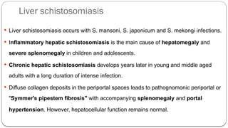
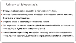
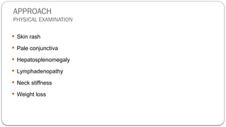
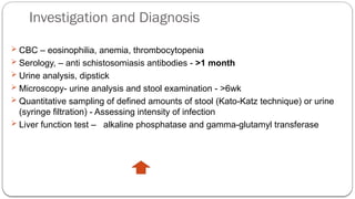
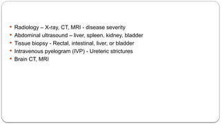
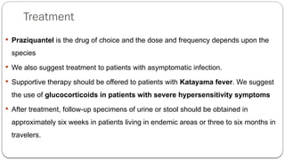
Prepared by: Hermela Seifu
C1 Student
Schistosomiasis
BETHEL MEDICAL COLLEGE
AUG 16/2023
CONTENT
๏‚— Definition
๏‚— Epidemiology
๏‚— Classification
๏‚— Pathogenesis
๏‚— Clinical features
๏‚— Approach
๏‚— Investigations
๏‚— Treatment
DEFINITION
๏‚— Schistosomiasis is sometimes referred to as "bilharziasis" after Theodor Bilharz who
identified the parasite first in 1852.
๏‚— Schistosomiasis is caused by infection with parasitic blood flukes known as
schistosomes.
๏‚— Schistosomiasis can be associated with serious morbidity and mortality.
๏‚— Chronic complications are generally seen in those with a high parasite load, in
individuals who live in endemic areas and have recurrent exposure. It can also occur in
travelers.
RISK FACTORS
๏‚— Bathing and swimming in fresh water
๏‚— Scuba diving
๏‚— Walking barefoot
๏‚— Traveling to endemic area
EPIDEMIOLOGY
๏‚— Schistosomiasis is prevalent in tropical and subtropical areas.
๏‚— According to WHO, an estimated 207 million people may have schistosomiasis
worldwide. It is responsible for more than 200,000 deaths annually.
๏‚— On a global scale, 1 in every 30 individuals has schistosomiasis.
๏‚— >90% live in sub-Saharan Africa.
๏‚— Currently in Ethiopia, about 5.1 million people are infected and 37.5 million people
are at an increased risk of infection.
SCHISTOSOME SPECIES
๏‚— Three major species and two less common species produce infection in humans:
๏‚— Of the major species, S. mansoni and S. japonicum, can provoke intestinal and hepatic
complications.
๏‚— S. haematobium predominantly leads to renal and bladder sequelae, although
occasionally, it results in liver disease.
๏‚— The minor species include S. mekongi and S. intercalatum, both of which can also
induce intestinal and liver disease.
๏‚— Mixed infections can also occur.
PATHOGENESIS
LIFE CYCLE
๏‚— The life cycle of schistosomiasis is complex and requires both intermediate and
definitive hosts
๏‚— The adult worms are approximately 1 to 2 cm in length with a cylindrical body, two
terminal suckers, a blind digestive tract, and reproductive organs.
๏‚— The male schistosome forms a groove or gynecophoric channel where the female
resides.
๏‚— The females produce hundreds to thousands of eggs per day, depending on
the species.
๏‚— After contact with water, each ovum releases a ciliated miracidium larva,
which seeks an intermediate host, the snail.
๏‚— After four to six weeks, the miracida multiply asexually into sporocysts and
later into cercarial larvae, which have a bifurcated tail.
๏‚— The cercaria leave the snail and seek a definitive host, where they develop
into adult worms.
PATHOGENESIS
ADULT WORMS
๏‚— Unless adult worms migrate to an unusual location (such as the spinal cord or
brain), adult worms do little damage to the host.
๏‚— They feed on circulating glucose and blood products, but usually do not cause
symptoms.
INTHE BOWEL
๏‚— Granulomatous inflammation around the invading eggs can result in intestinal
schistosomiasis characterized by ulceration and scarring.
๏‚— INTHE LIVER
๏‚— Eggs lodge in the presinusoidal radicals of the portal vein, where they elicit a
granulomatous fibrosing reaction that can eventually lead to blockade of venous blood
flow.
๏‚— Secondary portal venous hypertension results, with compensatory portosystemic
blood flow and progressive liver damage.
๏‚— The characteristic pathology seen within the liver is known as โ€˜Symmers pipestem
fibrosisโ€™.
๏‚— This pathology is distinct from cirrhosis, since there is no hepatocyte dysfunction;
instead, the portal hypertension results from fibrosis within blood vessels.
The eggs of S. haematobium lead to granulomatous inflammation, ulceration of the
vesical and ureteral walls with subsequent fibrosis.
๏‚— Functional bladder neck obstruction, hydroureter and hydronephrosis can ensue
๏‚— calcification of the urinary tract and bladder.
๏‚— These changes can eventually lead to renal failure.
๏‚— Occasionally, S. haematobium infection is also associated with hepatic damage.
Parasite-host immune response
๏‚— Adult worms absorb host proteins and coat themselves with host antigens so that
they can reside in the bloodstream for years without being attacked by the immune
system.
๏‚— It is the host immune response to invading egg, which is the major cause of clinical
disease.
๏‚— A host-mediated Type 2 T-helper cell-induced (Th2)- fibro-granulomatous
inflammatory response occurs in reaction to the parasitic eggs, which may result in
activation of hepatic stellate cells, the mediators of fibrosis.
CLINICAL FEATURES
๏‚— Most patients infected with schistosomes of all species, are asymptomatic.
๏‚— The clinical picture in individuals presenting in nonendemic countries tends to be
different from manifestations seen in the developing world because of differences in
immunity to the parasite and in intensity of infection.
๏‚— Acute symptoms tend to be more common in nonimmune individuals, such as
travelers, due to a more intense immune response to exposure.
๏‚— By contrast, chronic complications require a higher burden of infection and, thus, are
mainly seen in individuals from endemic areas.
๏‚— Symptoms can be localized to specific organs or can be generalized, including
fatigue.
๏‚— Symptoms can be divided into early and late stages of infection.
๏‚— The different species of Schistosoma cause varying clinical complications, and
different geographic strains within a species may have variable pathogenicity.
Acute infection
Acute symptoms may present as Swimmer's itch or Katayama fever.
Swimmer's itch โ€“ seen in one day
๏‚— The initial penetration of the skin by cercariae may be asymptomatic or may cause
the characteristic "swimmer's itch". This is a localized dermatitis that can result in
a Pruritic papular or urticarial rash at the site of larval entry, which is typically on
the lower legs or feet.
๏‚— Immediate tingling and itching at the site of entry may develop, followed by an
intensely pruritic papular eruption 12 to 24 hours later, which can last more than a
week.
๏‚— Prior sensitization to cercariae results in more rapid immune response and more
severe symptoms.
๏‚— This manifestation is most common with S. japonicum and occurs rarely with S.
haematobium.
Katayama fever
๏‚— Katayama fever is a systemic hypersensitivity reaction against the migrating
parasites, which occurs between 2-8 weeks after exposure.
๏‚— It is thought to be due to hypersensitivity to schistosome antigens and circulating
immune complexes and coincides with the first two weeks of egg production, a time
when the antigen burden grows dramatically.
๏‚— This acute manifestation is seen more frequently with S. mansoni or S. japonicum
infections in travelers and nonimmune hosts.
Symptoms of Katayama fever
๏‚— sudden onset of fever, chills, myalgias, arthralgias, dry cough, diarrhea and headache, often
resembling serum sickness.
๏‚— Lymphadenopathy and hepatosplenomegaly may be prominent findings on physical
examination.
๏‚— The symptoms usually resolve spontaneously over a period of a few weeks, but neck stiffness and
coma or even death can occur.
๏‚— Patients may develop eosinophilia and patchy infiltrates on chest x-ray.
๏‚— Some patients also develop persistent and more serious disease such as weight loss, dyspnea,
and chronic diarrhea.
๏‚— The diagnosis for Katayama fever can be complicated by the fact that eggs are
rarely excreted in detectable amounts during this stage and antibody tests are
usually negative.
๏‚— However, a peripheral eosinophilia will usually be present and often serves as
an important clue.
Chronic infection
๏‚— The severity of disease is related to ;
๏ƒ˜ The number of eggs trapped in tissues,
๏ƒ˜ Their anatomic distribution, and
๏ƒ˜ The duration and intensity of infection.
๏‚— Symptoms of chronic infection often begins insidiously but are frequently
progressive without treatment, particularly with a high intensity of disease.
๏‚— The major manifestations vary according to the infecting species.
Intestinal schistosomiasis
๏‚— Intestinal schistosomiasis is caused by S. mansoni, S. japonicum, S. intercalatum, S.
mekongi and occasionally, S. haematobium infections.
๏‚— The most common symptoms include chronic or intermittent abdominal pain, poor
appetite and diarrhea.
๏‚— Stools may be grossly bloody and chronic ulcerations may lead to secondary iron
deficiency anemia.
๏‚— Intestinal polyps can arise due to granulomatous inflammation surrounding eggs that
are deposited in the bowel wall. Bowel ulcers and strictures can also develop.
๏‚— Rarely, an inflammatory mass can lead to obstruction.
Liver schistosomiasis
๏‚— Liver schistosomiasis occurs with S. mansoni, S. japonicum and S. mekongi infections.
๏‚— Inflammatory hepatic schistosomiasis is the main cause of hepatomegaly and
severe splenomegaly in children and adolescents.
๏‚— Chronic hepatic schistosomiasis develops years later in young and middle aged
adults with a long duration of intense infection.
๏‚— Diffuse collagen deposits in the periportal spaces leads to pathognomonic periportal or
"Symmer's pipestem fibrosis" with accompanying splenomegaly and portal
hypertension. However, hepatocellular function remains normal.
Urinary schistosomiasis
๏‚— Urinary schistosomiasis is caused by S. haematobium infections.
๏‚— It may be asymptomatic or may cause microscopic or macroscopic terminal hematuria,
dysuria, and urinary frequency.
๏‚— Symptoms related to secondary anemia may be present.
๏‚— With progressive involvement, fibrosis and calcification of the bladder and ureters can
occur resulting in hydroureter and hydronephrosis.
๏‚— Obstruction leading to kidney damage and secondary bacterial infections may also
occure. however, treatment usually results in improvement in anatomic abnormalities.
๏‚— In addition, chronic inflammation may lead to an increased risk of developing
some forms of squamous cell carcinoma of the bladder.
๏‚— In addition to the direct invasion of the urinary system, infection with any
schistosomal species can be associated with immune complex deposition in the
kidneys, leading to nephortic syndrome which has protienuria.
Neurologic complications
๏‚— The two main clinical syndromes are ;
๏‚— Spinal cord neuroschistosomiasis (acute or subacute myelopathy) and
๏‚— Localized cerebral or cerebellar neuroschistosomiasis (focal CNS impairment,
seizures, increased intracranial pressure)
๏‚— Neurologic complications can occur even in individuals who are lightly infected;
prevention of this complication is one of the main reasons that infected but
asymptomatic travelers should be treated.
๏‚— These complications result in one to two ways:
๏‚— Eggs may enter the vertebral venous plexus and then embolize to the spinal cord
or brain, resulting in granulomas at these sites.
๏‚— Adult worms may migrate aberrantly to the spinal cord or brain.
Pulmonary complications
๏‚— In people with hepatosplenic disease due to heavy infections with S. mansoni, S.
japonicum or S. haematobium infection.
๏‚— Presinusoidal portal hypertension fosters the development of portosystemic collateral
vessels that allow schistosome eggs to embolize into the pulmonary circulation.
๏‚— Eggs lodge in pulmonary arterioles of 50 to 100 micrometer diameter and produce
a granulomatous pulmonary end arteritis.
๏‚— Pulmonary hypertension and cor pulmonale gradually arise.
Other
๏‚— Genital involvement can also occur, especially with S. haematobium.
๏‚— Symptoms in female patients include hypertrophic and ulcerative lesions of the
vulva, vagina, and cervix.
๏‚— Involvement on the ovaries or fallopian tubes can lead to infertility.
๏‚— In men, the epididymis, testicles, spermatic chord or prostate can be involved.
APPROACH
HISTORY
๏‚— What country they have traveled to
๏‚— if they walk bare foot
๏‚— if they have been to any open water
๏‚— Any rash, hematuria, diarrhea?
๏‚— Dry cough, dyspnea?
Fever, headache, fatigue?
๏‚— chills, myalgias, arthralgias, dry cough
๏‚— chronic or intermittent abdominal pain, poor appetite
APPROACH
PHYSICAL EXAMINATION
๏‚— Skin rash
๏‚— Pale conjunctiva
๏‚— Hepatosplenomegaly
๏‚— Lymphadenopathy
๏‚— Neck stiffness
๏‚— Weight loss
Investigation and Diagnosis
๏ƒ˜ CBC โ€“ eosinophilia, anemia, thrombocytopenia
๏ƒ˜ Serology, โ€“ anti schistosomiasis antibodies - >1 month
๏ƒ˜ Urine analysis, dipstick
๏ƒ˜ Microscopy- urine analysis and stool examination - >6wk
๏ƒ˜ Quantitative sampling of defined amounts of stool (Kato-Katz technique) or urine
(syringe filtration) - Assessing intensity of infection
๏ƒ˜ Liver function test โ€“ alkaline phosphatase and gamma-glutamyl transferase
๏‚— Radiology โ€“ X-ray, CT, MRI - disease severity
๏‚— Abdominal ultrasound โ€“ liver, spleen, kidney, bladder
๏‚— Tissue biopsy - Rectal, intestinal, liver, or bladder
๏‚— Intravenous pyelogram (IVP) - Ureteric strictures
๏‚— Brain CT, MRI
Treatment
๏‚— Praziquantel is the drug of choice and the dose and frequency depends upon the
species
๏‚— We also suggest treatment to patients with asymptomatic infection.
๏‚— Supportive therapy should be offered to patients with Katayama fever. We suggest
the use of glucocorticoids in patients with severe hypersensitivity symptoms
๏‚— After treatment, follow-up specimens of urine or stool should be obtained in
approximately six weeks in patients living in endemic areas or three to six months in
travelers.
THANK YOU

Schistosomiasis.pptx Schistosomiasis explanation

  • 1.
    Prepared by: HermelaSeifu C1 Student Schistosomiasis BETHEL MEDICAL COLLEGE AUG 16/2023
  • 2.
    CONTENT ๏‚— Definition ๏‚— Epidemiology ๏‚—Classification ๏‚— Pathogenesis ๏‚— Clinical features ๏‚— Approach ๏‚— Investigations ๏‚— Treatment
  • 3.
    DEFINITION ๏‚— Schistosomiasis issometimes referred to as "bilharziasis" after Theodor Bilharz who identified the parasite first in 1852. ๏‚— Schistosomiasis is caused by infection with parasitic blood flukes known as schistosomes. ๏‚— Schistosomiasis can be associated with serious morbidity and mortality. ๏‚— Chronic complications are generally seen in those with a high parasite load, in individuals who live in endemic areas and have recurrent exposure. It can also occur in travelers.
  • 4.
    RISK FACTORS ๏‚— Bathingand swimming in fresh water ๏‚— Scuba diving ๏‚— Walking barefoot ๏‚— Traveling to endemic area
  • 5.
    EPIDEMIOLOGY ๏‚— Schistosomiasis isprevalent in tropical and subtropical areas. ๏‚— According to WHO, an estimated 207 million people may have schistosomiasis worldwide. It is responsible for more than 200,000 deaths annually. ๏‚— On a global scale, 1 in every 30 individuals has schistosomiasis. ๏‚— >90% live in sub-Saharan Africa. ๏‚— Currently in Ethiopia, about 5.1 million people are infected and 37.5 million people are at an increased risk of infection.
  • 6.
    SCHISTOSOME SPECIES ๏‚— Threemajor species and two less common species produce infection in humans: ๏‚— Of the major species, S. mansoni and S. japonicum, can provoke intestinal and hepatic complications. ๏‚— S. haematobium predominantly leads to renal and bladder sequelae, although occasionally, it results in liver disease. ๏‚— The minor species include S. mekongi and S. intercalatum, both of which can also induce intestinal and liver disease. ๏‚— Mixed infections can also occur.
  • 8.
    PATHOGENESIS LIFE CYCLE ๏‚— Thelife cycle of schistosomiasis is complex and requires both intermediate and definitive hosts ๏‚— The adult worms are approximately 1 to 2 cm in length with a cylindrical body, two terminal suckers, a blind digestive tract, and reproductive organs. ๏‚— The male schistosome forms a groove or gynecophoric channel where the female resides.
  • 10.
    ๏‚— The femalesproduce hundreds to thousands of eggs per day, depending on the species. ๏‚— After contact with water, each ovum releases a ciliated miracidium larva, which seeks an intermediate host, the snail. ๏‚— After four to six weeks, the miracida multiply asexually into sporocysts and later into cercarial larvae, which have a bifurcated tail. ๏‚— The cercaria leave the snail and seek a definitive host, where they develop into adult worms.
  • 12.
    PATHOGENESIS ADULT WORMS ๏‚— Unlessadult worms migrate to an unusual location (such as the spinal cord or brain), adult worms do little damage to the host. ๏‚— They feed on circulating glucose and blood products, but usually do not cause symptoms.
  • 13.
    INTHE BOWEL ๏‚— Granulomatousinflammation around the invading eggs can result in intestinal schistosomiasis characterized by ulceration and scarring. ๏‚— INTHE LIVER ๏‚— Eggs lodge in the presinusoidal radicals of the portal vein, where they elicit a granulomatous fibrosing reaction that can eventually lead to blockade of venous blood flow. ๏‚— Secondary portal venous hypertension results, with compensatory portosystemic blood flow and progressive liver damage.
  • 14.
    ๏‚— The characteristicpathology seen within the liver is known as โ€˜Symmers pipestem fibrosisโ€™. ๏‚— This pathology is distinct from cirrhosis, since there is no hepatocyte dysfunction; instead, the portal hypertension results from fibrosis within blood vessels.
  • 15.
    The eggs ofS. haematobium lead to granulomatous inflammation, ulceration of the vesical and ureteral walls with subsequent fibrosis. ๏‚— Functional bladder neck obstruction, hydroureter and hydronephrosis can ensue ๏‚— calcification of the urinary tract and bladder. ๏‚— These changes can eventually lead to renal failure. ๏‚— Occasionally, S. haematobium infection is also associated with hepatic damage.
  • 16.
    Parasite-host immune response ๏‚—Adult worms absorb host proteins and coat themselves with host antigens so that they can reside in the bloodstream for years without being attacked by the immune system. ๏‚— It is the host immune response to invading egg, which is the major cause of clinical disease. ๏‚— A host-mediated Type 2 T-helper cell-induced (Th2)- fibro-granulomatous inflammatory response occurs in reaction to the parasitic eggs, which may result in activation of hepatic stellate cells, the mediators of fibrosis.
  • 17.
    CLINICAL FEATURES ๏‚— Mostpatients infected with schistosomes of all species, are asymptomatic. ๏‚— The clinical picture in individuals presenting in nonendemic countries tends to be different from manifestations seen in the developing world because of differences in immunity to the parasite and in intensity of infection. ๏‚— Acute symptoms tend to be more common in nonimmune individuals, such as travelers, due to a more intense immune response to exposure. ๏‚— By contrast, chronic complications require a higher burden of infection and, thus, are mainly seen in individuals from endemic areas.
  • 18.
    ๏‚— Symptoms canbe localized to specific organs or can be generalized, including fatigue. ๏‚— Symptoms can be divided into early and late stages of infection. ๏‚— The different species of Schistosoma cause varying clinical complications, and different geographic strains within a species may have variable pathogenicity.
  • 19.
    Acute infection Acute symptomsmay present as Swimmer's itch or Katayama fever. Swimmer's itch โ€“ seen in one day ๏‚— The initial penetration of the skin by cercariae may be asymptomatic or may cause the characteristic "swimmer's itch". This is a localized dermatitis that can result in a Pruritic papular or urticarial rash at the site of larval entry, which is typically on the lower legs or feet. ๏‚— Immediate tingling and itching at the site of entry may develop, followed by an intensely pruritic papular eruption 12 to 24 hours later, which can last more than a week.
  • 20.
    ๏‚— Prior sensitizationto cercariae results in more rapid immune response and more severe symptoms. ๏‚— This manifestation is most common with S. japonicum and occurs rarely with S. haematobium.
  • 21.
    Katayama fever ๏‚— Katayamafever is a systemic hypersensitivity reaction against the migrating parasites, which occurs between 2-8 weeks after exposure. ๏‚— It is thought to be due to hypersensitivity to schistosome antigens and circulating immune complexes and coincides with the first two weeks of egg production, a time when the antigen burden grows dramatically. ๏‚— This acute manifestation is seen more frequently with S. mansoni or S. japonicum infections in travelers and nonimmune hosts.
  • 22.
    Symptoms of Katayamafever ๏‚— sudden onset of fever, chills, myalgias, arthralgias, dry cough, diarrhea and headache, often resembling serum sickness. ๏‚— Lymphadenopathy and hepatosplenomegaly may be prominent findings on physical examination. ๏‚— The symptoms usually resolve spontaneously over a period of a few weeks, but neck stiffness and coma or even death can occur. ๏‚— Patients may develop eosinophilia and patchy infiltrates on chest x-ray. ๏‚— Some patients also develop persistent and more serious disease such as weight loss, dyspnea, and chronic diarrhea.
  • 23.
    ๏‚— The diagnosisfor Katayama fever can be complicated by the fact that eggs are rarely excreted in detectable amounts during this stage and antibody tests are usually negative. ๏‚— However, a peripheral eosinophilia will usually be present and often serves as an important clue.
  • 24.
    Chronic infection ๏‚— Theseverity of disease is related to ; ๏ƒ˜ The number of eggs trapped in tissues, ๏ƒ˜ Their anatomic distribution, and ๏ƒ˜ The duration and intensity of infection. ๏‚— Symptoms of chronic infection often begins insidiously but are frequently progressive without treatment, particularly with a high intensity of disease. ๏‚— The major manifestations vary according to the infecting species.
  • 25.
    Intestinal schistosomiasis ๏‚— Intestinalschistosomiasis is caused by S. mansoni, S. japonicum, S. intercalatum, S. mekongi and occasionally, S. haematobium infections. ๏‚— The most common symptoms include chronic or intermittent abdominal pain, poor appetite and diarrhea. ๏‚— Stools may be grossly bloody and chronic ulcerations may lead to secondary iron deficiency anemia. ๏‚— Intestinal polyps can arise due to granulomatous inflammation surrounding eggs that are deposited in the bowel wall. Bowel ulcers and strictures can also develop. ๏‚— Rarely, an inflammatory mass can lead to obstruction.
  • 26.
    Liver schistosomiasis ๏‚— Liverschistosomiasis occurs with S. mansoni, S. japonicum and S. mekongi infections. ๏‚— Inflammatory hepatic schistosomiasis is the main cause of hepatomegaly and severe splenomegaly in children and adolescents. ๏‚— Chronic hepatic schistosomiasis develops years later in young and middle aged adults with a long duration of intense infection. ๏‚— Diffuse collagen deposits in the periportal spaces leads to pathognomonic periportal or "Symmer's pipestem fibrosis" with accompanying splenomegaly and portal hypertension. However, hepatocellular function remains normal.
  • 27.
    Urinary schistosomiasis ๏‚— Urinaryschistosomiasis is caused by S. haematobium infections. ๏‚— It may be asymptomatic or may cause microscopic or macroscopic terminal hematuria, dysuria, and urinary frequency. ๏‚— Symptoms related to secondary anemia may be present. ๏‚— With progressive involvement, fibrosis and calcification of the bladder and ureters can occur resulting in hydroureter and hydronephrosis. ๏‚— Obstruction leading to kidney damage and secondary bacterial infections may also occure. however, treatment usually results in improvement in anatomic abnormalities.
  • 28.
    ๏‚— In addition,chronic inflammation may lead to an increased risk of developing some forms of squamous cell carcinoma of the bladder. ๏‚— In addition to the direct invasion of the urinary system, infection with any schistosomal species can be associated with immune complex deposition in the kidneys, leading to nephortic syndrome which has protienuria.
  • 29.
    Neurologic complications ๏‚— Thetwo main clinical syndromes are ; ๏‚— Spinal cord neuroschistosomiasis (acute or subacute myelopathy) and ๏‚— Localized cerebral or cerebellar neuroschistosomiasis (focal CNS impairment, seizures, increased intracranial pressure) ๏‚— Neurologic complications can occur even in individuals who are lightly infected; prevention of this complication is one of the main reasons that infected but asymptomatic travelers should be treated.
  • 30.
    ๏‚— These complicationsresult in one to two ways: ๏‚— Eggs may enter the vertebral venous plexus and then embolize to the spinal cord or brain, resulting in granulomas at these sites. ๏‚— Adult worms may migrate aberrantly to the spinal cord or brain.
  • 31.
    Pulmonary complications ๏‚— Inpeople with hepatosplenic disease due to heavy infections with S. mansoni, S. japonicum or S. haematobium infection. ๏‚— Presinusoidal portal hypertension fosters the development of portosystemic collateral vessels that allow schistosome eggs to embolize into the pulmonary circulation. ๏‚— Eggs lodge in pulmonary arterioles of 50 to 100 micrometer diameter and produce a granulomatous pulmonary end arteritis. ๏‚— Pulmonary hypertension and cor pulmonale gradually arise.
  • 32.
    Other ๏‚— Genital involvementcan also occur, especially with S. haematobium. ๏‚— Symptoms in female patients include hypertrophic and ulcerative lesions of the vulva, vagina, and cervix. ๏‚— Involvement on the ovaries or fallopian tubes can lead to infertility. ๏‚— In men, the epididymis, testicles, spermatic chord or prostate can be involved.
  • 33.
    APPROACH HISTORY ๏‚— What countrythey have traveled to ๏‚— if they walk bare foot ๏‚— if they have been to any open water ๏‚— Any rash, hematuria, diarrhea? ๏‚— Dry cough, dyspnea? Fever, headache, fatigue? ๏‚— chills, myalgias, arthralgias, dry cough ๏‚— chronic or intermittent abdominal pain, poor appetite
  • 34.
    APPROACH PHYSICAL EXAMINATION ๏‚— Skinrash ๏‚— Pale conjunctiva ๏‚— Hepatosplenomegaly ๏‚— Lymphadenopathy ๏‚— Neck stiffness ๏‚— Weight loss
  • 35.
    Investigation and Diagnosis ๏ƒ˜CBC โ€“ eosinophilia, anemia, thrombocytopenia ๏ƒ˜ Serology, โ€“ anti schistosomiasis antibodies - >1 month ๏ƒ˜ Urine analysis, dipstick ๏ƒ˜ Microscopy- urine analysis and stool examination - >6wk ๏ƒ˜ Quantitative sampling of defined amounts of stool (Kato-Katz technique) or urine (syringe filtration) - Assessing intensity of infection ๏ƒ˜ Liver function test โ€“ alkaline phosphatase and gamma-glutamyl transferase
  • 36.
    ๏‚— Radiology โ€“X-ray, CT, MRI - disease severity ๏‚— Abdominal ultrasound โ€“ liver, spleen, kidney, bladder ๏‚— Tissue biopsy - Rectal, intestinal, liver, or bladder ๏‚— Intravenous pyelogram (IVP) - Ureteric strictures ๏‚— Brain CT, MRI
  • 37.
    Treatment ๏‚— Praziquantel isthe drug of choice and the dose and frequency depends upon the species ๏‚— We also suggest treatment to patients with asymptomatic infection. ๏‚— Supportive therapy should be offered to patients with Katayama fever. We suggest the use of glucocorticoids in patients with severe hypersensitivity symptoms ๏‚— After treatment, follow-up specimens of urine or stool should be obtained in approximately six weeks in patients living in endemic areas or three to six months in travelers.
  • 38.