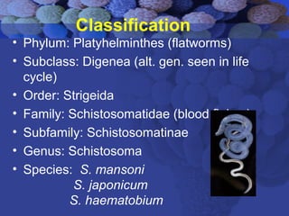
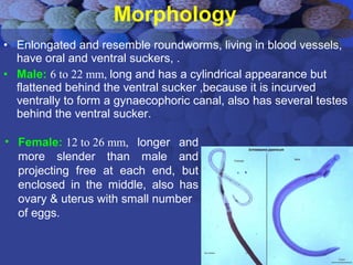
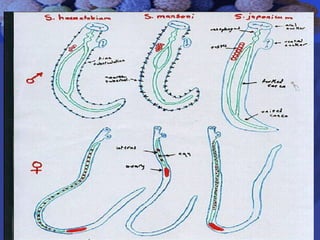
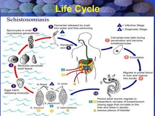
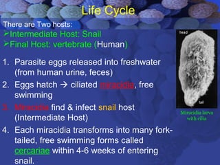
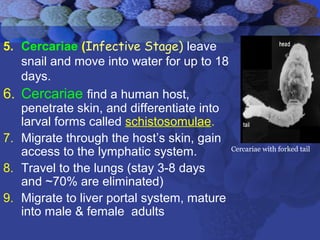
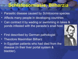
Schistosoma
&
Schistosomiasis
Dr. Rafal J. Al-Saigh
Medical Doctor
MB.Ch.B., MSc,
PhD Clin. Microb (Athens, Greece)
January 28th
, 2008
Gordana & Goldis
Classification
• Phylum: Platyhelminthes (flatworms)
• Subclass: Digenea (alt. gen. seen in life
cycle)
• Order: Strigeida
• Family: Schistosomatidae (blood flukes)
• Subfamily: Schistosomatinae
• Genus: Schistosoma
• Species: S. mansoni
S. japonicum
S. haematobium
Morphology
• Enlongated and resemble roundworms, living in blood vessels,
have oral and ventral suckers, .
• Male: 6 to 22 mm, long and has a cylindrical appearance but
flattened behind the ventral sucker ,because it is incurved
ventrally to form a gynaecophoric canal, also has several testes
behind the ventral sucker.
• Female: 12 to 26 mm, longer and
more slender than male and
projecting free at each end, but
enclosed in the middle, also has
ovary & uterus with small number
of eggs.
Life Cycle
Life Cycle
1. Parasite eggs released into freshwater
(from human urine, feces)
2. Eggs hatch  ciliated miracidia, free
swimming
3. Miracidia find & infect snail host
(Intermediate Host)
4. Each miracidia transforms into many fork-
tailed, free swimming forms called
cercariae within 4-6 weeks of entering
snail.
Miracidia larva
with cilia
There are Two hosts:
Intermediate Host: Snail
Final Host: vertebrate (Human)
5. Cercariae (Infective Stage) leave
snail and move into water for up to 18
days.
6. Cercariae find a human host,
penetrate skin, and differentiate into
larval forms called schistosomulae.
7. Migrate through the host’s skin, gain
access to the lymphatic system.
8. Travel to the lungs (stay 3-8 days
and ~70% are eliminated)
9. Migrate to liver portal system, mature
into male & female adults
Cercariae with forked tail
10. In liver, male & female pair up  female inserts
herself into the gynecophoral canal of male
 they are now ‘paired’.
11. Migrate to favoured sites:
S. mansoni – mesenteric venules of
large bowel & rectum
S. japonicum – mesenteric veins of the
small intestine
S. haematobium – perivesical venous plexus
surrounding the bladder
Lifespan of the adult worm ranges from 5 -10 years
Paired male &
female
12. Females release eggs.
Egg (Diagnostic Stage) characteristics
13. Eggs enter lumen of excretory organs
50%  passed out of body
50%  trapped in tissues, carried away
by blood circulation & lymph.
Eggs need 1-2 months to shed in stool or urine.
Sch. Japonicum
round egg
with small
lateral spine
Sch.mansoni
Oval egg with
lateral spine
Sch. Haematobium
oval egg with terminal spine
Schistosomiasis
Schistosomiasis, Bilharzia
• Parasitic disease caused by Schitosoma species
• Affects many people in developing countries
• Can contract it by wading or swimming in lakes &
ponds infested with the parasite’s snail host.
• First described by German pathologist
• Theodore Maximilian Bilharz
• In Egyptian patients who had died from the
disease (in their liver portal system &
bladder).
Acute Infection
(Early)
• Cercariae penetrate skin  rash
- called schistosome or swimmer’s itch.
• Eggs laid in target organs release antigens
 cause fever
- fever
- urticaria
- malaise
- diarrhea
Chronic Infection
(Late)
Symptoms of chronic infection caused by
eggs that travel to various parts of body
Eggs remain trapped in host tissues 
secrete Ags  granulomatous inflammatory
immune response
Fibroblasts mediate collagen deposition in
the granuloma, leading to fibrosis
Schistosomaiasis
Granuloma
• In S. mansoni infections
Wall of colon is damaged as eggs pass through it
• Inflamm. response  ulcers, inflammatory
polyps
• Clinically: diarrhea, abdominal pain
• Eggs can also accumulate in the appendix
• Can lead to appendicitis
Hepatosplenic schistosomiasis
•Eggs carried by portal circulation  liver
•Granulomatous response
•Granulomas are walled off with fibrous
tissue  fibrosis obstructs portal veins 
portal hypertension
• Esophageal varices bursting can cause
bleeding untill death.
• Splenomegaly (due to fibrosis)
Genitourinary complications
• Eggs lodge themselves in wall of bladder & can
develop into polyps
• Polyps can erode, ulcerate & cause hematuria
(blood cells in urine)
• Eggs lodge in ureters and urethra, cause lumps
and lesions  kidney failure
• Eggs lodge into ovaries, the uterus, cervix,
fallopian tubes  lumps  complications incl.
infertility
(For the men: eggs can also lodge into the testes
and the prostate )
CNS complications
S. haematobium and S. mansoni can
migrate to the spine
S. japonicum found in the brain and
causes encephalopathy
Diagnosis
1. Microscopic Detection of eggs in stool or
urine sample.
2. Serological tests
3. Antigen tests: Detect antigens in blood
with immunoelectrophoresis.
4. PCR
5. Intradermal skin test (schistosomian
skin test): become positive after few
months of infection.
Treatment
Praziquantel
• Extremely well tolerated, few side effects
• Broad-spectrum antihelminthic drug
• Resistance has been reported also
Metrifonate  against S. haematobium
Niridazole  against S. japonicum
Oxamniquine  against S. mansoni
• Recommend combination of 2 drugs
Prevention
For travelers it’s easy- don’t swim in fresh, stagnant
water (running water is better, still not safe).
Harder in endemic areas  people are dependent on
nearby freshwater.
Focused on education, eliminating snail nesting
grounds
Molluscicides can be used to eliminate snails.

Schistosomiasis dr.rafal

  • 1.
    Schistosoma & Schistosomiasis Dr. Rafal J.Al-Saigh Medical Doctor MB.Ch.B., MSc, PhD Clin. Microb (Athens, Greece)
  • 2.
  • 3.
    Classification • Phylum: Platyhelminthes(flatworms) • Subclass: Digenea (alt. gen. seen in life cycle) • Order: Strigeida • Family: Schistosomatidae (blood flukes) • Subfamily: Schistosomatinae • Genus: Schistosoma • Species: S. mansoni S. japonicum S. haematobium
  • 4.
    Morphology • Enlongated andresemble roundworms, living in blood vessels, have oral and ventral suckers, . • Male: 6 to 22 mm, long and has a cylindrical appearance but flattened behind the ventral sucker ,because it is incurved ventrally to form a gynaecophoric canal, also has several testes behind the ventral sucker. • Female: 12 to 26 mm, longer and more slender than male and projecting free at each end, but enclosed in the middle, also has ovary & uterus with small number of eggs.
  • 6.
  • 7.
    Life Cycle 1. Parasiteeggs released into freshwater (from human urine, feces) 2. Eggs hatch  ciliated miracidia, free swimming 3. Miracidia find & infect snail host (Intermediate Host) 4. Each miracidia transforms into many fork- tailed, free swimming forms called cercariae within 4-6 weeks of entering snail. Miracidia larva with cilia There are Two hosts: Intermediate Host: Snail Final Host: vertebrate (Human)
  • 8.
    5. Cercariae (InfectiveStage) leave snail and move into water for up to 18 days. 6. Cercariae find a human host, penetrate skin, and differentiate into larval forms called schistosomulae. 7. Migrate through the host’s skin, gain access to the lymphatic system. 8. Travel to the lungs (stay 3-8 days and ~70% are eliminated) 9. Migrate to liver portal system, mature into male & female adults Cercariae with forked tail
  • 9.
    10. In liver,male & female pair up  female inserts herself into the gynecophoral canal of male  they are now ‘paired’. 11. Migrate to favoured sites: S. mansoni – mesenteric venules of large bowel & rectum S. japonicum – mesenteric veins of the small intestine S. haematobium – perivesical venous plexus surrounding the bladder Lifespan of the adult worm ranges from 5 -10 years Paired male & female
  • 10.
    12. Females releaseeggs. Egg (Diagnostic Stage) characteristics 13. Eggs enter lumen of excretory organs 50%  passed out of body 50%  trapped in tissues, carried away by blood circulation & lymph. Eggs need 1-2 months to shed in stool or urine. Sch. Japonicum round egg with small lateral spine Sch.mansoni Oval egg with lateral spine Sch. Haematobium oval egg with terminal spine
  • 11.
  • 12.
    Schistosomiasis, Bilharzia • Parasiticdisease caused by Schitosoma species • Affects many people in developing countries • Can contract it by wading or swimming in lakes & ponds infested with the parasite’s snail host. • First described by German pathologist • Theodore Maximilian Bilharz • In Egyptian patients who had died from the disease (in their liver portal system & bladder).
  • 13.
    Acute Infection (Early) • Cercariaepenetrate skin  rash - called schistosome or swimmer’s itch. • Eggs laid in target organs release antigens  cause fever - fever - urticaria - malaise - diarrhea
  • 14.
    Chronic Infection (Late) Symptoms ofchronic infection caused by eggs that travel to various parts of body Eggs remain trapped in host tissues  secrete Ags  granulomatous inflammatory immune response Fibroblasts mediate collagen deposition in the granuloma, leading to fibrosis
  • 15.
  • 16.
    • In S.mansoni infections Wall of colon is damaged as eggs pass through it • Inflamm. response  ulcers, inflammatory polyps • Clinically: diarrhea, abdominal pain • Eggs can also accumulate in the appendix • Can lead to appendicitis
  • 17.
    Hepatosplenic schistosomiasis •Eggs carriedby portal circulation  liver •Granulomatous response •Granulomas are walled off with fibrous tissue  fibrosis obstructs portal veins  portal hypertension • Esophageal varices bursting can cause bleeding untill death. • Splenomegaly (due to fibrosis)
  • 18.
    Genitourinary complications • Eggslodge themselves in wall of bladder & can develop into polyps • Polyps can erode, ulcerate & cause hematuria (blood cells in urine) • Eggs lodge in ureters and urethra, cause lumps and lesions  kidney failure • Eggs lodge into ovaries, the uterus, cervix, fallopian tubes  lumps  complications incl. infertility (For the men: eggs can also lodge into the testes and the prostate )
  • 19.
    CNS complications S. haematobiumand S. mansoni can migrate to the spine S. japonicum found in the brain and causes encephalopathy
  • 20.
    Diagnosis 1. Microscopic Detectionof eggs in stool or urine sample. 2. Serological tests 3. Antigen tests: Detect antigens in blood with immunoelectrophoresis. 4. PCR 5. Intradermal skin test (schistosomian skin test): become positive after few months of infection.
  • 21.
    Treatment Praziquantel • Extremely welltolerated, few side effects • Broad-spectrum antihelminthic drug • Resistance has been reported also Metrifonate  against S. haematobium Niridazole  against S. japonicum Oxamniquine  against S. mansoni • Recommend combination of 2 drugs
  • 22.
    Prevention For travelers it’seasy- don’t swim in fresh, stagnant water (running water is better, still not safe). Harder in endemic areas  people are dependent on nearby freshwater. Focused on education, eliminating snail nesting grounds Molluscicides can be used to eliminate snails.

Editor's Notes

  • #9 Circariae need to find a human host within a day, or they die. Circariae find a human host and lose their tail as they’re penetrating the skin.
  • #11 Japonicum can release up to 2000 eggs/day!!
  • #14 Uticaria: itchy, raised, swollen bumps
  • #15 Uticaria: itchy, raised, swollen bumps
  • #18 gradient greater than 10 mmHg is considered portal hypertension. At gradients greater than 10 mmHg, blood flow though the hepatic portal system is redirected from the liver into areas with lower venous pressures  eg. The esophageal vein, which usually drains blood from the esophagus.