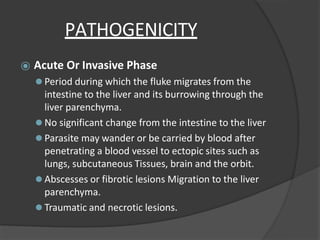
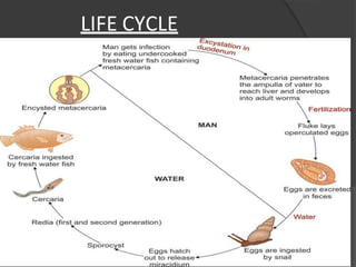
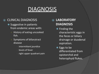
Characteristics
⦿Flat or fleshly, leaf-like unsegmented body.
⦿Incomplete alimentary canal.
⦿ Possess suckers with no hooks.
⦿Sexes are separate in Schistosomes, while the
others are hermaphroditic.
⦿ Oviparous
General Characteristics
⦿ Presence of two suckers.
⦿Has no body cavity, circulatory and
respiratory organs.
⦿ Alimentary system- mouth surrounded by
an oral sucker, muscular pharynx,
oesophagus which bifurcates into 2 blind
caeca (inverted Y shaped)
⦿Rudimentary nervous system – paired
ganglion cells.
Classification
BASED ON HABITAT:
⦿ BLOOD FLUKES
⦿ LIVER FLUKES
⦿ INTESTINAL FLUKES
⦿ LUNG FLUKES
Blood Flukes
⦿ Family : Schistosomatidae
⦿Genus : Schistosoma
⦿ Species:
1. S. haematobium
2. S. mansoni
3. S. japonicum
4. S. mekongi
5. S. intercalatum
SCHISTOSOMES
⦿ Schistosomiasis (bilharziasis)
⦿ Water-borne disease (Africa, Asia & Latin America).
⦿ Male worm is broader & lateral border is rolled
ventrally into a cylindrical shape, producing a long
groove – GYNECOPHORIC CANAL, inwhich
females are held.
⦿ Live in venous plexus in body of definitive host
Characteristics
⦿ Dioecious
⦿ Males are shorter and stouter than females
⦿ Lateral margins of males are folded ventrally to form a
gynecophoral canal in which females are received
⦿ Suckers are armed with delicate spines
⦿ There is no muscular pharynx
⦿ Eggs are non-operculated
⦿ Eggs are fully embryonated when laid
⦿ Embryonated eggs have a ciliated embryo called miracidium
⦿ Cercariae have bifid tails
⦿ There is no encysted metacerciarial stage
⦿ Infective Stage: cercaria penetrating the unbroken skin
Blood Flukes: Schistosomas
⦿Schistosoma japonicum: Oriental blood fluke
⦿Schistosoma haematobium: Vesical blood
fluke
⦿Schistosoma mansoni: Manson’s blood fluke
Features distinguishing Schistosomes
from other trematodes
⦿ Unisexual.
⦿Lack muscular pharynx
⦿Intestinal caeca reunite after bifurcation to
form a single canal.
⦿Produce non-operculated eggs.
⦿Cercariae have forked tails and infects by
penetrating unbroken skin of definitive
host.
SCHISTOSOMA HAEMATOBIUM
HISTORY & DISTRIBUTION
⦿ Bilharzia haematobium.
⦿ Bilharz described about the adult worm.
⦿Endemic in most parts of Africa, West Asia
some parts of India.
MORPHOLOGY
ADULT WORM FEMALE
20mm long,
0.25mm thick with
cuticular tubercles
confined to ends.
GYNECOPHORIC
CANAL
MALE
10-15mm long, 1mm
thick & covered by
finely tuberculated
cuticle
SUCKERS
EGG
Ovoid non-operculated Gravid worm has
20-30 eggs in
uterus at a time &
realises 300
Eggs/day
With a brownish yellow
transparent shell
carrying terminal spine
at one pole
Life Cycle
⦿ Definitive host : Humans
⦿ Intermediate host : freshwater snails
⦿Infective form : Cercaria larva
Clinical Features
⦿Classified Depending On Stages In
Evolution Of Infection:
o During Incubation Period
o During Oviposition
o During Tissue Proliferation & Repair
1.Skin penetration & Incubation period
⦿ Local cercarial
dermatitis/Swimmer’s itch –
transient itching and petechial
lesions at site of entry of cercariae.
o Often in visitors to endemic
areas than locals
⦿ Anaphylactic or toxic
symptoms – fever, malaise and
urticaria.
⦿ Accompanied by leucocytosis,
eosinophilia, enlarged tender liver
2.Oviposition
⦿Painless terminal hematuria – initially microscopic
later becomes gross.
⦿Develops frequency of micturition and burning.
⦿Cystoscopy – hyperplasia and inflammation of
bladder mucosa.
3.Tissue proliferation and repair
⦿ Generalised hyperplasia & fibrosis of vesical mucosa with
granular appearance – sandy patch.
⦿ Pseudo abscesses – d/t dense infiltration with
inflammatory cells at site of deposition of eggs.
⦿ Chronic cystitis – secondary bacterial infection.
⦿ Calculi formation d/t deposition of oxalate & uric acid
crystals around eggs.
⦿ Obstructive hyperplasia of ureters & urethra.
⦿ Associated Squamous cell carcinoma.
Lab Diagnosis
⦿ Detection of egg:
o Urine microscopy
o Bladder mucosal biopsy
⦿ Detection of antigen:
o Circulating anodic antigen & Circulating cathodic antigen by
ELISA
⦿ Detection of antibody:
o Complement Fixation Test (CFT)
o Immunofluorescence
o Indirect Hemagglutination
o Bentonite flocculation test
o Enzyme linked immunoelectrotransfer blot
o FAST/ELISA
⦿ Intradermal skin test(Fairley’s test):
o Group specific test gives positive
to all Schistosomiasis
⦿ Imaging:
o X-Ray- bladder and ureteral
calcification.
o USG-hydroureter & hydronephrosis
o Indirect diagnosis: IVP & Cystoscopy
Treatment & Prophylaxis
⦿ DOC – Praziquantel (40mg/kg for 1 day)
⦿Alternative DOC – Metriphonate
⦿ Prophylaxis:
o Eradication of intermediate molluscan hosts.
o Prevention of environmental pollution with urine and
faeces.
o Effective treated of infected.
o Avoid swimming, bathing and washing in infected water.
SCHISTOSOMA MANSONI
HISTORY & DISTRIBUTION
⦿Manson discovered eggs with lateral spines in the feces
of a west indian patient that led to the recognition of
this second species of human schistosomes.
⦿It is widely distributed in Africa, South America and
the Carribean islands.
HABIT MORPHOLOGY
⦿ Adult worm lives in the
inferiormesentericvein.
⦿ S. mansoni resembles S.
haematobium in
morphology and life cycle,
except:
⚫ Adult worms are smaller
and their integuments
studded with prominet
carse tubercles.
⚫ The prepatent period is 4-5
weeks.
Life Cycle
⦿ DEFINITIVE HOST: Humans are the only natural
definitive hosts.
⦿INTERMEDIATE HOST: Planorbid freshwater
snails of the genus Biomphalaria.
⦿ INFECTIVE FORM: Fork-tailed cercaria.
PathogenicityAnd Clinical Features
⦿ CERCARIAL DERMATITIS:
⚫Following skin penetration by cercariae
⦿ KATAYAMA FEVER:
⚫Formation of immune complexes. Signs and symptoms
include high fever, rash, arthralgia, etc..
⦿ INTESTINAL BILHARZIASIS:
⚫Microabscesses.
⚫Portal hypertension.
Laboratory Diagnosis
⦿ Stool microscopy.
⦿Rectal biopsy.
⦿ Serological diagnosis.
⦿ Imaging.
⦿Blood examination.
TREATMENT & PROPHYLAXIS
⦿ DOC – Praziquantel (40mg/kg for 1 day)
⦿Oxamniquine(single oral dose 15mg/kg) is
also effective.
⦿ Prophylaxis:
o Eradication of intermediate molluscan hosts.
o Prevention of environmental pollution with urine and
faeces.
o Effective treated of infected.
o Avoid swimming, bathing and washing in infected
water.
SCHISTOSOMA JAPONICUM
⦿ COMMON NAME: Oriental blood fluke
⦿DISTRIBUTION: Far east, Japan, China..
⦿HABITAT: superior mesenteric vein, also seen
in the intrahepatic portal venules and
hemorrhoidal plexus of veins
⦿ MORPHOLOGY:
⚫Adult male is comparatively slender(0.5mm
thick).
⚫Female lays upto 3500 eggs at one time.
⚫The prepatent period is 4-5 weeks.
⚫Eggs are smaller as compared to S.
haematobium and S. mansoni.
LIFE CYCLE
⦿Similar to S. haematobium, except;
⚫DEFINITIVE HOST: man
⚫INTERMEDIATE HOST: Amphibian snails of
the genus Oncomelania.
⚫INFECTIVE FORM FOR HUMAN: Fork
tailed cercaria.
PATHOGENICITYAND CLINICAL FEATURES
⦿ Katayama Fever.
⦿ Chronic Illness.
⦿Central Nervous System and Lungs Involved.
TREATMENT & PROPHYLAXIS
⦿ DOC – Praziquantel (40mg/kg for 1 day)
⦿ Prophylaxis:
o Eradication of intermediate molluscan hosts.
o Prevention of environmental pollution with
urine and faeces.
o Effective treated of infected.
o Avoid swimming, bathing and washing in
infected water.
Schistosoma Intercalatum
⦿First noted in 1934 in West-Central Africa.
⦿Eggs are fully embryonated without any operculum.
Eggs are acid-fast.
⦿Produces few symptoms involveing the mesentric
portal system.
⦿Diagnosis is established by detecting the egg in
feces and rectal biopsy.
⦿Praziquantel is the drug of choice.
Schistosoma Mekongi
⦿ First recognized in 1978 is found in Thailand and
Cambodia along the Mekong river.
⦿ Closely related to S. japonicum but is slightly
smaller and round.
⦿ Man and dog are the definitive host.
⦿ Man acquires infection in the same way as in S.
japonicum.
⦿ Hepatosplenomegaly and ascites are the common
clinical finding.
HERMAPHRODITIC FLUKES:
LIVER FLUKES
Fasciola Hepatica
⦿ COMMON NAME: Sheep liver fluke.
⦿ HISTORY & DISTRIBUTION:
⚫ First trematode that was found more than 600
years ago in 1379 by Jehan de brie.
⚫ Named by Linnaeus in 1758.
⚫ Largest and most common liver fluke.
⚫ Cause economically important disease “liver rot” in
sheep.
⚫ F.gigantia is more prevalent in india than
F.hepatica.
⦿ HABITAT: parasite resides in the liver and
biliary passages of the definitive host.
⦿ MORPHAOLOGY:
⚫ Large in size, flat leaf-shaped fluke measuring 30mm long
and 15mm broad, gray or brown in color.
⚫ Adult worm lives 5 years in sheep and 10 years in
humans.
⚫ Has a conical projection anteriorly containing an oral
sucker and is rounded posteriorly.
⚫ EGG:
○ Are large, ovoid, operculated,bile-stained and about
140µm by 80µm in size.
○ Contain an immature larva, the miracidium.
○ Do not float in saturated solution of common salt.
○ Eggs of F.hepatica and Fasciolopsis Buski cannot be
differentiated.
○ Eggs are unembryonated when freshly passed.
LIFE CYCLE
PATHOGENICITY
⦿ Acute Or Invasive Phase
⚫ Period during which the fluke migrates from the
intestine to the liver and its burrowing through the
liver parenchyma.
⚫ No significant change from the intestine to the liver
⚫ Parasite may wander or be carried by blood after
penetrating a blood vessel to ectopic sites such as
lungs, subcutaneous Tissues, brain and the orbit.
⚫ Abscesses or fibrotic lesions Migration to the liver
parenchyma.
⚫ Traumatic and necrotic lesions.
⦿ Chronic Or Latent Phase
⚫ period when the parasite has already reached the bile
ducts
⚫ obstruction in the vessel
⚫ inflammatory and adenomatous changes of the biliary
epithelium
⚫ fibrosis of the ducts
⚫ pressure atrophy of the liver parenchyma
⚫ intensive periductal fibrosis
⚫ Heavy infections
○ erosion of the epithelium
○ young worms wander back into the liver to produce
abscess pockets and to seed the vital liver tissue with their
eggs
DIAGNOSIS
⦿Clinical-based on:
○ Biliary symptoms.
○ Moderate to high eosinophilia.
○ Eating watercress as a green
salad(metacercaria in vegetation)
⦿ Laboratory
○ Recovery of the eggs in the patient’s stool or
from duodenal or biliary tract drainage.
○ Serodiagnosis-helpful but not adapted
for routine diagnosis.
TREATMENTAND PROPHYLAXIS
⦿ Oral triclabendazole(10mg/kg once) is the
treatment of choice.
⦿ Alternative drug is bithionol(30-50mg for 10-
15days).
⦿ Prednisolone at a dose of 10-20mg/kg is used to
control toxemia.
⦿ PHROPHYLAXIS
⚫ Health education.
⚫ Control of snails.
⚫ Proper disposal of human, sheep and cattle feces.
⚫ Proper disinfection of watercresses and other water
vegetations before consumption.
DICROCOELIUM DENTRITICUM
⦿Morphology
⚫ slender, lancet shaped, flat transparent, aspinous body 5 to 15 mm
by 1.5 to2.5 mm
⚫ Acetabulum lies at the beginning of the second fifth of the
body
⚫ two large, slightly lobed testes situated obliquely to each other
anterior to the small subglobose ovary just behind the ventral
sucker
⚫ voluminous uterine coils in the posterior thirds of the worm
⚫ Subglobose ovary lies to the right of the midline and
somewhat anterior to the equator of the worm
⚫ discrete vitelline follicles occupy lateral fields in the middle of
the body.
⚫ Eggs are deep golden-brown thick shelled, distinctly
operculated, measuring38 to 45u by 22 to 30u containing a fully
developed miracidium.
LIFE CYCLE
TREATMENTAND PROPHYLAXIS
⦿ Praziquantel -25 mg per kg TID for 2
consecutive days
⦿ PROPHYLAXIS:
⚫ no effective measures of control
⚫ Fresh herbs collected from grazing areas for use as food
for humans should be washed to remove the ants.
CLONORCHIS SINENSIS
⦿COMMON NAME: Chinese liver fluke
⦿HABITAT: Biliary tract and sometimes
pancreatic duct.
⦿ MORPHOLOGY:
⚫ADULT WORM:
○ 10-25mm long and 3-5mm broad.
○ Can survive for 15 years in biliary tract.
⚫EGGS:
○ Flask shaped, 35µm by 20µm with a yellowish
brown shell.
LIFE CYCLE
PATHOLOGY
⦿ Distal bile ducts are irritated mechanically and by its
toxic secretions
⦿ Slight leukocytosis and eosinophilia in early infection
⦿ Enlarged tender liver
⦿ Bile ducts thicken and become dilated and tortuous
⦿ Adenomatous proliferation of the biliary epithelium
⦿ Fibrosis and destruction of hepatic parenchyma
⦿ Liver function is impaired although SGPT and SGOT are
normal
DIAGNOSIS
⦿ CLINICALDIAGNOSIS
⚫ Suggestive in patients
from endemic areas with:
○ History of eating uncooked
fish.
○ Symptoms of biliarytract
disease
- intermittent jaundice
- bouts of fever
- right upper quadrant pain
⦿ LABORATORY
DIAGNOSIS
⚫ Finding the
characteristic eggs in
the feces or biliary
drainage or duodenal
aspiration.
⚫ Eggs to be
differentiated from
opistorchid and
heterophyid flukes.
TREATMENTAND PROPHYLAXIS
⦿ Praziquantel -25 mg per kg TID for 2
consecutive days
⦿ PROPHYLAXIS:
⚫ Proper cooking of fish.
⚫ Proper disposal of feces.
⚫ Control of snails.
INTESTINAL FLUKES
FASCIOLOPSIS BUSKI
⦿ COMMON NAME: Giant intestinal fluke.
⦿ First described by busk in 1843.
⦿ HABITAT: Duodenum or jejunum of pigs and man.
⦿ MORPHOLOGY:
⚫ Largest fluke. Thick, fleshy, ovate.Flesh-coloured. 2-7.5
x 1-2 cm.
⚫ Oral sucker and ventral suckers present.Typical trematode
alimentary canal with unbranched intestinal caeca.
⚫ Two dendritic testes.
⚫ Single branched ovary.
⚫ Vitellaria on lateral sides and a convoluted uterus and
genital pore that is anterior to theventral sucker.
⦿ EGG: Yellowish and oval. 130-140 x 80-85 μ
.
Thin walled
with small operculum. Undeveloped ovum.
PATHOLOGY
⦿ Attached to intestine by suckers.
⦿ Mucosa inflamed, ulcerated and abscesses
form. Epigastric pain, nausea, diarrhoea.
⦿ Heavy infection leads to oedema, ascites and
anarsaca.
⦿ Toxic products of worm.
⦿ Anaemia, leukocytosis, lymphocytosis, and
esinophilia.
⦿ Good prognosis if worm treated early.
⦿ 1000-2000 worms affect intestinal secretions
and food passage and
⦿ intoxication and sensitization.
LIFE CYCLE
DIAGNOSIS, TREATMENTAND
PROPHYLAXIS
⦿ DIAGNOSIS:
⚫ Eggs in feces. Differentiate from F.hepatica, Gastro discoides
and Echinostoma sp.
⦿ Adult worms vomited or passed in stool.
⦿ TREATMENT:
⚫ Praziquantel(15 mg/kg) body weight, single dose is the drug of
choice .
⦿ PROPHYLAXIS:
⚫ Sanitary disposal of human feces.
⚫ Discourage use of night soil.
⚫ Restrict pigs.
⚫ Snail control.
⚫ Health education.
⚫ Cooking or steeping of water plants.
HETEROPHYES HETEROPHYES
⦿ MORPHOLOGY:
⚫ Pyriform, greyish, 1.3 x 0.5 mm.Oral, Ventral and Genital suckers.
⦿ EGG:
⚫ Light brown in colour, thick shell, operculated, 29 x 16μm,Fully
developed miracidium.
⦿ TREATMENT:
⚫ Praziquantel(15 mg/kg) body weight, single dose is the drug of
choice .
⦿ PROPHYLAXIS:
⚫ Sanitary disposal of human feces.
⚫ Discourage use of night soil.
⚫ Restrict pigs.
⚫ Snail control.
⚫ Health education.
⚫ Cooking or steeping of water plants.
METAGONIMUS YOKOGAWAI
⦿ MORPHOLOGY:
⚫ Small, 1.4 x 0.6 mm. Pyriform shape with rounded posterior
and tapering anterior. Large ventral sucker.
⦿ EGG:
⚫ Light yellow-brown, thin-shelled, operculated, 28x17 μm,
mature miracidium.
⦿ TREATMENT:
⚫ Praziquantel(15 mg/kg) body weight, single dose is the drug of
choice .
⦿ PROPHYLAXIS:
⚫ Sanitary disposal of human feces.
⚫ Discourage use of night soil.
⚫ Restrict pigs.
⚫ Snail control.
⚫ Health education.
⚫ Cooking or steeping of water plants.
LUNG FLUKES
PARAGONIMUS WSTERMANI
⦿ Common name: Oriental lung fluke
⦿ History and Distribution:
⚫ P.westermani was discovered in 1878 by Kerbert in
the lungs of Bengal tigers that died in the zoological
gardens at Hamberg and Amsterdam.
⚫ The parasite is endemic in the Far East—Japan,
Korea, Taiwan, China, and south east asia—Sri Lanka
and India. Cases have been reported from Assam,
Bengal, TamilNadu, and Kerala.
⚫ P.mexicanus is an important human pathogen in
Central and South America.
MORPHOLOGY
ADULT WORM:
⦿ The adult worm is egg-shaped about 10 mm long,
5 mm
⦿ broad and 4 mm thick and reddish brown in color.
⦿ The integument is covered with scale like spines.
⦿ It has 2 unbranched intestinal caeca which end
blindly in the caudal area.
⦿ They have a lifespan of up to 20 years in humans.
EGG:
⦿ The eggs are operculated, golden brown in color and
about 100μm by 50μmin size.
⦿ They are unembryonated when freshly laid.
LIFE CYCLE
CLINICAL FEATURES, DIAGNOSIS,
TREATMENTAND PROPHYLAXIS
⦿ Clinical features:
⚫ Peribronchial granuloma and cystic dilation of
bronchi. Dyspnea, hemoptysis, pneumonitis,
bronchiectasis, abcess, pneumothorax.
Extrapulmonary lesions in brain and intestine.
⦿ Diagnosis:
⚫ Ova in sputum, X-ray and CT scan of chest, CFT, IHA,
and ELISA.
⦿ Treatment:
⚫ Praziquantel is the drug of choice.
⦿ Prophylaxis:
⚫ Adequate cooking of crabs and crayfish, eradication of
molluscan hosts and treatment of infected persons.
THANK YOU

trematodes.pptx

  • 2.
    Characteristics ⦿Flat or fleshly,leaf-like unsegmented body. ⦿Incomplete alimentary canal. ⦿ Possess suckers with no hooks. ⦿Sexes are separate in Schistosomes, while the others are hermaphroditic. ⦿ Oviparous
  • 3.
    General Characteristics ⦿ Presenceof two suckers. ⦿Has no body cavity, circulatory and respiratory organs. ⦿ Alimentary system- mouth surrounded by an oral sucker, muscular pharynx, oesophagus which bifurcates into 2 blind caeca (inverted Y shaped) ⦿Rudimentary nervous system – paired ganglion cells.
  • 5.
    Classification BASED ON HABITAT: ⦿BLOOD FLUKES ⦿ LIVER FLUKES ⦿ INTESTINAL FLUKES ⦿ LUNG FLUKES
  • 6.
    Blood Flukes ⦿ Family: Schistosomatidae ⦿Genus : Schistosoma ⦿ Species: 1. S. haematobium 2. S. mansoni 3. S. japonicum 4. S. mekongi 5. S. intercalatum
  • 7.
    SCHISTOSOMES ⦿ Schistosomiasis (bilharziasis) ⦿Water-borne disease (Africa, Asia & Latin America). ⦿ Male worm is broader & lateral border is rolled ventrally into a cylindrical shape, producing a long groove – GYNECOPHORIC CANAL, inwhich females are held. ⦿ Live in venous plexus in body of definitive host
  • 8.
    Characteristics ⦿ Dioecious ⦿ Malesare shorter and stouter than females ⦿ Lateral margins of males are folded ventrally to form a gynecophoral canal in which females are received ⦿ Suckers are armed with delicate spines ⦿ There is no muscular pharynx ⦿ Eggs are non-operculated ⦿ Eggs are fully embryonated when laid ⦿ Embryonated eggs have a ciliated embryo called miracidium ⦿ Cercariae have bifid tails ⦿ There is no encysted metacerciarial stage ⦿ Infective Stage: cercaria penetrating the unbroken skin
  • 9.
    Blood Flukes: Schistosomas ⦿Schistosomajaponicum: Oriental blood fluke ⦿Schistosoma haematobium: Vesical blood fluke ⦿Schistosoma mansoni: Manson’s blood fluke
  • 10.
    Features distinguishing Schistosomes fromother trematodes ⦿ Unisexual. ⦿Lack muscular pharynx ⦿Intestinal caeca reunite after bifurcation to form a single canal. ⦿Produce non-operculated eggs. ⦿Cercariae have forked tails and infects by penetrating unbroken skin of definitive host.
  • 11.
  • 12.
    HISTORY & DISTRIBUTION ⦿Bilharzia haematobium. ⦿ Bilharz described about the adult worm. ⦿Endemic in most parts of Africa, West Asia some parts of India.
  • 13.
  • 14.
    ADULT WORM FEMALE 20mmlong, 0.25mm thick with cuticular tubercles confined to ends. GYNECOPHORIC CANAL MALE 10-15mm long, 1mm thick & covered by finely tuberculated cuticle SUCKERS
  • 15.
    EGG Ovoid non-operculated Gravidworm has 20-30 eggs in uterus at a time & realises 300 Eggs/day With a brownish yellow transparent shell carrying terminal spine at one pole
  • 16.
    Life Cycle ⦿ Definitivehost : Humans ⦿ Intermediate host : freshwater snails ⦿Infective form : Cercaria larva
  • 19.
    Clinical Features ⦿Classified DependingOn Stages In Evolution Of Infection: o During Incubation Period o During Oviposition o During Tissue Proliferation & Repair
  • 20.
    1.Skin penetration &Incubation period ⦿ Local cercarial dermatitis/Swimmer’s itch – transient itching and petechial lesions at site of entry of cercariae. o Often in visitors to endemic areas than locals ⦿ Anaphylactic or toxic symptoms – fever, malaise and urticaria. ⦿ Accompanied by leucocytosis, eosinophilia, enlarged tender liver
  • 21.
    2.Oviposition ⦿Painless terminal hematuria– initially microscopic later becomes gross. ⦿Develops frequency of micturition and burning. ⦿Cystoscopy – hyperplasia and inflammation of bladder mucosa.
  • 22.
    3.Tissue proliferation andrepair ⦿ Generalised hyperplasia & fibrosis of vesical mucosa with granular appearance – sandy patch. ⦿ Pseudo abscesses – d/t dense infiltration with inflammatory cells at site of deposition of eggs. ⦿ Chronic cystitis – secondary bacterial infection. ⦿ Calculi formation d/t deposition of oxalate & uric acid crystals around eggs. ⦿ Obstructive hyperplasia of ureters & urethra. ⦿ Associated Squamous cell carcinoma.
  • 23.
    Lab Diagnosis ⦿ Detectionof egg: o Urine microscopy o Bladder mucosal biopsy ⦿ Detection of antigen: o Circulating anodic antigen & Circulating cathodic antigen by ELISA ⦿ Detection of antibody: o Complement Fixation Test (CFT) o Immunofluorescence o Indirect Hemagglutination o Bentonite flocculation test o Enzyme linked immunoelectrotransfer blot o FAST/ELISA
  • 24.
    ⦿ Intradermal skintest(Fairley’s test): o Group specific test gives positive to all Schistosomiasis ⦿ Imaging: o X-Ray- bladder and ureteral calcification. o USG-hydroureter & hydronephrosis o Indirect diagnosis: IVP & Cystoscopy
  • 25.
    Treatment & Prophylaxis ⦿DOC – Praziquantel (40mg/kg for 1 day) ⦿Alternative DOC – Metriphonate ⦿ Prophylaxis: o Eradication of intermediate molluscan hosts. o Prevention of environmental pollution with urine and faeces. o Effective treated of infected. o Avoid swimming, bathing and washing in infected water.
  • 26.
  • 27.
    HISTORY & DISTRIBUTION ⦿Mansondiscovered eggs with lateral spines in the feces of a west indian patient that led to the recognition of this second species of human schistosomes. ⦿It is widely distributed in Africa, South America and the Carribean islands.
  • 28.
    HABIT MORPHOLOGY ⦿ Adultworm lives in the inferiormesentericvein. ⦿ S. mansoni resembles S. haematobium in morphology and life cycle, except: ⚫ Adult worms are smaller and their integuments studded with prominet carse tubercles. ⚫ The prepatent period is 4-5 weeks.
  • 29.
    Life Cycle ⦿ DEFINITIVEHOST: Humans are the only natural definitive hosts. ⦿INTERMEDIATE HOST: Planorbid freshwater snails of the genus Biomphalaria. ⦿ INFECTIVE FORM: Fork-tailed cercaria.
  • 30.
    PathogenicityAnd Clinical Features ⦿CERCARIAL DERMATITIS: ⚫Following skin penetration by cercariae ⦿ KATAYAMA FEVER: ⚫Formation of immune complexes. Signs and symptoms include high fever, rash, arthralgia, etc.. ⦿ INTESTINAL BILHARZIASIS: ⚫Microabscesses. ⚫Portal hypertension.
  • 31.
    Laboratory Diagnosis ⦿ Stoolmicroscopy. ⦿Rectal biopsy. ⦿ Serological diagnosis. ⦿ Imaging. ⦿Blood examination.
  • 32.
    TREATMENT & PROPHYLAXIS ⦿DOC – Praziquantel (40mg/kg for 1 day) ⦿Oxamniquine(single oral dose 15mg/kg) is also effective. ⦿ Prophylaxis: o Eradication of intermediate molluscan hosts. o Prevention of environmental pollution with urine and faeces. o Effective treated of infected. o Avoid swimming, bathing and washing in infected water.
  • 33.
  • 34.
    ⦿ COMMON NAME:Oriental blood fluke ⦿DISTRIBUTION: Far east, Japan, China.. ⦿HABITAT: superior mesenteric vein, also seen in the intrahepatic portal venules and hemorrhoidal plexus of veins ⦿ MORPHOLOGY: ⚫Adult male is comparatively slender(0.5mm thick). ⚫Female lays upto 3500 eggs at one time. ⚫The prepatent period is 4-5 weeks. ⚫Eggs are smaller as compared to S. haematobium and S. mansoni.
  • 35.
    LIFE CYCLE ⦿Similar toS. haematobium, except; ⚫DEFINITIVE HOST: man ⚫INTERMEDIATE HOST: Amphibian snails of the genus Oncomelania. ⚫INFECTIVE FORM FOR HUMAN: Fork tailed cercaria.
  • 36.
    PATHOGENICITYAND CLINICAL FEATURES ⦿Katayama Fever. ⦿ Chronic Illness. ⦿Central Nervous System and Lungs Involved.
  • 37.
    TREATMENT & PROPHYLAXIS ⦿DOC – Praziquantel (40mg/kg for 1 day) ⦿ Prophylaxis: o Eradication of intermediate molluscan hosts. o Prevention of environmental pollution with urine and faeces. o Effective treated of infected. o Avoid swimming, bathing and washing in infected water.
  • 38.
    Schistosoma Intercalatum ⦿First notedin 1934 in West-Central Africa. ⦿Eggs are fully embryonated without any operculum. Eggs are acid-fast. ⦿Produces few symptoms involveing the mesentric portal system. ⦿Diagnosis is established by detecting the egg in feces and rectal biopsy. ⦿Praziquantel is the drug of choice.
  • 39.
    Schistosoma Mekongi ⦿ Firstrecognized in 1978 is found in Thailand and Cambodia along the Mekong river. ⦿ Closely related to S. japonicum but is slightly smaller and round. ⦿ Man and dog are the definitive host. ⦿ Man acquires infection in the same way as in S. japonicum. ⦿ Hepatosplenomegaly and ascites are the common clinical finding.
  • 40.
  • 41.
    Fasciola Hepatica ⦿ COMMONNAME: Sheep liver fluke. ⦿ HISTORY & DISTRIBUTION: ⚫ First trematode that was found more than 600 years ago in 1379 by Jehan de brie. ⚫ Named by Linnaeus in 1758. ⚫ Largest and most common liver fluke. ⚫ Cause economically important disease “liver rot” in sheep. ⚫ F.gigantia is more prevalent in india than F.hepatica. ⦿ HABITAT: parasite resides in the liver and biliary passages of the definitive host.
  • 42.
    ⦿ MORPHAOLOGY: ⚫ Largein size, flat leaf-shaped fluke measuring 30mm long and 15mm broad, gray or brown in color. ⚫ Adult worm lives 5 years in sheep and 10 years in humans. ⚫ Has a conical projection anteriorly containing an oral sucker and is rounded posteriorly. ⚫ EGG: ○ Are large, ovoid, operculated,bile-stained and about 140µm by 80µm in size. ○ Contain an immature larva, the miracidium. ○ Do not float in saturated solution of common salt. ○ Eggs of F.hepatica and Fasciolopsis Buski cannot be differentiated. ○ Eggs are unembryonated when freshly passed.
  • 44.
  • 46.
    PATHOGENICITY ⦿ Acute OrInvasive Phase ⚫ Period during which the fluke migrates from the intestine to the liver and its burrowing through the liver parenchyma. ⚫ No significant change from the intestine to the liver ⚫ Parasite may wander or be carried by blood after penetrating a blood vessel to ectopic sites such as lungs, subcutaneous Tissues, brain and the orbit. ⚫ Abscesses or fibrotic lesions Migration to the liver parenchyma. ⚫ Traumatic and necrotic lesions.
  • 47.
    ⦿ Chronic OrLatent Phase ⚫ period when the parasite has already reached the bile ducts ⚫ obstruction in the vessel ⚫ inflammatory and adenomatous changes of the biliary epithelium ⚫ fibrosis of the ducts ⚫ pressure atrophy of the liver parenchyma ⚫ intensive periductal fibrosis ⚫ Heavy infections ○ erosion of the epithelium ○ young worms wander back into the liver to produce abscess pockets and to seed the vital liver tissue with their eggs
  • 48.
    DIAGNOSIS ⦿Clinical-based on: ○ Biliarysymptoms. ○ Moderate to high eosinophilia. ○ Eating watercress as a green salad(metacercaria in vegetation) ⦿ Laboratory ○ Recovery of the eggs in the patient’s stool or from duodenal or biliary tract drainage. ○ Serodiagnosis-helpful but not adapted for routine diagnosis.
  • 49.
    TREATMENTAND PROPHYLAXIS ⦿ Oraltriclabendazole(10mg/kg once) is the treatment of choice. ⦿ Alternative drug is bithionol(30-50mg for 10- 15days). ⦿ Prednisolone at a dose of 10-20mg/kg is used to control toxemia. ⦿ PHROPHYLAXIS ⚫ Health education. ⚫ Control of snails. ⚫ Proper disposal of human, sheep and cattle feces. ⚫ Proper disinfection of watercresses and other water vegetations before consumption.
  • 50.
    DICROCOELIUM DENTRITICUM ⦿Morphology ⚫ slender,lancet shaped, flat transparent, aspinous body 5 to 15 mm by 1.5 to2.5 mm ⚫ Acetabulum lies at the beginning of the second fifth of the body ⚫ two large, slightly lobed testes situated obliquely to each other anterior to the small subglobose ovary just behind the ventral sucker ⚫ voluminous uterine coils in the posterior thirds of the worm ⚫ Subglobose ovary lies to the right of the midline and somewhat anterior to the equator of the worm ⚫ discrete vitelline follicles occupy lateral fields in the middle of the body. ⚫ Eggs are deep golden-brown thick shelled, distinctly operculated, measuring38 to 45u by 22 to 30u containing a fully developed miracidium.
  • 51.
  • 52.
    TREATMENTAND PROPHYLAXIS ⦿ Praziquantel-25 mg per kg TID for 2 consecutive days ⦿ PROPHYLAXIS: ⚫ no effective measures of control ⚫ Fresh herbs collected from grazing areas for use as food for humans should be washed to remove the ants.
  • 53.
    CLONORCHIS SINENSIS ⦿COMMON NAME:Chinese liver fluke ⦿HABITAT: Biliary tract and sometimes pancreatic duct. ⦿ MORPHOLOGY: ⚫ADULT WORM: ○ 10-25mm long and 3-5mm broad. ○ Can survive for 15 years in biliary tract. ⚫EGGS: ○ Flask shaped, 35µm by 20µm with a yellowish brown shell.
  • 54.
  • 55.
    PATHOLOGY ⦿ Distal bileducts are irritated mechanically and by its toxic secretions ⦿ Slight leukocytosis and eosinophilia in early infection ⦿ Enlarged tender liver ⦿ Bile ducts thicken and become dilated and tortuous ⦿ Adenomatous proliferation of the biliary epithelium ⦿ Fibrosis and destruction of hepatic parenchyma ⦿ Liver function is impaired although SGPT and SGOT are normal
  • 56.
    DIAGNOSIS ⦿ CLINICALDIAGNOSIS ⚫ Suggestivein patients from endemic areas with: ○ History of eating uncooked fish. ○ Symptoms of biliarytract disease - intermittent jaundice - bouts of fever - right upper quadrant pain ⦿ LABORATORY DIAGNOSIS ⚫ Finding the characteristic eggs in the feces or biliary drainage or duodenal aspiration. ⚫ Eggs to be differentiated from opistorchid and heterophyid flukes.
  • 57.
    TREATMENTAND PROPHYLAXIS ⦿ Praziquantel-25 mg per kg TID for 2 consecutive days ⦿ PROPHYLAXIS: ⚫ Proper cooking of fish. ⚫ Proper disposal of feces. ⚫ Control of snails.
  • 58.
  • 59.
    FASCIOLOPSIS BUSKI ⦿ COMMONNAME: Giant intestinal fluke. ⦿ First described by busk in 1843. ⦿ HABITAT: Duodenum or jejunum of pigs and man. ⦿ MORPHOLOGY: ⚫ Largest fluke. Thick, fleshy, ovate.Flesh-coloured. 2-7.5 x 1-2 cm. ⚫ Oral sucker and ventral suckers present.Typical trematode alimentary canal with unbranched intestinal caeca. ⚫ Two dendritic testes. ⚫ Single branched ovary. ⚫ Vitellaria on lateral sides and a convoluted uterus and genital pore that is anterior to theventral sucker. ⦿ EGG: Yellowish and oval. 130-140 x 80-85 μ . Thin walled with small operculum. Undeveloped ovum.
  • 60.
    PATHOLOGY ⦿ Attached tointestine by suckers. ⦿ Mucosa inflamed, ulcerated and abscesses form. Epigastric pain, nausea, diarrhoea. ⦿ Heavy infection leads to oedema, ascites and anarsaca. ⦿ Toxic products of worm. ⦿ Anaemia, leukocytosis, lymphocytosis, and esinophilia. ⦿ Good prognosis if worm treated early. ⦿ 1000-2000 worms affect intestinal secretions and food passage and ⦿ intoxication and sensitization.
  • 61.
  • 62.
    DIAGNOSIS, TREATMENTAND PROPHYLAXIS ⦿ DIAGNOSIS: ⚫Eggs in feces. Differentiate from F.hepatica, Gastro discoides and Echinostoma sp. ⦿ Adult worms vomited or passed in stool. ⦿ TREATMENT: ⚫ Praziquantel(15 mg/kg) body weight, single dose is the drug of choice . ⦿ PROPHYLAXIS: ⚫ Sanitary disposal of human feces. ⚫ Discourage use of night soil. ⚫ Restrict pigs. ⚫ Snail control. ⚫ Health education. ⚫ Cooking or steeping of water plants.
  • 63.
    HETEROPHYES HETEROPHYES ⦿ MORPHOLOGY: ⚫Pyriform, greyish, 1.3 x 0.5 mm.Oral, Ventral and Genital suckers. ⦿ EGG: ⚫ Light brown in colour, thick shell, operculated, 29 x 16μm,Fully developed miracidium. ⦿ TREATMENT: ⚫ Praziquantel(15 mg/kg) body weight, single dose is the drug of choice . ⦿ PROPHYLAXIS: ⚫ Sanitary disposal of human feces. ⚫ Discourage use of night soil. ⚫ Restrict pigs. ⚫ Snail control. ⚫ Health education. ⚫ Cooking or steeping of water plants.
  • 64.
    METAGONIMUS YOKOGAWAI ⦿ MORPHOLOGY: ⚫Small, 1.4 x 0.6 mm. Pyriform shape with rounded posterior and tapering anterior. Large ventral sucker. ⦿ EGG: ⚫ Light yellow-brown, thin-shelled, operculated, 28x17 μm, mature miracidium. ⦿ TREATMENT: ⚫ Praziquantel(15 mg/kg) body weight, single dose is the drug of choice . ⦿ PROPHYLAXIS: ⚫ Sanitary disposal of human feces. ⚫ Discourage use of night soil. ⚫ Restrict pigs. ⚫ Snail control. ⚫ Health education. ⚫ Cooking or steeping of water plants.
  • 65.
  • 66.
    PARAGONIMUS WSTERMANI ⦿ Commonname: Oriental lung fluke ⦿ History and Distribution: ⚫ P.westermani was discovered in 1878 by Kerbert in the lungs of Bengal tigers that died in the zoological gardens at Hamberg and Amsterdam. ⚫ The parasite is endemic in the Far East—Japan, Korea, Taiwan, China, and south east asia—Sri Lanka and India. Cases have been reported from Assam, Bengal, TamilNadu, and Kerala. ⚫ P.mexicanus is an important human pathogen in Central and South America.
  • 67.
    MORPHOLOGY ADULT WORM: ⦿ Theadult worm is egg-shaped about 10 mm long, 5 mm ⦿ broad and 4 mm thick and reddish brown in color. ⦿ The integument is covered with scale like spines. ⦿ It has 2 unbranched intestinal caeca which end blindly in the caudal area. ⦿ They have a lifespan of up to 20 years in humans. EGG: ⦿ The eggs are operculated, golden brown in color and about 100μm by 50μmin size. ⦿ They are unembryonated when freshly laid.
  • 68.
  • 69.
    CLINICAL FEATURES, DIAGNOSIS, TREATMENTANDPROPHYLAXIS ⦿ Clinical features: ⚫ Peribronchial granuloma and cystic dilation of bronchi. Dyspnea, hemoptysis, pneumonitis, bronchiectasis, abcess, pneumothorax. Extrapulmonary lesions in brain and intestine. ⦿ Diagnosis: ⚫ Ova in sputum, X-ray and CT scan of chest, CFT, IHA, and ELISA. ⦿ Treatment: ⚫ Praziquantel is the drug of choice. ⦿ Prophylaxis: ⚫ Adequate cooking of crabs and crayfish, eradication of molluscan hosts and treatment of infected persons.
  • 70.