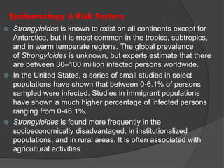
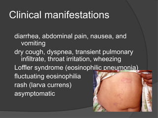
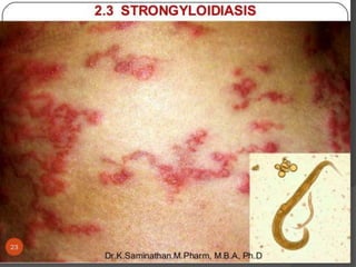
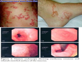
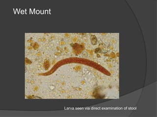
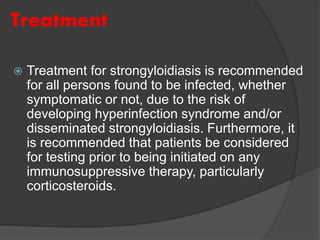
 Strongyloidiasis was first described in French troops
stationed in modern day Vietnam during the late
19th century who were suffering from severe,
persistent diarrhea. It is a parasitic disease caused
by nematodes, or roundworms, in the
genus Strongyloides that enter the body through
exposed skin, such as bare feet. Strongyloides is
most common in tropical or subtropical climates.
 Most people who are infected with Strongyloides do
not know they are infected and have no symptoms.
Others may develop a severe form and, if untreated,
become critically ill and potentially die.
strongyloides
 Strongyloides is known to exist on all continents except for
Antarctica, but it is most common in the tropics, subtropics,
and in warm temperate regions. The global prevalence
of Strongyloides is unknown, but experts estimate that there
are between 30–100 million infected persons worldwide.
 In the United States, a series of small studies in select
populations have shown that between 0-6.1% of persons
sampled were infected. Studies in immigrant populations
have shown a much higher percentage of infected persons
ranging from 0-46.1%.
 Strongyloides is found more frequently in the
socioeconomically disadvantaged, in institutionalized
populations, and in rural areas. It is often associated with
agricultural activities.
Epidemiology & Risk Factors
 The most common way of becoming infected
with Strongyloides is by contacting soil that is contaminated
with Strongyloides larvae. Therefore, activities that increase
contact with the soil increase the risk of becoming infected,
such as:
1. walking with bare feet
2. contact with human waste or sewage
3. occupations that increase contact with contaminated soil
such as farming and coal mining.
 Furthermore, many studies have shown an association
with Strongyloides and infection with Human T-Cell
Lymphotropic Virus-1 (HTLV-1). These studies have shown
that people infected with HTLV-1 are more likely to become
infected with Strongyloides, and that once infected, are more
likely to develop severe cases of strongyloidiasis.
 Of note, being infected with HIV/AIDS has not been shown to
be a risk factor for developingStrongyloides or having a
worse clinical course.
Life cycle
 Free-living cycle: The rhabditiform larvae passed
in the stool can either molt twice and become
infective filariform larvae (direct development) or
molt four times and become free living adult males
and females that mate and produce eggs from
which rhabditiform larvae hatch . The latter in turn
can either develop into a new generation of free-
living adults (as represented in ), or into infective
filariform larvae . The filariform larvae penetrate the
human host skin to initiate the parasitic cycle .
Two types of cycles exist:
 Parasitic cycle: Filariform larvae in contaminated
soil penetrate the human skin , and are
transported to the lungs where they penetrate the
alveolar spaces; they are carried through the
bronchial tree to the pharynx, are swallowed and
then reach the small intestine . In the small
intestine they molt twice and become adult
female worms . The females live threaded in the
epithelium of the small intestine and by
parthenogenesis produce eggs , which yield
rhabditiform larvae. The rhabditiform larvae can
either be passed in the stool or can cause
*autoinfection
 The infection of a primary host with a parasite, particularly
a helminth, in such a way that the complete life cycle of the
parasite happens in a single organism, without the
 In autoinfection, the rhabditiform larvae become
infective filariform larvae, which can penetrate either
the intestinal mucosa (internal autoinfection) or the
skin of the perianal area (external autoinfection); in
either case, the filariform larvae may follow the
previously described route, being carried
successively to the lungs, the bronchial tree, the
pharynx, and the small intestine where they mature
into adults; or they may disseminate widely in the
body. To date, occurrence of autoinfection in
humans with helminthic infections is recognized
only in Strongyloides stercoralis and Capillaria
philippinensis infections. In the case
of Strongyloides, autoinfection may explain the
possibility of persistent infections for many years in
persons who have not been in an endemic area and
of hyperinfections in immunosuppressed individuals.
Pathology
Invasive : Skin Penetration.
Pulmonary: During Cycle or Immigration.
Intestinal: Tissue Destruction
Disease
 Most people infected with Strongyloides do not know they’re
infected. If they do feel sick the most common complaints are
the following:
1. Abdominal
 stomachache, bloating, and heartburn
 intermittent episodes of diarrhea and constipation
 nausea and loss of appetite
2. Respiratory
 dry cough
 throat irritation
3. Skin
 an itchy, red rash that occurs where the worm entered the skin
 recurrent raised red rash typically along the thighs and
buttocks.
Life Cycle
Life Cycle
Life Cycle
Life Cycle
Clinical manifestations
diarrhea, abdominal pain, nausea, and
vomiting
dry cough, dyspnea, transient pulmonary
infiltrate, throat irritation, wheezing
Loffler syndrome (eosinophilic pneumonia)
fluctuating eosinophilia
rash (larva currens)
asymptomatic
Symptoms and Signs of
Hyperinfection
anemia (for example, pale skin)
constipation
cough
diarrhea
eosinophilic pneumonitis (during larvae
migration through the lungs)
Nausea
vomiting
weight loss.
Symptoms of Immunosuppressed patients
(organ transplant) or immunocompromised
patients (HIV):
Death
neurological and pulmonary complications
shock.
Diagnosis
 Strongyloides is usually diagnosed by seeing
larvae in stool when examined under the
microscope. This may require that you provide
multiple stool samples to your doctor or the
laboratory. Some laboratories are capable of
diagnosing Strongyloides with blood tests.
Wet Mount
Larva seen via direct examination of stool
Laboratory Diagnosis
 Direct stool smears (larvae)
 Cultivation of stool. (Damp charcoal or Harada-Mori
mediums).
 Histological examination of duodenal or jejunal biopsy
specimens obtained by endoscopy can demonstrate adult
worms embedded in the mucosa.
 Eosinophilia, is present in uncomplicated strongyloidiasis,
but is lost in hyper infection
 For population screening in endemic areas, an ELISA for
IgG anfi-Strongyloides antibodies is effective.
Serology
ELISA
Most sensitive method (88-95%)
May be lower in immunocompromised patients
Cannot distinguish between past and present
infections
Can cross-react with other nematode
infections
If results are positive, can move on to try and
establish a microscopic dx
Imaging
CXR – patchy alveolar infiltrates, diffuse
interstitial infiltrates, pleural effusions
AXR – Loops of dilated small bowel, ileus
Barium swallow – stenosis, ulceration, bowel
dilitation
Small bowel follow-through – worms in the
instestine
CT abdomen/pelvis – nonspecific thickening
of the bowel wall
Histology
Larvae typically found in proximal portion
of small intestine
Embedded in lamina propria
Cause edema, cellular infiltration, villous
atrophy, ulcerations
In-long standing infections, may see
fibrosis
Treatment
 Treatment for strongyloidiasis is recommended
for all persons found to be infected, whether
symptomatic or not, due to the risk of
developing hyperinfection syndrome and/or
disseminated strongyloidiasis. Furthermore, it
is recommended that patients be considered
for testing prior to being initiated on any
immunosuppressive therapy, particularly
corticosteroids.
Treatment
Antihelminitic therapy
Ivermectin
Albendazole
Thiabendazole
Abx directed toward enteric pathogens if bacteremia
or meningitis (2-4wks)
Minimize immunosuppression as possible
Directed supportive tx
Transfusions if GI bleed, antihistamines for itching, surgery if
bowel perf, etc
Repeat course of antihelminitic therapy if
immunocompromised, as relapse common
Follow-Up
Repeat stool exams or duodenal aspirations
in 2-3 mos to document cure
Repeat serologies 4-8 mos after therapy
Ab titer should be low or undetectable 6-18
mos after successful tx
If titer not falling, additional antihelminitic tx
Precautions for travelers to endemic areas,
but no prophylaxis or vaccine available
Strongyloides

Strongyloides

  • 2.
     Strongyloidiasis wasfirst described in French troops stationed in modern day Vietnam during the late 19th century who were suffering from severe, persistent diarrhea. It is a parasitic disease caused by nematodes, or roundworms, in the genus Strongyloides that enter the body through exposed skin, such as bare feet. Strongyloides is most common in tropical or subtropical climates.  Most people who are infected with Strongyloides do not know they are infected and have no symptoms. Others may develop a severe form and, if untreated, become critically ill and potentially die. strongyloides
  • 3.
     Strongyloides isknown to exist on all continents except for Antarctica, but it is most common in the tropics, subtropics, and in warm temperate regions. The global prevalence of Strongyloides is unknown, but experts estimate that there are between 30–100 million infected persons worldwide.  In the United States, a series of small studies in select populations have shown that between 0-6.1% of persons sampled were infected. Studies in immigrant populations have shown a much higher percentage of infected persons ranging from 0-46.1%.  Strongyloides is found more frequently in the socioeconomically disadvantaged, in institutionalized populations, and in rural areas. It is often associated with agricultural activities. Epidemiology & Risk Factors
  • 4.
     The mostcommon way of becoming infected with Strongyloides is by contacting soil that is contaminated with Strongyloides larvae. Therefore, activities that increase contact with the soil increase the risk of becoming infected, such as: 1. walking with bare feet 2. contact with human waste or sewage 3. occupations that increase contact with contaminated soil such as farming and coal mining.  Furthermore, many studies have shown an association with Strongyloides and infection with Human T-Cell Lymphotropic Virus-1 (HTLV-1). These studies have shown that people infected with HTLV-1 are more likely to become infected with Strongyloides, and that once infected, are more likely to develop severe cases of strongyloidiasis.  Of note, being infected with HIV/AIDS has not been shown to be a risk factor for developingStrongyloides or having a worse clinical course.
  • 5.
    Life cycle  Free-livingcycle: The rhabditiform larvae passed in the stool can either molt twice and become infective filariform larvae (direct development) or molt four times and become free living adult males and females that mate and produce eggs from which rhabditiform larvae hatch . The latter in turn can either develop into a new generation of free- living adults (as represented in ), or into infective filariform larvae . The filariform larvae penetrate the human host skin to initiate the parasitic cycle . Two types of cycles exist:
  • 6.
     Parasitic cycle:Filariform larvae in contaminated soil penetrate the human skin , and are transported to the lungs where they penetrate the alveolar spaces; they are carried through the bronchial tree to the pharynx, are swallowed and then reach the small intestine . In the small intestine they molt twice and become adult female worms . The females live threaded in the epithelium of the small intestine and by parthenogenesis produce eggs , which yield rhabditiform larvae. The rhabditiform larvae can either be passed in the stool or can cause *autoinfection  The infection of a primary host with a parasite, particularly a helminth, in such a way that the complete life cycle of the parasite happens in a single organism, without the
  • 8.
     In autoinfection,the rhabditiform larvae become infective filariform larvae, which can penetrate either the intestinal mucosa (internal autoinfection) or the skin of the perianal area (external autoinfection); in either case, the filariform larvae may follow the previously described route, being carried successively to the lungs, the bronchial tree, the pharynx, and the small intestine where they mature into adults; or they may disseminate widely in the body. To date, occurrence of autoinfection in humans with helminthic infections is recognized only in Strongyloides stercoralis and Capillaria philippinensis infections. In the case of Strongyloides, autoinfection may explain the possibility of persistent infections for many years in persons who have not been in an endemic area and of hyperinfections in immunosuppressed individuals.
  • 9.
    Pathology Invasive : SkinPenetration. Pulmonary: During Cycle or Immigration. Intestinal: Tissue Destruction
  • 10.
    Disease  Most peopleinfected with Strongyloides do not know they’re infected. If they do feel sick the most common complaints are the following: 1. Abdominal  stomachache, bloating, and heartburn  intermittent episodes of diarrhea and constipation  nausea and loss of appetite 2. Respiratory  dry cough  throat irritation 3. Skin  an itchy, red rash that occurs where the worm entered the skin  recurrent raised red rash typically along the thighs and buttocks.
  • 11.
  • 12.
  • 13.
  • 14.
  • 16.
    Clinical manifestations diarrhea, abdominalpain, nausea, and vomiting dry cough, dyspnea, transient pulmonary infiltrate, throat irritation, wheezing Loffler syndrome (eosinophilic pneumonia) fluctuating eosinophilia rash (larva currens) asymptomatic
  • 17.
    Symptoms and Signsof Hyperinfection anemia (for example, pale skin) constipation cough diarrhea eosinophilic pneumonitis (during larvae migration through the lungs) Nausea vomiting weight loss.
  • 18.
    Symptoms of Immunosuppressedpatients (organ transplant) or immunocompromised patients (HIV): Death neurological and pulmonary complications shock.
  • 21.
    Diagnosis  Strongyloides isusually diagnosed by seeing larvae in stool when examined under the microscope. This may require that you provide multiple stool samples to your doctor or the laboratory. Some laboratories are capable of diagnosing Strongyloides with blood tests.
  • 22.
    Wet Mount Larva seenvia direct examination of stool
  • 23.
    Laboratory Diagnosis  Directstool smears (larvae)  Cultivation of stool. (Damp charcoal or Harada-Mori mediums).  Histological examination of duodenal or jejunal biopsy specimens obtained by endoscopy can demonstrate adult worms embedded in the mucosa.  Eosinophilia, is present in uncomplicated strongyloidiasis, but is lost in hyper infection  For population screening in endemic areas, an ELISA for IgG anfi-Strongyloides antibodies is effective.
  • 24.
    Serology ELISA Most sensitive method(88-95%) May be lower in immunocompromised patients Cannot distinguish between past and present infections Can cross-react with other nematode infections If results are positive, can move on to try and establish a microscopic dx
  • 25.
    Imaging CXR – patchyalveolar infiltrates, diffuse interstitial infiltrates, pleural effusions AXR – Loops of dilated small bowel, ileus Barium swallow – stenosis, ulceration, bowel dilitation Small bowel follow-through – worms in the instestine CT abdomen/pelvis – nonspecific thickening of the bowel wall
  • 26.
    Histology Larvae typically foundin proximal portion of small intestine Embedded in lamina propria Cause edema, cellular infiltration, villous atrophy, ulcerations In-long standing infections, may see fibrosis
  • 27.
    Treatment  Treatment forstrongyloidiasis is recommended for all persons found to be infected, whether symptomatic or not, due to the risk of developing hyperinfection syndrome and/or disseminated strongyloidiasis. Furthermore, it is recommended that patients be considered for testing prior to being initiated on any immunosuppressive therapy, particularly corticosteroids.
  • 28.
    Treatment Antihelminitic therapy Ivermectin Albendazole Thiabendazole Abx directedtoward enteric pathogens if bacteremia or meningitis (2-4wks) Minimize immunosuppression as possible Directed supportive tx Transfusions if GI bleed, antihistamines for itching, surgery if bowel perf, etc Repeat course of antihelminitic therapy if immunocompromised, as relapse common
  • 29.
    Follow-Up Repeat stool examsor duodenal aspirations in 2-3 mos to document cure Repeat serologies 4-8 mos after therapy Ab titer should be low or undetectable 6-18 mos after successful tx If titer not falling, additional antihelminitic tx Precautions for travelers to endemic areas, but no prophylaxis or vaccine available