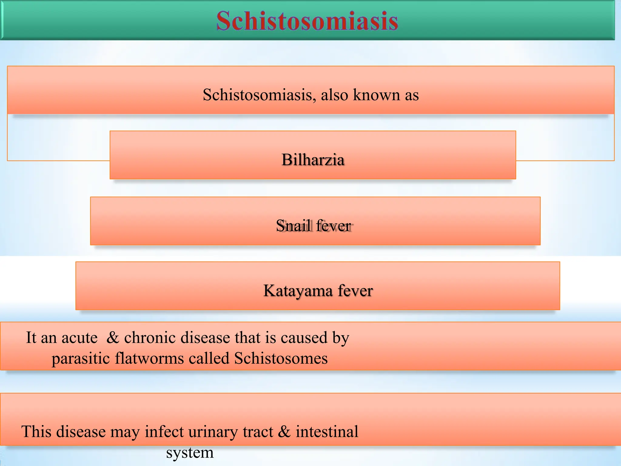
Presenter – Insha jan
Schistosomiasis
Schistosomiasis, also known as
Bilharzia
Snail fever
Katayama fever
It an acute & chronic disease that is caused by
parasitic flatworms called Schistosomes
This disease may infect urinary tract & intestinal
system
Trematodes
Intestinal
trematodes
F. buski
Lung
trematodes
P.
westermani
Blood
trematodes
Schistosome
s
Hepatic
trematodes
Clonorchi
s
Phylum : Platyhelminthes
Class : Trematoda
Order : Prosostomata
Superfamily: Schistosomatoidea
Genus : Schistosoma
Species : S.haematobium
S. mansoni
S. japonicum
S. intercalatum
S. menkongi
Obligate intravascular parasites
Unisexual (dioecious) - males are
shorter & stouter than females
Males possess a gynaecophoric
canal
Muscular Pharynx is lacking.
Suckers are armed with delicate
spines
Intestinal caeca reunite behind the
ventral sucker to form a single
canal.
Females laurer’s canal
(vestigial vagina) is absent, in
males 4-8 testes present.
Eggs are non – operculated & fully
embryonated when laid.
Cercariae have bifid tails &
penetrate into
the definitive host through the
unbroken skin.
Encysted meta cercarial stage is absent. Adult
1847
Funjii mentioned about
the katayama disease
1851
Bilharz discovered the adult
worm of S. haematobium
from the mesenteric vein of a
native of Cairo.
1903
Manson observed lateral –
spined eggs in the faeces
1904
Funjiami recovered an
adult female of S.
japonicum in the portal
vein of a man at autopsy.
1904
Katsurada observed the eggs
of S. japonicum in the human
faeces.
1907
Sambon pointed out that the
lateral – spined eggs belonged
to a separate species , S.
mansoni
1913-1914
Miyairi and suzuki
worked out the life
cycle of S. japonicum
1915
Leiper worked out the life
cycle of s. haematobium
in bulinus in Egypt .
1910
Schistosome eggs were found by
Ruffer in the renal pelvis of a
mummy of the twentieth dynasty
( 1250-1000 B.C).
Common name : Vesical blood fluke
Geographical distribution :Various parts of Africa and middle East.
 Gadgil and Shah (1952) reported a few cases from India ( Ratnagiri in
Maharashtra state).
 200 million persons are at a risk of infection & 90 million are infected by
S.
haematobium globally.
Adult worm (Males)
 10–15 mm long & 1mm thick.
 Covered by a finely tuberculated cuticle.
Possesses two muscular suckers-
oral sucker & ventral sucker
Gynecophoric canal is located behind
the ventral sucker and extending to a
caudal end.
 Long and slender, 20 mm × 0.25
mm with the cuticular tubercles
confined to the two ends.
 Gravid worm contains 20–30 eggs in its
uterus at one time & may pass up to 300
eggs a day.
Adult female
Egg
 Ovoid
 120–170 μm in length & 40–70 μm
in breadth.
 Non-operculated with a brownish yellow
transparent shell carrying a terminal
spine at one pole.
Larva
 S. haematobium has many larval stages
 miracidium
 sporocyst and cercaria.
Miracidium
 First larval stage
 Ciliated larva develops in all trematode eggs & is
infective to molluscs only
Sporocyst
 Thin walled sac containing masses of proliferating
cells (germ cells)
 Asexual reproduction
 Primary Sporocyst & secondary Sporocyst
Cercaria
 Elongated & oval with 400 μm length (including tail) & 60 μm
breadth.
 Body is covered with minute spine like projection on the surface.
 Two suckers, i.e. anterior & ventral & has bifurcated tail.
 life span of 24–72 hours
 S. heamatobium passes its life cycle in 2 hosts.
 Definitive host: Humans. No animal reservoir is
known.
 Intermediate host: Fresh water snails.
 Infective form: Cercarial larva
cercariae
An individual bathing in an
infected pool or coming in
contact with contaminated
water
Cercariae stick to the
surface of the skin of the
swimmers or bathers,
by means of their
ventral suckers
Terminal – spined
eggs of S.
haematobium may
erode blood vessels
& cause
haemorrhages
Tissue reaction in connective
tissue hyperplasia produces
“pseudotubercle ’’ around the
egg (egg granuloma).
P
aPtahtohgoegne
ni
ci
ci
ti
tyy ooff SS..
hhaaeemmaat
otboibuimu
m
 Disease caused by infection with S.haematobium is referred as
schistosomiasis
haematobia ( urinary schistosomiasis or bilharziasis )
 Clinical features depend on the stages in the evolution of the
infection,
 By the cercariae at the site of entrance.
 By the toxic metabolites.
 At the time of laying eggs.
Cercarial Dermatitis
 After 2 or 3 days of cercarial invasion, an itchy
maculopapular rash develops on the affected areas
of the skin called as cercarial dermatitis
(swimmer’s itch).
 Particularly seen with the cercariae of non human
Schistosomes.
 Liberated during the growth of schistosomulae in the portal blood of the
liver
 General anaphylactic reaction characterised by
 Fever, urticaria
 Eosinophilic Leucocytosis
 Enlarged tender liver
 Palpable spleen
 Symptoms appear between the 4th and the 5th week of the infection
 Regarded as a localising symptom, generally occurring within 3 to 9 months of
the
infection.
 Characteristic manifestation is a painless terminal haematuria.
 Adjacent structures of uro-genital apparatus are involved
 At first by the reversible granulomatous inflammatory reaction to eggs
 Later by the irreversible fibrosis and calcification.
 Squamous cell carcinoma of urinary bladder has been seen to be associated
with urinary schistosomiasis.
 Geographical distribution: various parts of Africa and South America. No
cases
are reported from India so far.
 Habitat: Adult worm lives in the inferior mesenteric vein.
 S. mansoni produces intestinal schistosomiasis in
humans.
Common name: Manson’s blood fluke
,
Morphology
 Adult worms are similar to other Schistosomes
 Gravid female - the uterus contains very few
eggs
usually 1–3 only.
 Prepatent period is 4–5 weeks.
 Nonoperculated eggs have characteristic
lateral spine.
 Measures 110–175 μm × 45–70 μm
 Definitive host: Humans are the only natural definitive hosts, though in
endemic areas monkeys and baboons have also been found infected.
 Intermediate host: Planorbid fresh-water snails of the genus Biomphalaria.
 Infective form: Fork-tailed cercaria.
Life cycle
 In humans, the schistosomulae mature in the liver & the adult worms move against
the blood stream into the venules of the inferior mesenteric group in
the
sigmoidorectal area.
 Eggs penetrate the gut wall, reach the colonic lumen, & are shed in
feces.
Life cycle
Pathogenesis of mansonian schistosomiasis occurs in three
stages.
 Cercarial Dermatitis
 Acute Schistosomiasis (Katayama Fever)
 Chronic schistosomiasis
Acute Schistosomiasis (Katayama Fever)
 Acute phase of disease occurs within 4–8 weeks of infection, especially when the
Schistosomes start producing eggs.
 Antigens (released from eggs) & adult worms stimulate the host humoral
response, leading to the formation of immune complexes & serum sickness like
illness called Katayama fever.
 Characterized by
 Fever
 Generalized lymphadenopathy and
 Hepatosplenomegaly.
Chronic Schistosomiasis
 After eggs are produced, they are trapped in the small venules & are carried into
the intestine (or less commonly to bladder) & are excreted in feces. Some are
carried through portal circulation into liver and other parts of the body.
 Intestinal disease
 The eggs are deposited in the intestinal wall.
 Soluble antigens liberated from eggs induce inflammatory reactions that lead to
granuloma formation around the eggs in the intestine.
(Symmers pipe stem fibrosis)
Hepatosplenic disease
 Granuloma formation and fibrosis in
liver seriously impedes the portal blood flow
leading
 Portal hypertension,
 Hepatomegaly (seen in 15–
20%),
 Splenomegaly and
 Gastric varices.
Other body sites
 Pulmonary involvement occurs when eggs are carried & lodged in the lungs
by collateral circulation.
 Egg sequestration and granuloma formation may cause
 pulmonary emboli formation,
 Pulmonary hypertension and
 right sided heart failure(cor pulmonale).
 Spinal cord schistosomiasis & myelopathy
 Nephrosclerosis & kidney failure due to circulating immune complexes
deposited in glomerular membrane
 Secondary bacterial infection especially with Salmonella species.
 Habitat
 Adult worms are seen typically in the venules of the superior mesenteric
vein draining the ileocecal region.
 Seen in the intrahepatic portal venules and hemorrhoidal plexus of veins.
 Common name: Oriental blood fluke
 Distribution
 S. japonicum is found in the far east, Japan, China, Taiwan, Philippines, and
Sulawesi.
Morphology
 similar to the schistosomes except
 Adult male is comparatively slender (0.5
mm thick) & does not have cuticular
tuberculation's.
 Gravid female - the uterus contains as many as 100 eggs at one time & up to
3,500 eggs may be passed daily by a single worm.
 Prepatent period is 4–5 weeks. Eggs are smaller & more spherical than those
of S.
haematobium and S. mansoni.
Life Cycle
 Life cycle of S. japonicum is similar to S. haematobium with the
following
exceptions.
 Definitive host: Man is the definitive host but in endemic areas, natural infection
occurs widely in several domestic animals & rodents,
 Intermediate host: Amphibian snails of the genus Oncomelania.
 Infective form for humans: Fork tailed cercaria.
 Eggs deposited in the superior mesenteric venules penetrate the gut wall and
are passed in feces.
 They hatch in water and the miracidia infect the intermediate hosts, amphibian
snails of the genus Oncomelania.
 Fork-tailed cercaria, which escapes from the snails is the infective form for
men and other definitive hosts.
Life Cycle
 Pathogenesis is similar to that caused by S. mansoni.
 Disease is more severe because of the higher egg production and smaller
size of the eggs (easy dissemination).
 Cercarial dermatitis
 Katayama fever: Seen after 40 days of infection.
 More severe and sometimes leads to death.
Intestinal disease: Deposition of egg granulomas in the intestinal wall leads to
 Mucosal hyperplasia,
 Ulcers,
 Micro abscess formation &
 Pseudopolyposis with blood loss
Hepatosplenic disease: Seen due to granulomatous response surrounding the
eggs
 Parietal lobe is the most common site.
 Symptoms include Jacksonian convulsions & grand mal seizures
 Carcinoma: Both colorectal carcinoma & liver carcinoma (& cirrhosis) are
reported from people of China and Japan infected with S. japonicum
 Chronic secondary infection with Salmonella species and hepatitis B virus has
been associated with S. japonicum.
Central nervous system (CNS) infection: Occurs in 2–4% of
cases.
 First recognized in 1934 is found in West-
Central Africa.
 Eggs are fully embroyonated without any
operculum having terminal spines, but are
passed exclusively in stools. The eggs are acid
fast.
 Produces few symptoms involving the
mesenteric portal system.
 Diagnosis is established by detection of the egg in feces and rectal
biopsy.
 First recognized in 1978 is found in Thailand
and Cambodia, along the Mekong river.
 Closely related to S. japonicum but are slightly
smaller and round.
 Man and dog are the definitive host.
 Man acquires infection in the same way as in S. Japonicum.
 Hepatosplenomegaly and ascites are the common clinical finding.
Urine Microscopy
 Detection of nonoperculated terminal spined eggs in the urine or rarely in
feces.
 Terminal hematuria portion of urine is collected between 12 pm & 3 pm,
concentrated by centrifugation or by membrane filtration.
 Observed under microscope
 Eggs with lateral spine can be demonstrated in stool or rarely in
urine.
 Chronic cases or in patients with low worm
burden, the number of eggs excreted in stool is less
& intermittent.
 Multiple stool specimens should be examined.
 Stool concentration techniques - Gravity or
centrifugal sedimentation
 Hatching test
 Quantitation of eggs in stool specimens can be done by Kato thick smear
technique.
Rectal Biopsy Specimen
 Lateral spined eggs in biopsy material from rectal mucosa confirms the
diagnosis of schistosomiasis.
 Egg shell of S. mansoni is acid fast & can be stained by modified ZiehlNeelsen
stain.
Antigen Detection
 Detection of circulating antigen indicates recent infection
 Used for monitoring the treatment response.
 Useful when urine microscopy fails to detect eggs (chronic & ectopic cases).
 Circulating cathodic antigen (CCA) & circulating anodic antigen (CAA) in
serum and soluble egg antigen (SEA) in serum.
Antibody Detection
 ELISA
 Complement fixation test
 Immunofluorescence
 Indirect heamagglutination test
 Latex agglutination test
 RIA
Intradermal skin test ( fairley’s test )
 Allergic reaction , positive in all the varieties of schistosomiasis
Imaging
 X-ray –bladder and ureteral calcification
USG- hydroureter & hydronephrosis.
Other tests
 Blood examination
 Eosinophilic count – increased in early cases
 Aldehyde test – often positive (due to globulin value)
 Treatment of schistosomiasis depends
 stage of infection
 clinical presentation.
 Therapy for acute schistosomiasis or Katayama syndrome needs to be
adjusted appropriately for each case.
 Severe acute schistosomiasis management in an acute-care setting is necessary,
with supportive measures & consideration of glucocorticoid treatment to reduce
inflammation.
 Once the acute critical phase is over, specific chemotherapy is indicated for
parasite elimination.
 Drug of choice is praziquantel, depending on the infecting species is
administered PO as a total of 40 or 60 mg/kg in two or three doses over a single
day.
 Praziquantel treatment results in parasitologic cure in ~85% of cases and reduces
egg counts by >90%.
 Metriphonate is the alternative drug of choice in schistosomiasis due to S.
haematobium. (7.5 mg/kg. weekly for 3 weeks).
 Oxamniquine ( 15- 20 mg/kg as a single dose) is also effective for S.mansoni
 Prophylactic measures include:
 Eradication of the intermediate molluscan hosts.
 Prevention of environmental pollution with urine and
feces.
 Effective treatment of infected persons.
 Avoid swimming, bathing, and washing in infected water.
 Vaccination.
1. Antigenic modification
2. Production of blocking antibodies
3. Inhibition of Immune Factors
1. Antigenic modification
 Antigen disguise:
 Adult Schistosoma cover themselves with host proteins to be considered as self
&
will not be attacked by the immune factors.
 Surface turnover :
 Schistosoma mansoni shed their teguments in abundance can neutralize antibody
response at a distance away from the parasite.
 Antigen mimicry:
 Schistosoma produce antigens similar to host antigens so they are not
recognized by the host’s immune system.
2- Production of blocking
antibodies

Antibodies of little protective effect.
 Schistosoma produce blocking Abs that combine with Schistosoma Ags making
them unavailable for antibodies of high protective effect .
3- Inhibition of Immune Factors
Schistosoma larva , inactivates the complement system through protease
activity .The mechanism of immune evasion is not resolved completely till now.
 Vaccine strategies represent an essential component as an adjunct to chemotherapy
for the future control of Schistosomiasis.
 An improving understanding of immune response to Schistosome infection suggest
the development of vaccine is possible .
 Vaccination against Schistosome can be targeted towards prevention of infection and
reduction in worm burden.
 Existance of immunity in the presence of active adult
infection is considered by Smithers and Terry (1969),to be an example of
Concomitant immunity.
 Phenomena of concomitant immunity is studied in baboon, mice, rats and humans.
 Live cercariae or Schistosomulae as well as attenuated forms are used to immunize
experimental animals in which they induce immunity.
 Such immunized animals are substantially protected from pathogenic effect
and
shows 60-70% lower worm count and tissue egg count than non –vaccinated
animals.
Tetraspanin (Sm-tsp-1& Sm-tsp-2)
 Important vaccine candidate .
 Present at apical syncytial surface of S. mansoni.
 Used in a defined vaccine formulation & upon administration
provided :
 Protection 29-61 %
 Reduction in egg burden 50-61 %
 Sm-28-GST (Glutathione S- transferase)
Expressed in subtegumental tissues of developmental stages of
parasite.
 Used as the potential vaccine candidate against human Schistosome
infection.
 Successfully tested at primate level and ready for human
trial.
 Sm-p80 (calpain)
 First vaccine antigen identified on the basis of T-cell activity.
 Sm-p80 – based vaccine formulation have three protective
effects:
 Worm reduction .
 Antifecundity effect .
 Protection against acute Schistosomiasis .
 worm reduction 60-70%
 A number of recent studies, have utilised plasmid DNA
vaccines.
 Generate both T-cell and B-cell immune responses.
 Particularly appealing for Schistosome vaccines
development.
 An effective, protective Schistosomiasis vaccine would be of immense public
importance.
 The apical membrane proteins, help in immune evasion are the logical
vaccine targets.
 All mentioned candidate vaccines are based on this idea.
 One successful human trial is yet to be completed, the vaccine will be
available for commercial product in next few years time.
Schistosomiasis_Explaination in parasitology

Schistosomiasis_Explaination in parasitology

  • 1.
    Presenter – Inshajan Schistosomiasis
  • 2.
    Schistosomiasis, also knownas Bilharzia Snail fever Katayama fever It an acute & chronic disease that is caused by parasitic flatworms called Schistosomes This disease may infect urinary tract & intestinal system
  • 3.
    Trematodes Intestinal trematodes F. buski Lung trematodes P. westermani Blood trematodes Schistosome s Hepatic trematodes Clonorchi s Phylum :Platyhelminthes Class : Trematoda Order : Prosostomata Superfamily: Schistosomatoidea Genus : Schistosoma Species : S.haematobium S. mansoni S. japonicum S. intercalatum S. menkongi
  • 4.
    Obligate intravascular parasites Unisexual(dioecious) - males are shorter & stouter than females Males possess a gynaecophoric canal Muscular Pharynx is lacking. Suckers are armed with delicate spines
  • 5.
    Intestinal caeca reunitebehind the ventral sucker to form a single canal. Females laurer’s canal (vestigial vagina) is absent, in males 4-8 testes present. Eggs are non – operculated & fully embryonated when laid. Cercariae have bifid tails & penetrate into the definitive host through the unbroken skin. Encysted meta cercarial stage is absent. Adult
  • 6.
    1847 Funjii mentioned about thekatayama disease 1851 Bilharz discovered the adult worm of S. haematobium from the mesenteric vein of a native of Cairo. 1903 Manson observed lateral – spined eggs in the faeces 1904 Funjiami recovered an adult female of S. japonicum in the portal vein of a man at autopsy. 1904 Katsurada observed the eggs of S. japonicum in the human faeces.
  • 7.
    1907 Sambon pointed outthat the lateral – spined eggs belonged to a separate species , S. mansoni 1913-1914 Miyairi and suzuki worked out the life cycle of S. japonicum 1915 Leiper worked out the life cycle of s. haematobium in bulinus in Egypt . 1910 Schistosome eggs were found by Ruffer in the renal pelvis of a mummy of the twentieth dynasty ( 1250-1000 B.C).
  • 8.
    Common name :Vesical blood fluke Geographical distribution :Various parts of Africa and middle East.  Gadgil and Shah (1952) reported a few cases from India ( Ratnagiri in Maharashtra state).  200 million persons are at a risk of infection & 90 million are infected by S. haematobium globally.
  • 9.
    Adult worm (Males) 10–15 mm long & 1mm thick.  Covered by a finely tuberculated cuticle. Possesses two muscular suckers- oral sucker & ventral sucker Gynecophoric canal is located behind the ventral sucker and extending to a caudal end.
  • 10.
     Long andslender, 20 mm × 0.25 mm with the cuticular tubercles confined to the two ends.  Gravid worm contains 20–30 eggs in its uterus at one time & may pass up to 300 eggs a day. Adult female
  • 11.
    Egg  Ovoid  120–170μm in length & 40–70 μm in breadth.  Non-operculated with a brownish yellow transparent shell carrying a terminal spine at one pole.
  • 12.
    Larva  S. haematobiumhas many larval stages  miracidium  sporocyst and cercaria. Miracidium  First larval stage  Ciliated larva develops in all trematode eggs & is infective to molluscs only Sporocyst  Thin walled sac containing masses of proliferating cells (germ cells)  Asexual reproduction  Primary Sporocyst & secondary Sporocyst
  • 13.
    Cercaria  Elongated &oval with 400 μm length (including tail) & 60 μm breadth.  Body is covered with minute spine like projection on the surface.  Two suckers, i.e. anterior & ventral & has bifurcated tail.  life span of 24–72 hours
  • 14.
     S. heamatobiumpasses its life cycle in 2 hosts.  Definitive host: Humans. No animal reservoir is known.  Intermediate host: Fresh water snails.  Infective form: Cercarial larva
  • 16.
    cercariae An individual bathingin an infected pool or coming in contact with contaminated water Cercariae stick to the surface of the skin of the swimmers or bathers, by means of their ventral suckers Terminal – spined eggs of S. haematobium may erode blood vessels & cause haemorrhages Tissue reaction in connective tissue hyperplasia produces “pseudotubercle ’’ around the egg (egg granuloma). P aPtahtohgoegne ni ci ci ti tyy ooff SS.. hhaaeemmaat otboibuimu m
  • 17.
     Disease causedby infection with S.haematobium is referred as schistosomiasis haematobia ( urinary schistosomiasis or bilharziasis )  Clinical features depend on the stages in the evolution of the infection,  By the cercariae at the site of entrance.  By the toxic metabolites.  At the time of laying eggs.
  • 18.
    Cercarial Dermatitis  After2 or 3 days of cercarial invasion, an itchy maculopapular rash develops on the affected areas of the skin called as cercarial dermatitis (swimmer’s itch).  Particularly seen with the cercariae of non human Schistosomes.
  • 19.
     Liberated duringthe growth of schistosomulae in the portal blood of the liver  General anaphylactic reaction characterised by  Fever, urticaria  Eosinophilic Leucocytosis  Enlarged tender liver  Palpable spleen  Symptoms appear between the 4th and the 5th week of the infection
  • 20.
     Regarded asa localising symptom, generally occurring within 3 to 9 months of the infection.  Characteristic manifestation is a painless terminal haematuria.  Adjacent structures of uro-genital apparatus are involved  At first by the reversible granulomatous inflammatory reaction to eggs  Later by the irreversible fibrosis and calcification.  Squamous cell carcinoma of urinary bladder has been seen to be associated with urinary schistosomiasis.
  • 21.
     Geographical distribution:various parts of Africa and South America. No cases are reported from India so far.  Habitat: Adult worm lives in the inferior mesenteric vein.  S. mansoni produces intestinal schistosomiasis in humans. Common name: Manson’s blood fluke
  • 22.
    , Morphology  Adult wormsare similar to other Schistosomes  Gravid female - the uterus contains very few eggs usually 1–3 only.  Prepatent period is 4–5 weeks.  Nonoperculated eggs have characteristic lateral spine.  Measures 110–175 μm × 45–70 μm
  • 23.
     Definitive host:Humans are the only natural definitive hosts, though in endemic areas monkeys and baboons have also been found infected.  Intermediate host: Planorbid fresh-water snails of the genus Biomphalaria.  Infective form: Fork-tailed cercaria. Life cycle
  • 24.
     In humans,the schistosomulae mature in the liver & the adult worms move against the blood stream into the venules of the inferior mesenteric group in the sigmoidorectal area.  Eggs penetrate the gut wall, reach the colonic lumen, & are shed in feces. Life cycle
  • 25.
    Pathogenesis of mansonianschistosomiasis occurs in three stages.  Cercarial Dermatitis  Acute Schistosomiasis (Katayama Fever)  Chronic schistosomiasis
  • 26.
    Acute Schistosomiasis (KatayamaFever)  Acute phase of disease occurs within 4–8 weeks of infection, especially when the Schistosomes start producing eggs.  Antigens (released from eggs) & adult worms stimulate the host humoral response, leading to the formation of immune complexes & serum sickness like illness called Katayama fever.  Characterized by  Fever  Generalized lymphadenopathy and  Hepatosplenomegaly.
  • 27.
    Chronic Schistosomiasis  Aftereggs are produced, they are trapped in the small venules & are carried into the intestine (or less commonly to bladder) & are excreted in feces. Some are carried through portal circulation into liver and other parts of the body.  Intestinal disease  The eggs are deposited in the intestinal wall.  Soluble antigens liberated from eggs induce inflammatory reactions that lead to granuloma formation around the eggs in the intestine.
  • 28.
    (Symmers pipe stemfibrosis) Hepatosplenic disease  Granuloma formation and fibrosis in liver seriously impedes the portal blood flow leading  Portal hypertension,  Hepatomegaly (seen in 15– 20%),  Splenomegaly and  Gastric varices.
  • 29.
    Other body sites Pulmonary involvement occurs when eggs are carried & lodged in the lungs by collateral circulation.  Egg sequestration and granuloma formation may cause  pulmonary emboli formation,  Pulmonary hypertension and  right sided heart failure(cor pulmonale).  Spinal cord schistosomiasis & myelopathy  Nephrosclerosis & kidney failure due to circulating immune complexes deposited in glomerular membrane  Secondary bacterial infection especially with Salmonella species.
  • 30.
     Habitat  Adultworms are seen typically in the venules of the superior mesenteric vein draining the ileocecal region.  Seen in the intrahepatic portal venules and hemorrhoidal plexus of veins.  Common name: Oriental blood fluke  Distribution  S. japonicum is found in the far east, Japan, China, Taiwan, Philippines, and Sulawesi.
  • 31.
    Morphology  similar tothe schistosomes except  Adult male is comparatively slender (0.5 mm thick) & does not have cuticular tuberculation's.  Gravid female - the uterus contains as many as 100 eggs at one time & up to 3,500 eggs may be passed daily by a single worm.  Prepatent period is 4–5 weeks. Eggs are smaller & more spherical than those of S. haematobium and S. mansoni.
  • 32.
    Life Cycle  Lifecycle of S. japonicum is similar to S. haematobium with the following exceptions.  Definitive host: Man is the definitive host but in endemic areas, natural infection occurs widely in several domestic animals & rodents,  Intermediate host: Amphibian snails of the genus Oncomelania.  Infective form for humans: Fork tailed cercaria.
  • 33.
     Eggs depositedin the superior mesenteric venules penetrate the gut wall and are passed in feces.  They hatch in water and the miracidia infect the intermediate hosts, amphibian snails of the genus Oncomelania.  Fork-tailed cercaria, which escapes from the snails is the infective form for men and other definitive hosts. Life Cycle
  • 34.
     Pathogenesis issimilar to that caused by S. mansoni.  Disease is more severe because of the higher egg production and smaller size of the eggs (easy dissemination).  Cercarial dermatitis  Katayama fever: Seen after 40 days of infection.  More severe and sometimes leads to death.
  • 35.
    Intestinal disease: Depositionof egg granulomas in the intestinal wall leads to  Mucosal hyperplasia,  Ulcers,  Micro abscess formation &  Pseudopolyposis with blood loss Hepatosplenic disease: Seen due to granulomatous response surrounding the eggs
  • 36.
     Parietal lobeis the most common site.  Symptoms include Jacksonian convulsions & grand mal seizures  Carcinoma: Both colorectal carcinoma & liver carcinoma (& cirrhosis) are reported from people of China and Japan infected with S. japonicum  Chronic secondary infection with Salmonella species and hepatitis B virus has been associated with S. japonicum. Central nervous system (CNS) infection: Occurs in 2–4% of cases.
  • 37.
     First recognizedin 1934 is found in West- Central Africa.  Eggs are fully embroyonated without any operculum having terminal spines, but are passed exclusively in stools. The eggs are acid fast.  Produces few symptoms involving the mesenteric portal system.  Diagnosis is established by detection of the egg in feces and rectal biopsy.
  • 38.
     First recognizedin 1978 is found in Thailand and Cambodia, along the Mekong river.  Closely related to S. japonicum but are slightly smaller and round.  Man and dog are the definitive host.  Man acquires infection in the same way as in S. Japonicum.  Hepatosplenomegaly and ascites are the common clinical finding.
  • 39.
    Urine Microscopy  Detectionof nonoperculated terminal spined eggs in the urine or rarely in feces.  Terminal hematuria portion of urine is collected between 12 pm & 3 pm, concentrated by centrifugation or by membrane filtration.  Observed under microscope
  • 40.
     Eggs withlateral spine can be demonstrated in stool or rarely in urine.  Chronic cases or in patients with low worm burden, the number of eggs excreted in stool is less & intermittent.  Multiple stool specimens should be examined.  Stool concentration techniques - Gravity or centrifugal sedimentation
  • 41.
     Hatching test Quantitation of eggs in stool specimens can be done by Kato thick smear technique. Rectal Biopsy Specimen  Lateral spined eggs in biopsy material from rectal mucosa confirms the diagnosis of schistosomiasis.  Egg shell of S. mansoni is acid fast & can be stained by modified ZiehlNeelsen stain.
  • 42.
    Antigen Detection  Detectionof circulating antigen indicates recent infection  Used for monitoring the treatment response.  Useful when urine microscopy fails to detect eggs (chronic & ectopic cases).  Circulating cathodic antigen (CCA) & circulating anodic antigen (CAA) in serum and soluble egg antigen (SEA) in serum.
  • 43.
    Antibody Detection  ELISA Complement fixation test  Immunofluorescence  Indirect heamagglutination test  Latex agglutination test  RIA
  • 44.
    Intradermal skin test( fairley’s test )  Allergic reaction , positive in all the varieties of schistosomiasis Imaging  X-ray –bladder and ureteral calcification USG- hydroureter & hydronephrosis. Other tests  Blood examination  Eosinophilic count – increased in early cases  Aldehyde test – often positive (due to globulin value)
  • 45.
     Treatment ofschistosomiasis depends  stage of infection  clinical presentation.  Therapy for acute schistosomiasis or Katayama syndrome needs to be adjusted appropriately for each case.  Severe acute schistosomiasis management in an acute-care setting is necessary, with supportive measures & consideration of glucocorticoid treatment to reduce inflammation.  Once the acute critical phase is over, specific chemotherapy is indicated for parasite elimination.
  • 46.
     Drug ofchoice is praziquantel, depending on the infecting species is administered PO as a total of 40 or 60 mg/kg in two or three doses over a single day.  Praziquantel treatment results in parasitologic cure in ~85% of cases and reduces egg counts by >90%.  Metriphonate is the alternative drug of choice in schistosomiasis due to S. haematobium. (7.5 mg/kg. weekly for 3 weeks).  Oxamniquine ( 15- 20 mg/kg as a single dose) is also effective for S.mansoni
  • 47.
     Prophylactic measuresinclude:  Eradication of the intermediate molluscan hosts.  Prevention of environmental pollution with urine and feces.  Effective treatment of infected persons.  Avoid swimming, bathing, and washing in infected water.  Vaccination.
  • 48.
    1. Antigenic modification 2.Production of blocking antibodies 3. Inhibition of Immune Factors 1. Antigenic modification  Antigen disguise:  Adult Schistosoma cover themselves with host proteins to be considered as self & will not be attacked by the immune factors.  Surface turnover :  Schistosoma mansoni shed their teguments in abundance can neutralize antibody response at a distance away from the parasite.  Antigen mimicry:  Schistosoma produce antigens similar to host antigens so they are not recognized by the host’s immune system.
  • 49.
    2- Production ofblocking antibodies  Antibodies of little protective effect.  Schistosoma produce blocking Abs that combine with Schistosoma Ags making them unavailable for antibodies of high protective effect . 3- Inhibition of Immune Factors Schistosoma larva , inactivates the complement system through protease activity .The mechanism of immune evasion is not resolved completely till now.
  • 50.
     Vaccine strategiesrepresent an essential component as an adjunct to chemotherapy for the future control of Schistosomiasis.  An improving understanding of immune response to Schistosome infection suggest the development of vaccine is possible .  Vaccination against Schistosome can be targeted towards prevention of infection and reduction in worm burden.
  • 51.
     Existance ofimmunity in the presence of active adult infection is considered by Smithers and Terry (1969),to be an example of Concomitant immunity.  Phenomena of concomitant immunity is studied in baboon, mice, rats and humans.
  • 52.
     Live cercariaeor Schistosomulae as well as attenuated forms are used to immunize experimental animals in which they induce immunity.  Such immunized animals are substantially protected from pathogenic effect and shows 60-70% lower worm count and tissue egg count than non –vaccinated animals.
  • 53.
    Tetraspanin (Sm-tsp-1& Sm-tsp-2) Important vaccine candidate .  Present at apical syncytial surface of S. mansoni.  Used in a defined vaccine formulation & upon administration provided :  Protection 29-61 %  Reduction in egg burden 50-61 %
  • 54.
     Sm-28-GST (GlutathioneS- transferase) Expressed in subtegumental tissues of developmental stages of parasite.  Used as the potential vaccine candidate against human Schistosome infection.  Successfully tested at primate level and ready for human trial.
  • 55.
     Sm-p80 (calpain) First vaccine antigen identified on the basis of T-cell activity.  Sm-p80 – based vaccine formulation have three protective effects:  Worm reduction .  Antifecundity effect .  Protection against acute Schistosomiasis .  worm reduction 60-70%
  • 56.
     A numberof recent studies, have utilised plasmid DNA vaccines.  Generate both T-cell and B-cell immune responses.  Particularly appealing for Schistosome vaccines development.
  • 57.
     An effective,protective Schistosomiasis vaccine would be of immense public importance.  The apical membrane proteins, help in immune evasion are the logical vaccine targets.  All mentioned candidate vaccines are based on this idea.  One successful human trial is yet to be completed, the vaccine will be available for commercial product in next few years time.