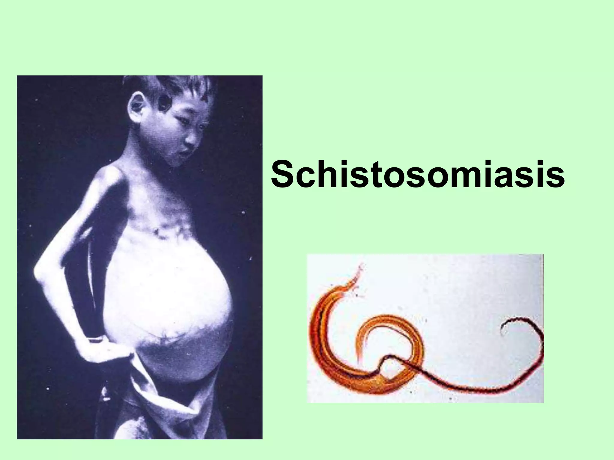
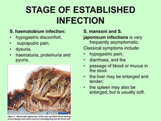
Schistosomiasis
Schistosomiasis (also known as bilharzia,
bilharziosis or snail fever) is a chronic parasitic
disease caused by several species of flatworm of the
genus Schistosoma and can cause liver, intestinal and
urinary damage.
• Schistosomiasis is known in many countries, after
Theodor Bilharz, who first described the cause of
urinary schistosomiasis in 1851.
• It is most commonly found in Asia, Africa, and
South America, especially in areas with water that
is contaminated with freshwater snails, which may
carry the parasite.
Types
There are five species of flatworms that cause
schistosomiasis.
• Schistosoma mansoni and Schistosoma
intercalatum cause intestinal schistosomiasis
• Schistosoma haematobium causes urinary
schistosomiasis
• Schistosoma japonicum and Schistosoma
mekongi cause Asian intestinal schistosomiasis.
Each causes a different clinical presentation of the
disease.
Epidemiology
• The definitive vertebrate host is man.
• The «intermediate» invertebrate host are
the snails (usually of the Biomphalaria,
Bulinus, or Oncomelania genus) that are
the natural reservoirs of the Schistosoma
pathogen.
• The mechanism of transfer is contact with
contaminated water.
• The reservoir hosts for the S. japonicum
are cattle, water buffalo, pigs, dogs, and
rodents serve as major reservoirs;
• For the S. mansoni are possible but less
important non-human primates and some
rodents can sometimes act as reservoirs.
• Adult worms live for 3 to 7 years on
average (in an endemic area up to 30
years previously), during which time the
female worm produces eggs at the rate of
300 a day (S. mansoni) to 3000 a day (S.
japonicum).
Schistosoma mansoni - is cause of
intestinal schistosomiasis
Morphology
• The male is 10 to 12 mm long, and has
gynaecophoric canal or schist in which
may be tubercules or spikes.
• The shorter and flatter male envelopes
the female in schist.
• The female is to 15 mm long. In a short
uterus usually is on one egg.
• The eggs are ellipsoidal with a lateral
spine.
• The size of eggs is 0,14 – 0,16 mm х
0,06 – 0,07 mm.
Geographical distribution
• Schistosoma mansoni is found in parts of South
America and the Caribbean, Africa, and the
Middle East;
Schistosoma haematobium causes
urinary schistosomiasis
Morphology
• The male is 10 to 15 mm long, and has
gynaecophoric canal or schist in which
may be tubercules or spikes. There are
many papillars on the helminthes body.
• The female is more longer (to 20 mm)
and thin than male worm. The uterus
contains from 20 to 30 eggs.
• The eggs are large, colorless, fusiform
and have a terminal spine.
• The size of eggs is 0,12 – 0,19 mm х
0,05 – 0,073 mm.
Geographical distribution
• S. haematobium is found in Africa and the Middle East;
Schistosoma japonicum causes
intestinal schistosomiasis
Morphology
• The male is 9,5 to 20 mm long, its
schist is without papillars.
• The female is 15 to 20 mm long.
The uterus contains many eggs.
• The eggs are more spherical form
and have small knob nearly at one
of poles. The size of eggs is 0,075 –
0,106 mm х 0,055 – 0,08 mm.
Geographical distribution
• S. japonicum is found in the Far East
Life cycle
The life cycles of all human
schistosomes are broadly similar:
• parasite eggs are released into the
environment from infected individuals,
hatching on contact with fresh water to
release the free-swimming miracidium.
• Miracidia infect fresh-water snails by
penetrating the snail's foot. After
infection, close to the site of penetration,
the miracidium transforms at first into a
primary (mother) sporocyst and then
into secondary (daughter) sporocysts,
which migrate to the snail's
hepatopancreas.
• Once at the hepatopancreas the
secondary sporocyst begin to divide
again and producing thousands of new
parasites, known as cercariae, which are
the larvae capable of infecting
mammals.
Life cycle
Life cycle
• During 2 days after penetration
schistosomulum may remain in
the skin before locating a post-
capillary venule;
• Then it travels to the lungs where
it changes to next form and
migrates to the liver sinusoids (8-
10 days after penetration).
• Juvenile S. mansoni and S.
japonicum worms develop an oral
sucker after arriving at the liver,
and it begins to feed on red blood
cells.
• Worm pairs of S. mansoni and S.
japonicum relocate to the
mesenteric or rectal veins. S.
haematobium schistosomula
migrate from the liver to the
perivesical venous plexus of the
bladder, ureters, and kidneys
through the hemorrhoidal plexus.
• Parasites reach maturity in six to
eight weeks, at which time they
begin to produce eggs.
• Many of the eggs pass through the
walls of the blood vessels, and
through the intestinal wall, to be
passed out of the body in faeces.
S. haematobium eggs pass
through the ureteral or bladder wall
and into the urine.
• Up to half the eggs released by the
worm pairs become trapped in the
mesenteric veins, or will be
washed back into the liver, where
they will become lodged. Worm
pairs can live in the body for an
average of four and a half years,
but may persist up to 20 years.
Clinical features
• Above all, schistosomiasis is a chronic disease. Many infections are
subclinically symptomatic, with mild anemia and malnutrition being
common in endemic areas. Acute schistosomiasis (Katayama's
fever) may occur weeks after the initial infection, especially by S.
mansoni and S. japonicum. Manifestations include:
• Abdominal pain
• Cough
• Diarrhea
• Eosinophilia - extremely high eosinophil granulocyte count.
• Fever
• Fatigue
• Hepatosplenomegaly - the enlargement of both the liver and the
spleen.
Clinical features
There are 4 stages
1. STAGE OF INVASION: CERCARIAL
DERMATITIS
2. STAGE OF MATURATION: ACUTE
SCHISTOSOMIASIS
3. STAGE OF ESTABLISHED INFECTION
4. STAGE OF LATE INFECTION AND
SEQUELAE
STAGE OF INVASION: CERCARIAL
DERMATITIS
• Penetration of the skin by cercariae, leads to
occurrence of dermatitis (cercarial dermatitis
or «swimmer's itch») and can be associated
the death of cercariae in the skin.
• A transient immediate hypersensitivity
reaction that occurs 10 to 15 min after
exposure is followed by a more prolonged,
delayed reaction, which develops after 12 to
24 h and may persist for up to 15 days.
• The lesion is characterized by a small, red,
pruritic, macular rash, which progresses to
papules, possibly accompanied by vesicle
formation and oedema. Pustules may form if
secondary infection occurs, and residual
pigmentation may persist for months.
• The next symptoms develops a few days
after exposure and can be associated with
transient fever, cough and pulmonary
infiltrates, as well as myalgia and abdominal
pain.
STAGE OF MATURATION: ACUTE
SCHISTOSOMIASIS
• fever,
• rigors,
• sweating,
• headache,
• malaise,
• muscular aches,
• profound weakness,
• an unproductive,
• irritating cough,
• abdominal pain or swelling,
• nausea, vomiting,
• diarrhoea, and loss of weight.
• pyrexia (intermittent or remittent with
evening peaks),
• oedema,
• a generalized soft lymphadenopathy,
• a tender enlarged liver, enlarged and soft
spleen, stuporose, or show visual
impairment or papilloedema.
• Severe central-nervous manifestations
• Eggs become detectable in the faeces
about 6 weeks after exposure.
STAGE OF ESTABLISHED
INFECTION
S. haematobium infection:
• hypogastric discomfort,
• suprapubic pain,
• dysuria,
• haematuria, proteinuria and
pyuria.
S. mansoni and S.
japonicum infections is very
frequently asymptomatic.
Classical symptoms include:
• hypogastric pain,
• diarrhoea, and the
• passage of blood or mucus in
the stool.
• the liver may be enlarged and
tender;
• the spleen may also be
enlarged, but is usually soft.
STAGE OF LATE INFECTION AND
SEQUELAE
Urinary schistosomiasis,
in the bladder:
• calcification,
• ulceration,
• papillomas,
• nocturia,
• precipitancy,
• retention of urine,
• dribbling,
• severe pain.
• The ureteric pathology is usually
asymptomatic, but may lead to
ureteric colic.
• uraemia.
S. mansoni and S. japonicum
infections, is associated with:
• intermittent diarrhoea, with or
without the passage of blood or
mucus;
• the colon may be tender.
• In between the episodes of
diarrhoea, the stools are normal.
• bleeding from oesophageal
varices
• haematemesis,
• melaena.
• Blood loss is frequently
massive, and exsanguination is
the usual cause of death rather
than hepatic coma.
Laboratory diagnosis
• Microscopic exam:
detection the eggs in the
feces or urine.
• Pelvic x-ray.
• Photomicrography of
bladder in S.
hematobium infection,
showing clusters of the
parasite eggs with
intense eosinophilia
Laboratory diagnosis
• Tissue biopsy (rectal
biopsy and biopsy of
the bladder) may
demonstrate eggs
when stool or urine
examinations are
negative.
• Serology test –
Antibody detection.
Treatment
• Praziquantel - is safe and highly
effective in curing an infected
patient, it does not prevent re-
infection by cercariae and is thus
not an optimum treatment for
people living in endemic areas.
Praziquantel is universally used.
• Antimony has been used in the
past to treat the disease. In low
doses, this toxic metalloid bonds to
sulfur atoms in enzymes used by
the parasite and kills it without
harming the host.
• Oxamniquine has been used for
treating Schistosoma mansoni.
• Niridazole, is active against all
species of parasite.
• Metrifonate is an inexpensive
organophosphorus compound
that is active only against S.
haematobium and that can be
given orally(usually 7.5 mg/kg
body weight, once every 2
weeks on up to three
occasions each year).
Preventation
• Individual protection against infection is
achieved by avoiding contact with all
potentially contaminated fresh water in
endemic areas; for example, by wearing
boots and other waterproof clothing.
• Such water should be boiled before
drinking, or left to stand for at least 2 days
before use for other purposes such as
washing. Should contact with water occur,
the skin should be immediately rubbed
vigorously with a towel; the water should
not be allowed to evaporate, as this aids
cercarial penetration.
• Soap and alcohol also kill cercariae, and
cercaricidal barrier creams can be used.
Leishmaniasis is a parasitic disease caused by the
protozoa belonging to the genus, Leishmania .
Human leishmaniasis is not a disease, but a group of diseases.
While several ways to classify leishmaniasis (eg, by
geography or taxonomy) are available, clinically, it can present
itself in various ways, and is more easily classified as
cutaneous, mucocutaneous, and visceral leishmaniasis.
Classification
Eukaryota (organisms
with nucleated cells),
• Kingdom Protista,
• Phylum Protozoa,
• Class Flagellates,
• Genus Leishmania.
Species, Reservoirs, and Clinical Diseases
Clinical Disease Leishmaniasis Species (Possible
reservoir)
Geographic Location
Cutaneous leishmaniasis
L. tropica complex
L. tropica (dog)
L. aethiopica (rock hyrax)
L. major (gerbils & rodents)
Old World
L. mexicana complex
L. mexicana (woodrats, cat, and
others)
L. pifanoi
L. amazonensis (small forest
mammals, rodents, marsupials, and
foxes)
L. garnhami
L. venezuelensis
New World
L. braziliensis complex
L. peruviana (domestic dog and
probably a wild rodent)
L. guyanensis (arboreal sloths and
anteaters)
L. panamensis (sloths, rodents,
monkeys, procyonids)
L. lainsoni (agouti)
L. colombiensis (sloth)
New World
Cutaneous leishmaniasis L. infantum Old World
L. chagasi New World
Mucocutaneous
leishmaniasis
L. braziliensis complex
L. braziliensis
L. guyanensis
L. panamensis
New World
L. mexicana New World
L. tropica Old World
L. major Old World
Species, Reservoirs, and Clinical Diseases
Visceral leishmaniasis L. donovani complex
L. donovani (no
reservoir in Indian or
Kenyan area, various
rodents in Sudan , dogs
in China )
L. infantum (human is
accidental host, natural
infection in dogs, other
Canidae, and
porcupines)
Old World
Old World
L. chagasi (domestic
dogs and cats, foxes)
New World
L. tropica Old World
L. amazonensis New World
• During blood meal, infected sandflies inject the infective stage,
the so-called promastigote parasite, into the human host.
• Injected promastigotes are first phagocytized by macrophages
and transform into so-called amastigote parasites.
• These multiply in the infected cells and also affect different
tissues, depending on the Leishmania species, which causes
the corresponding clinical manifestation of the disease.
• When sandflies sandflies take blood meals from an
infected host, they take up parasitized macrophages.
• In the vector fly's midgut, these parasites differentiate
into the so-called promastigote form, which multiplies
and finally migrates to the fly's proboscis.
Clinical forms
VISCERAL
MUCOCUTANEOUS
CUTANEOUS
Visceral Leishmaniasis
(Kala-azar)
Etiology
The L. donovani species
complex includes
several species:
 L. infantum and
 L. chagasi
Epidemiology
• The species of visceral
leishmaniasis are endemic in
areas of India, China, Central
and South America, East and
West Africa, and the countries
surrounding the Mediterranean.
• In India, no extrahuman
reservoirs are known, but in
other regions, infection may
involve several mammalian
species, including dogs, foxes,
and wild rodents.
• Sandflies of the genus
Phlebotomus are the insect
vectors that spread L.
donovani.
Pathogenesis
• The flagellated promastigotes of L. donovani are
introduced by an insect bite.
• After entering macrophages of the
reticuloendothelial system, these forms change
into amastigotes, which multiply in phagocytic
cells.
• Released amastigotes disseminate
hematogenously and invade reticuloendothelial
cells in the spleen, liver, lymph nodes, bone
marrow, and skin.
Incubation and Clinical Symptoms
• Incubation period is 6-8 months.
Symptoms:
• weakness, dizziness, weight
loss, diarrhea, and
constipation.
• Fever, may spike twice daily;
• chills and sweating.
• hepatosplenomegaly
• anemia and leukopenia.
• bleeding from the gingivae,
nose, or GI tract,
• ecchymoses and petechiae on
the skin.
Cutaneous and Mucocutaneous Leishmaniasis
Etiology and Epidemiology
Old World cutaneous leishmaniasis is
caused by three species of Leishmania
that belong to the L. tropica complex:
 L. tropica is present in the Middle East
and the Mediterranean littoral;
 L. major is found in the Middle East,
Arabia, India, and sub-Saharan Africa;
 L. aethiopica is found principally in
Ethiopia and Kenya.
Phlebotomus sandflies are the
principal vectors.
• Infections that are caused by
Leishmania can be acquired by
travelers, as well as by military and other
personnel residing in endemic areas.
• Military personnel in the Middle East
have acquired cutaneous leishmaniasis
with L. major and viscerotropic
infections with L. tropica.
Cutaneous and Mucocutaneous Leishmaniasis
• New World cutaneous leishmaniasis arises from infection with
parasites belonging to the L. mexicana group or the L.
braziliensis (Viannia subgenus) group.
• The patterns of illness vary with the nature of the infecting
leishmanial organisms, which are found in different regions of
North, Central, and South America.
• Infections with strains of L. viannia, which are endemic in
various areas of South America, cause cutaneous
leishmaniasis and, in a small percentage of those infected,
result in the later development of mucocutaneous
leishmaniasis. Such mucocutaneous disease (espundia)
involves the nasal or oropharyngeal mucosa, or both, and may
prove fatal.
• All of these New World leishmanial parasites are transmitted
principally by sandfly vectors, although direct human contact
may also bring about infection.
• Various mammals are naturally infected reservoirs of the
organisms.
Pathogenesis
• Both Old World and New World forms of
leishmaniasis are initiated when the bite of an
infected sandfly injects promastigotes into the
human host.
• The organisms enter tissue macrophages and
capillary endothelial cells, become amastigotes,
and multiply.
• A granulomatous inflammatory response
develops at the bite site.
• With local ischemia, the lesion ulcerates; a
bacterial infection of the necrotic area may
extend the ulceration.
Incubation and Clinical Symptoms
Incubation period is from 2-8
months to 1,5 years and more.
In Old World symptoms of
cutaneous leishmaniasis:
• a papule (at the inoculation
site).
• papule ulcerates and a shallow
circular lesion appears that is
several centimeters in diameter
and has a raised margin.
• lymphadenopathy.
• Healing of the lesions is slow,
sometimes requiring more than
a year.
Clinical Symptoms of New World
leishmaniasis
L. mexicana
• single lesion or a few lesions on
exposed surfaces of the body such
as the face and ear, which heals
spontaneously over 6 months.
• extensive destruction of the pinna.
L. viannia
• lesions on the skin or mucous
membranes.
• progressive ulcerations of
lymphatic nodes and mucous
membranes.
• the infection metastasizes to
the nasal or oral mucosa.
• Metastatic lesions can erode
the nasal septum or the hard
palate or soft palate.
• Some patients die of
malnutrition or bacterial
infection.
Incubation period is 2-3 weeks to 1-3 mounths.
Immunity
• In visceral leishmaniasis (Kala-Azar) cellular
immunity is responsible for resolving mild
disease. High levels of antibodies are found.
• In cutaneous and mucocutaneous leishmaniasis
host defense relies on cell-mediated immunity;
antibody titers are low. The response ranges
from a local granuloma with few parasites to a
histiocytoma with many parasites.
Laboratory Diagnostics of visceral
leishmaniasis
• Demonstration of the organism in host tissues cultured
on a Novy-MacNeal-Nicolle (NNN) or other medium or
detection of Leishman-Donovan bodies (amastigotes) in
stained tissue samples.
• PCR can be performed using genus- or species-specific
oligonucleotides.
• Established by examining bone marrow aspirates.
• Splenic aspirates have the highest yields but may be
risky.
• Liver biopsy or aspiration of enlarged lymph nodes can
also provide diagnostic material.
Laboratory Diagnostics of cutaneous and
mucocutaneous leishmaniasis
• Demonstrating amastigotes on
stained smears of a biopsy or of
scrapings from the border of an
ulcer.
• Culturing amastigotes on NNN
medium inoculated with lesion
material.
• PCR targeting parasite kinetoplast
DNA has allowed detection of
organisms that might be missed
on histologic section or culturing.
Laboratory Diagnostics of cutaneous and
mucocutaneous leishmaniasis
• Except in diffuse cutaneous
leishmaniasis, the
leishmanin skin test is
usually positive.
Treatment
• There are two common therapies containing antimony,
meglumine antimoniate (Glucantim®) and sodium
stibogluconate (Pentostam®). Unfortunately, in many
parts of the world, the parasite has become resistant to
antimony and for visceral or mucocutaneous
leishmaniasis, amphotericin is now the treatment of
choice.
• Miltefosine (Impavido®), is a new drug for visceral,
mucocutaneous and cutaneous leishmaniasis.
• Drug-resistant leishmaniasis may respond to
immunotherapy (inoculation with parasite antigens plus
an adjuvant) which aims to stimulate the body's own
immune system to kill the parasite.
Prevention:
• Preventing sandfly bites is the
most immediate form of
protection. Insect repellent,
appropriate clothing, screening
of windows, and fine mesh
netting around the bed (in
endemic areas) will reduce
exposure.
• Public health measures to
reduce the sandfly population
and animal reservoirs are
important. There are no
preventive vaccines or drugs for
leishmaniasis.

Schistosomiasisleishmaniasis.ppt

  • 1.
  • 2.
    Schistosomiasis (also knownas bilharzia, bilharziosis or snail fever) is a chronic parasitic disease caused by several species of flatworm of the genus Schistosoma and can cause liver, intestinal and urinary damage. • Schistosomiasis is known in many countries, after Theodor Bilharz, who first described the cause of urinary schistosomiasis in 1851. • It is most commonly found in Asia, Africa, and South America, especially in areas with water that is contaminated with freshwater snails, which may carry the parasite.
  • 3.
    Types There are fivespecies of flatworms that cause schistosomiasis. • Schistosoma mansoni and Schistosoma intercalatum cause intestinal schistosomiasis • Schistosoma haematobium causes urinary schistosomiasis • Schistosoma japonicum and Schistosoma mekongi cause Asian intestinal schistosomiasis. Each causes a different clinical presentation of the disease.
  • 4.
    Epidemiology • The definitivevertebrate host is man. • The «intermediate» invertebrate host are the snails (usually of the Biomphalaria, Bulinus, or Oncomelania genus) that are the natural reservoirs of the Schistosoma pathogen. • The mechanism of transfer is contact with contaminated water. • The reservoir hosts for the S. japonicum are cattle, water buffalo, pigs, dogs, and rodents serve as major reservoirs; • For the S. mansoni are possible but less important non-human primates and some rodents can sometimes act as reservoirs. • Adult worms live for 3 to 7 years on average (in an endemic area up to 30 years previously), during which time the female worm produces eggs at the rate of 300 a day (S. mansoni) to 3000 a day (S. japonicum).
  • 5.
    Schistosoma mansoni -is cause of intestinal schistosomiasis Morphology • The male is 10 to 12 mm long, and has gynaecophoric canal or schist in which may be tubercules or spikes. • The shorter and flatter male envelopes the female in schist. • The female is to 15 mm long. In a short uterus usually is on one egg. • The eggs are ellipsoidal with a lateral spine. • The size of eggs is 0,14 – 0,16 mm х 0,06 – 0,07 mm.
  • 6.
    Geographical distribution • Schistosomamansoni is found in parts of South America and the Caribbean, Africa, and the Middle East;
  • 7.
    Schistosoma haematobium causes urinaryschistosomiasis Morphology • The male is 10 to 15 mm long, and has gynaecophoric canal or schist in which may be tubercules or spikes. There are many papillars on the helminthes body. • The female is more longer (to 20 mm) and thin than male worm. The uterus contains from 20 to 30 eggs. • The eggs are large, colorless, fusiform and have a terminal spine. • The size of eggs is 0,12 – 0,19 mm х 0,05 – 0,073 mm.
  • 8.
    Geographical distribution • S.haematobium is found in Africa and the Middle East;
  • 9.
    Schistosoma japonicum causes intestinalschistosomiasis Morphology • The male is 9,5 to 20 mm long, its schist is without papillars. • The female is 15 to 20 mm long. The uterus contains many eggs. • The eggs are more spherical form and have small knob nearly at one of poles. The size of eggs is 0,075 – 0,106 mm х 0,055 – 0,08 mm.
  • 10.
    Geographical distribution • S.japonicum is found in the Far East
  • 11.
    Life cycle The lifecycles of all human schistosomes are broadly similar: • parasite eggs are released into the environment from infected individuals, hatching on contact with fresh water to release the free-swimming miracidium. • Miracidia infect fresh-water snails by penetrating the snail's foot. After infection, close to the site of penetration, the miracidium transforms at first into a primary (mother) sporocyst and then into secondary (daughter) sporocysts, which migrate to the snail's hepatopancreas. • Once at the hepatopancreas the secondary sporocyst begin to divide again and producing thousands of new parasites, known as cercariae, which are the larvae capable of infecting mammals.
  • 12.
  • 13.
    Life cycle • During2 days after penetration schistosomulum may remain in the skin before locating a post- capillary venule; • Then it travels to the lungs where it changes to next form and migrates to the liver sinusoids (8- 10 days after penetration). • Juvenile S. mansoni and S. japonicum worms develop an oral sucker after arriving at the liver, and it begins to feed on red blood cells. • Worm pairs of S. mansoni and S. japonicum relocate to the mesenteric or rectal veins. S. haematobium schistosomula migrate from the liver to the perivesical venous plexus of the bladder, ureters, and kidneys through the hemorrhoidal plexus. • Parasites reach maturity in six to eight weeks, at which time they begin to produce eggs. • Many of the eggs pass through the walls of the blood vessels, and through the intestinal wall, to be passed out of the body in faeces. S. haematobium eggs pass through the ureteral or bladder wall and into the urine. • Up to half the eggs released by the worm pairs become trapped in the mesenteric veins, or will be washed back into the liver, where they will become lodged. Worm pairs can live in the body for an average of four and a half years, but may persist up to 20 years.
  • 14.
    Clinical features • Aboveall, schistosomiasis is a chronic disease. Many infections are subclinically symptomatic, with mild anemia and malnutrition being common in endemic areas. Acute schistosomiasis (Katayama's fever) may occur weeks after the initial infection, especially by S. mansoni and S. japonicum. Manifestations include: • Abdominal pain • Cough • Diarrhea • Eosinophilia - extremely high eosinophil granulocyte count. • Fever • Fatigue • Hepatosplenomegaly - the enlargement of both the liver and the spleen.
  • 15.
    Clinical features There are4 stages 1. STAGE OF INVASION: CERCARIAL DERMATITIS 2. STAGE OF MATURATION: ACUTE SCHISTOSOMIASIS 3. STAGE OF ESTABLISHED INFECTION 4. STAGE OF LATE INFECTION AND SEQUELAE
  • 16.
    STAGE OF INVASION:CERCARIAL DERMATITIS • Penetration of the skin by cercariae, leads to occurrence of dermatitis (cercarial dermatitis or «swimmer's itch») and can be associated the death of cercariae in the skin. • A transient immediate hypersensitivity reaction that occurs 10 to 15 min after exposure is followed by a more prolonged, delayed reaction, which develops after 12 to 24 h and may persist for up to 15 days. • The lesion is characterized by a small, red, pruritic, macular rash, which progresses to papules, possibly accompanied by vesicle formation and oedema. Pustules may form if secondary infection occurs, and residual pigmentation may persist for months. • The next symptoms develops a few days after exposure and can be associated with transient fever, cough and pulmonary infiltrates, as well as myalgia and abdominal pain.
  • 17.
    STAGE OF MATURATION:ACUTE SCHISTOSOMIASIS • fever, • rigors, • sweating, • headache, • malaise, • muscular aches, • profound weakness, • an unproductive, • irritating cough, • abdominal pain or swelling, • nausea, vomiting, • diarrhoea, and loss of weight. • pyrexia (intermittent or remittent with evening peaks), • oedema, • a generalized soft lymphadenopathy, • a tender enlarged liver, enlarged and soft spleen, stuporose, or show visual impairment or papilloedema. • Severe central-nervous manifestations • Eggs become detectable in the faeces about 6 weeks after exposure.
  • 19.
    STAGE OF ESTABLISHED INFECTION S.haematobium infection: • hypogastric discomfort, • suprapubic pain, • dysuria, • haematuria, proteinuria and pyuria. S. mansoni and S. japonicum infections is very frequently asymptomatic. Classical symptoms include: • hypogastric pain, • diarrhoea, and the • passage of blood or mucus in the stool. • the liver may be enlarged and tender; • the spleen may also be enlarged, but is usually soft.
  • 20.
    STAGE OF LATEINFECTION AND SEQUELAE Urinary schistosomiasis, in the bladder: • calcification, • ulceration, • papillomas, • nocturia, • precipitancy, • retention of urine, • dribbling, • severe pain. • The ureteric pathology is usually asymptomatic, but may lead to ureteric colic. • uraemia. S. mansoni and S. japonicum infections, is associated with: • intermittent diarrhoea, with or without the passage of blood or mucus; • the colon may be tender. • In between the episodes of diarrhoea, the stools are normal. • bleeding from oesophageal varices • haematemesis, • melaena. • Blood loss is frequently massive, and exsanguination is the usual cause of death rather than hepatic coma.
  • 21.
    Laboratory diagnosis • Microscopicexam: detection the eggs in the feces or urine. • Pelvic x-ray. • Photomicrography of bladder in S. hematobium infection, showing clusters of the parasite eggs with intense eosinophilia
  • 22.
    Laboratory diagnosis • Tissuebiopsy (rectal biopsy and biopsy of the bladder) may demonstrate eggs when stool or urine examinations are negative. • Serology test – Antibody detection.
  • 23.
    Treatment • Praziquantel -is safe and highly effective in curing an infected patient, it does not prevent re- infection by cercariae and is thus not an optimum treatment for people living in endemic areas. Praziquantel is universally used. • Antimony has been used in the past to treat the disease. In low doses, this toxic metalloid bonds to sulfur atoms in enzymes used by the parasite and kills it without harming the host. • Oxamniquine has been used for treating Schistosoma mansoni. • Niridazole, is active against all species of parasite. • Metrifonate is an inexpensive organophosphorus compound that is active only against S. haematobium and that can be given orally(usually 7.5 mg/kg body weight, once every 2 weeks on up to three occasions each year).
  • 24.
    Preventation • Individual protectionagainst infection is achieved by avoiding contact with all potentially contaminated fresh water in endemic areas; for example, by wearing boots and other waterproof clothing. • Such water should be boiled before drinking, or left to stand for at least 2 days before use for other purposes such as washing. Should contact with water occur, the skin should be immediately rubbed vigorously with a towel; the water should not be allowed to evaporate, as this aids cercarial penetration. • Soap and alcohol also kill cercariae, and cercaricidal barrier creams can be used.
  • 26.
    Leishmaniasis is aparasitic disease caused by the protozoa belonging to the genus, Leishmania . Human leishmaniasis is not a disease, but a group of diseases. While several ways to classify leishmaniasis (eg, by geography or taxonomy) are available, clinically, it can present itself in various ways, and is more easily classified as cutaneous, mucocutaneous, and visceral leishmaniasis.
  • 27.
    Classification Eukaryota (organisms with nucleatedcells), • Kingdom Protista, • Phylum Protozoa, • Class Flagellates, • Genus Leishmania.
  • 28.
    Species, Reservoirs, andClinical Diseases Clinical Disease Leishmaniasis Species (Possible reservoir) Geographic Location Cutaneous leishmaniasis L. tropica complex L. tropica (dog) L. aethiopica (rock hyrax) L. major (gerbils & rodents) Old World L. mexicana complex L. mexicana (woodrats, cat, and others) L. pifanoi L. amazonensis (small forest mammals, rodents, marsupials, and foxes) L. garnhami L. venezuelensis New World L. braziliensis complex L. peruviana (domestic dog and probably a wild rodent) L. guyanensis (arboreal sloths and anteaters) L. panamensis (sloths, rodents, monkeys, procyonids) L. lainsoni (agouti) L. colombiensis (sloth) New World
  • 29.
    Cutaneous leishmaniasis L.infantum Old World L. chagasi New World Mucocutaneous leishmaniasis L. braziliensis complex L. braziliensis L. guyanensis L. panamensis New World L. mexicana New World L. tropica Old World L. major Old World Species, Reservoirs, and Clinical Diseases
  • 30.
    Species, Reservoirs, andClinical Diseases Visceral leishmaniasis L. donovani complex L. donovani (no reservoir in Indian or Kenyan area, various rodents in Sudan , dogs in China ) L. infantum (human is accidental host, natural infection in dogs, other Canidae, and porcupines) Old World Old World L. chagasi (domestic dogs and cats, foxes) New World L. tropica Old World L. amazonensis New World
  • 31.
    • During bloodmeal, infected sandflies inject the infective stage, the so-called promastigote parasite, into the human host. • Injected promastigotes are first phagocytized by macrophages and transform into so-called amastigote parasites. • These multiply in the infected cells and also affect different tissues, depending on the Leishmania species, which causes the corresponding clinical manifestation of the disease.
  • 32.
    • When sandfliessandflies take blood meals from an infected host, they take up parasitized macrophages. • In the vector fly's midgut, these parasites differentiate into the so-called promastigote form, which multiplies and finally migrates to the fly's proboscis.
  • 33.
  • 34.
    Visceral Leishmaniasis (Kala-azar) Etiology The L.donovani species complex includes several species:  L. infantum and  L. chagasi
  • 35.
    Epidemiology • The speciesof visceral leishmaniasis are endemic in areas of India, China, Central and South America, East and West Africa, and the countries surrounding the Mediterranean. • In India, no extrahuman reservoirs are known, but in other regions, infection may involve several mammalian species, including dogs, foxes, and wild rodents. • Sandflies of the genus Phlebotomus are the insect vectors that spread L. donovani.
  • 36.
    Pathogenesis • The flagellatedpromastigotes of L. donovani are introduced by an insect bite. • After entering macrophages of the reticuloendothelial system, these forms change into amastigotes, which multiply in phagocytic cells. • Released amastigotes disseminate hematogenously and invade reticuloendothelial cells in the spleen, liver, lymph nodes, bone marrow, and skin.
  • 37.
    Incubation and ClinicalSymptoms • Incubation period is 6-8 months. Symptoms: • weakness, dizziness, weight loss, diarrhea, and constipation. • Fever, may spike twice daily; • chills and sweating. • hepatosplenomegaly • anemia and leukopenia. • bleeding from the gingivae, nose, or GI tract, • ecchymoses and petechiae on the skin.
  • 39.
    Cutaneous and MucocutaneousLeishmaniasis Etiology and Epidemiology Old World cutaneous leishmaniasis is caused by three species of Leishmania that belong to the L. tropica complex:  L. tropica is present in the Middle East and the Mediterranean littoral;  L. major is found in the Middle East, Arabia, India, and sub-Saharan Africa;  L. aethiopica is found principally in Ethiopia and Kenya. Phlebotomus sandflies are the principal vectors. • Infections that are caused by Leishmania can be acquired by travelers, as well as by military and other personnel residing in endemic areas. • Military personnel in the Middle East have acquired cutaneous leishmaniasis with L. major and viscerotropic infections with L. tropica.
  • 40.
    Cutaneous and MucocutaneousLeishmaniasis • New World cutaneous leishmaniasis arises from infection with parasites belonging to the L. mexicana group or the L. braziliensis (Viannia subgenus) group. • The patterns of illness vary with the nature of the infecting leishmanial organisms, which are found in different regions of North, Central, and South America. • Infections with strains of L. viannia, which are endemic in various areas of South America, cause cutaneous leishmaniasis and, in a small percentage of those infected, result in the later development of mucocutaneous leishmaniasis. Such mucocutaneous disease (espundia) involves the nasal or oropharyngeal mucosa, or both, and may prove fatal. • All of these New World leishmanial parasites are transmitted principally by sandfly vectors, although direct human contact may also bring about infection. • Various mammals are naturally infected reservoirs of the organisms.
  • 41.
    Pathogenesis • Both OldWorld and New World forms of leishmaniasis are initiated when the bite of an infected sandfly injects promastigotes into the human host. • The organisms enter tissue macrophages and capillary endothelial cells, become amastigotes, and multiply. • A granulomatous inflammatory response develops at the bite site. • With local ischemia, the lesion ulcerates; a bacterial infection of the necrotic area may extend the ulceration.
  • 42.
    Incubation and ClinicalSymptoms Incubation period is from 2-8 months to 1,5 years and more. In Old World symptoms of cutaneous leishmaniasis: • a papule (at the inoculation site). • papule ulcerates and a shallow circular lesion appears that is several centimeters in diameter and has a raised margin. • lymphadenopathy. • Healing of the lesions is slow, sometimes requiring more than a year.
  • 43.
    Clinical Symptoms ofNew World leishmaniasis L. mexicana • single lesion or a few lesions on exposed surfaces of the body such as the face and ear, which heals spontaneously over 6 months. • extensive destruction of the pinna. L. viannia • lesions on the skin or mucous membranes. • progressive ulcerations of lymphatic nodes and mucous membranes. • the infection metastasizes to the nasal or oral mucosa. • Metastatic lesions can erode the nasal septum or the hard palate or soft palate. • Some patients die of malnutrition or bacterial infection. Incubation period is 2-3 weeks to 1-3 mounths.
  • 46.
    Immunity • In visceralleishmaniasis (Kala-Azar) cellular immunity is responsible for resolving mild disease. High levels of antibodies are found. • In cutaneous and mucocutaneous leishmaniasis host defense relies on cell-mediated immunity; antibody titers are low. The response ranges from a local granuloma with few parasites to a histiocytoma with many parasites.
  • 47.
    Laboratory Diagnostics ofvisceral leishmaniasis • Demonstration of the organism in host tissues cultured on a Novy-MacNeal-Nicolle (NNN) or other medium or detection of Leishman-Donovan bodies (amastigotes) in stained tissue samples. • PCR can be performed using genus- or species-specific oligonucleotides. • Established by examining bone marrow aspirates. • Splenic aspirates have the highest yields but may be risky. • Liver biopsy or aspiration of enlarged lymph nodes can also provide diagnostic material.
  • 48.
    Laboratory Diagnostics ofcutaneous and mucocutaneous leishmaniasis • Demonstrating amastigotes on stained smears of a biopsy or of scrapings from the border of an ulcer. • Culturing amastigotes on NNN medium inoculated with lesion material. • PCR targeting parasite kinetoplast DNA has allowed detection of organisms that might be missed on histologic section or culturing.
  • 49.
    Laboratory Diagnostics ofcutaneous and mucocutaneous leishmaniasis • Except in diffuse cutaneous leishmaniasis, the leishmanin skin test is usually positive.
  • 50.
    Treatment • There aretwo common therapies containing antimony, meglumine antimoniate (Glucantim®) and sodium stibogluconate (Pentostam®). Unfortunately, in many parts of the world, the parasite has become resistant to antimony and for visceral or mucocutaneous leishmaniasis, amphotericin is now the treatment of choice. • Miltefosine (Impavido®), is a new drug for visceral, mucocutaneous and cutaneous leishmaniasis. • Drug-resistant leishmaniasis may respond to immunotherapy (inoculation with parasite antigens plus an adjuvant) which aims to stimulate the body's own immune system to kill the parasite.
  • 51.
    Prevention: • Preventing sandflybites is the most immediate form of protection. Insect repellent, appropriate clothing, screening of windows, and fine mesh netting around the bed (in endemic areas) will reduce exposure. • Public health measures to reduce the sandfly population and animal reservoirs are important. There are no preventive vaccines or drugs for leishmaniasis.